

Dexamethasone disrupts intercellular junction formation and cytoskeleton organization in human trabecular meshwork cells
- PMID: 20090922
- PMCID: PMC2807616
Dexamethasone disrupts intercellular junction formation and cytoskeleton organization in human trabecular meshwork cells
Abstract
Purpose: Patients reproduce symptoms of primary open-angle glaucoma (POAG) when treated with glucocorticoids (GCs) topically on the eyes. Here we investigated the effects of GCs on junctional protein expression and cytoskeleton organization in primary human trabecular meshwork (TM) cultures to understand the molecular pathologies of POAG.
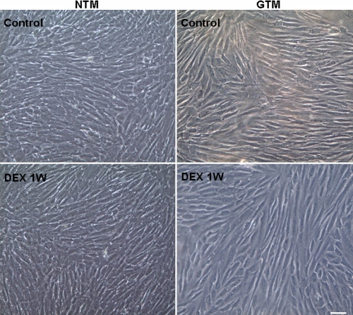
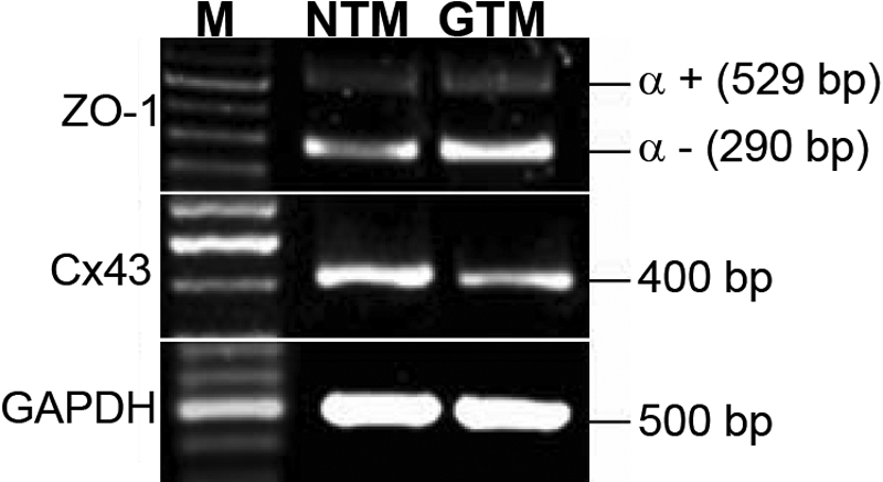
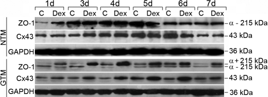
Methods: Human TM cells from POAG (GTM) and age-matched nondiseased (NTM) individuals were obtained by standard surgical trabeculectomy. Some of the cultures were treated with dexamethasone (DEX), a synthetic GC, at 1-5 x 10(-7) mol/l for 1-7 days. The expression levels of zonula occluden-1 (ZO-1) and connexin43 (Cx43) in TM cells with or without DEX treatment were measured using reverse transcription (RT)-PCR, immunocytochemistry, and western blot analysis.
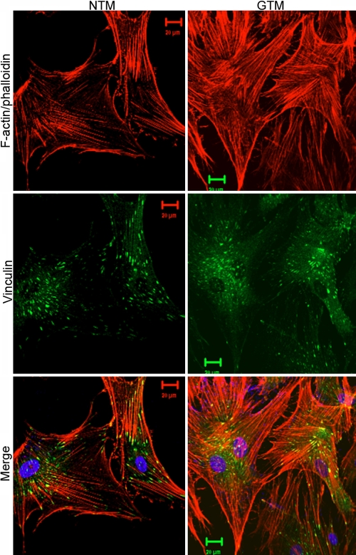
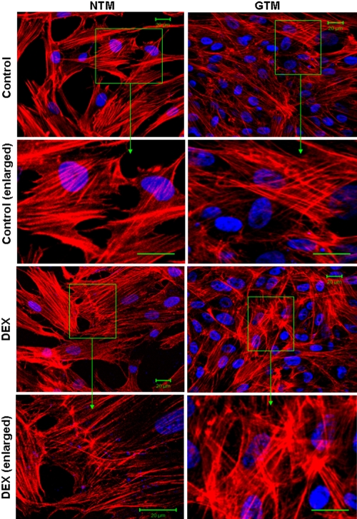
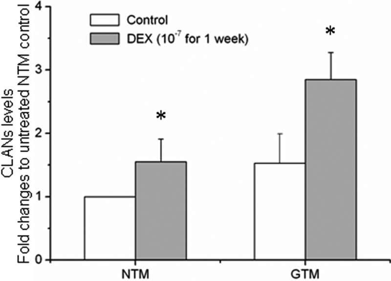
Results: mRNA and proteins of ZO-1 and Cx43 were found in both NTM and GTM cells. ZO-1 and Cx43 were located on the plasma membrane, especially along the border of adjacent cells. ZO-1 had no marked changes in localization in NTM and GTM cells after treatment with 10(-7) mol/l DEX for 48 h, whereas Cx43 appeared to increase in the cytoplasm. mRNA of two ZO-1 isoforms, alpha+ and alpha-, were present in TM cells, and the former was expressed less than the latter. Only ZO-1 alpha- isoform protein was expressed in NTM cells, whereas proteins of both isoforms were found in GTM cells. DEX increased the protein levels of ZO-1 and Cx43 in both NTM and GTM cells. DEX also altered the F-actin architecture and promoted cross-linked actin network formation, the effects of which were more pronounced in GTM cells.
Conclusions: Our findings not only provide molecular insights to the pathogenesis of GC-induced glaucoma but also suggest that junctional proteins ZO-1 and Cx43 as well as F-actin are targets for developing new modalities in glaucoma therapy.
Figures

Similar articles
-
Elevated pressure downregulates ZO-1 expression and disrupts cytoskeleton and focal adhesion in human trabecular meshwork cells.Mol Vis. 2011;17:2978-85. Epub 2011 Nov 16. Mol Vis. 2011. PMID: 22128243 Free PMC article.
-
Interactions of endothelin-1 with dexamethasone in primary cultured human trabecular meshwork cells.Invest Ophthalmol Vis Sci. 2003 Dec;44(12):5301-8. doi: 10.1167/iovs.03-0463. Invest Ophthalmol Vis Sci. 2003. PMID: 14638730
-
Down-regulation of GRP78 in human glaucomatous trabecular meshwork cells.Mol Vis. 2010 Jun 17;16:1122-31. Mol Vis. 2010. PMID: 20664690 Free PMC article.
-
Role of the alternatively spliced glucocorticoid receptor isoform GRβ in steroid responsiveness and glaucoma.J Ocul Pharmacol Ther. 2014 Mar-Apr;30(2-3):121-7. doi: 10.1089/jop.2013.0239. Epub 2014 Feb 7. J Ocul Pharmacol Ther. 2014. PMID: 24506296 Free PMC article. Review.
-
Cx43 and the Actin Cytoskeleton: Novel Roles and Implications for Cell-Cell Junction-Based Barrier Function Regulation.Biomolecules. 2020 Dec 10;10(12):1656. doi: 10.3390/biom10121656. Biomolecules. 2020. PMID: 33321985 Free PMC article. Review.
Cited by
-
Cytoskeletal dependence of adenosine triphosphate release by human trabecular meshwork cells.Invest Ophthalmol Vis Sci. 2011 Oct 10;52(11):7996-8005. doi: 10.1167/iovs.11-8170. Invest Ophthalmol Vis Sci. 2011. PMID: 21896846 Free PMC article.
-
Downregulation of secreted protein acidic and rich in cysteine in human trabecular meshwork cells.Exp Ther Med. 2021 Oct;22(4):1126. doi: 10.3892/etm.2021.10560. Epub 2021 Aug 5. Exp Ther Med. 2021. PMID: 34504578 Free PMC article.
-
Minireview: Steroid/nuclear receptor-regulated dynamics of occluding and anchoring junctions.Mol Endocrinol. 2014 Nov;28(11):1769-84. doi: 10.1210/me.2014-1037. Epub 2014 Sep 9. Mol Endocrinol. 2014. PMID: 25203673 Free PMC article. Review.
-
Glucocorticoid therapy and ocular hypertension.Eur J Pharmacol. 2016 Sep 15;787:57-71. doi: 10.1016/j.ejphar.2016.06.018. Epub 2016 Jul 5. Eur J Pharmacol. 2016. PMID: 27388141 Free PMC article. Review.
-
Effects of dexamethasone and HA1077 on actin cytoskeleton and β-catenin in cultured human trabecular meshwork cells.Int J Ophthalmol. 2016 Oct 18;9(10):1376-1380. doi: 10.18240/ijo.2016.10.02. eCollection 2016. Int J Ophthalmol. 2016. PMID: 27803851 Free PMC article.
References
-
- Sherif Z, Pleyer U. Corticosteroids in Ophthalmology: past, present and future. Ophthalmologica. 2002;216:305–15. - PubMed
-
- Underwood JL, Murphy GM, Chen J, Franse-Carman L, Wood I, Epstein DL, Alvarado JA. Glucocorticoids regulate transendothelial fluid flow resistance and formation of intercellular junctions. Am J Physiol. 1999;277:C330–42. - PubMed
-
- Jones R, 3rd, Rhee DJ. Corticosteroid-induced ocular hypertension and glaucoma: a brief review and update of the literature. Curr Opin Ophthalmol. 2006;17:163–7. - PubMed
-
- Kersey JP, Broadway DC. Corticosteroid-induced glaucoma: a review of the literature. Eye. 2006;20:407–16. - PubMed
-
- Kubota T, Okabe H, Hisatomi T, Yamakiri K, Sakamoto T, Tawara A. Ultrastructure of the trabecular meshwork in secondary glaucoma eyes after intravitreal triamcinolone acetonide. J Glaucoma. 2006;15:117–9. - PubMed
Publication types
MeSH terms
Substances
LinkOut - more resources
Full Text Sources
Miscellaneous