. 2010 Jan 15;5(1):e8732.
doi: 10.1371/journal.pone.0008732.

Hypoxia activates a Ca2+-permeable cation conductance sensitive to carbon monoxide and to GsMTx-4 in human and mouse sickle erythrocytes

Affiliations

Hypoxia activates a Ca2+-permeable cation conductance sensitive to carbon monoxide and to GsMTx-4 in human and mouse sickle erythrocytes

David H Vandorpe et al. PLoS One. .

Abstract

Background: Deoxygenation of sickle erythrocytes activates a cation permeability of unknown molecular identity (Psickle), leading to elevated intracellular [Ca(2+)] ([Ca(2+)](i)) and subsequent activation of K(Ca) 3.1. The resulting erythrocyte volume decrease elevates intracellular hemoglobin S (HbSS) concentration, accelerates deoxygenation-induced HbSS polymerization, and increases the likelihood of cell sickling. Deoxygenation-induced currents sharing some properties of Psickle have been recorded from sickle erythrocytes in whole cell configuration.

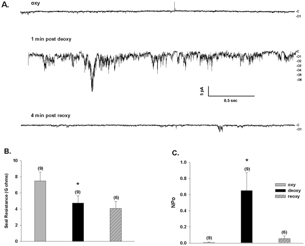
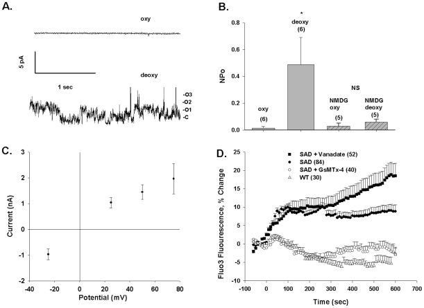
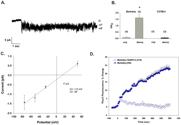
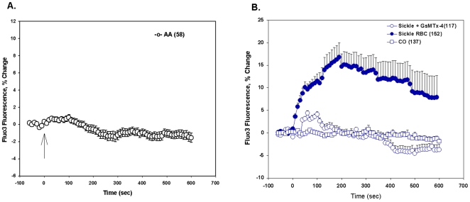
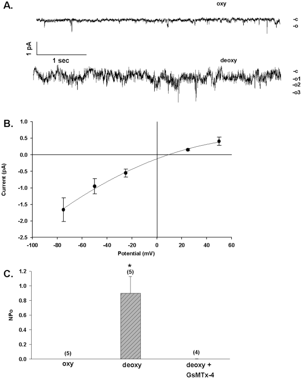
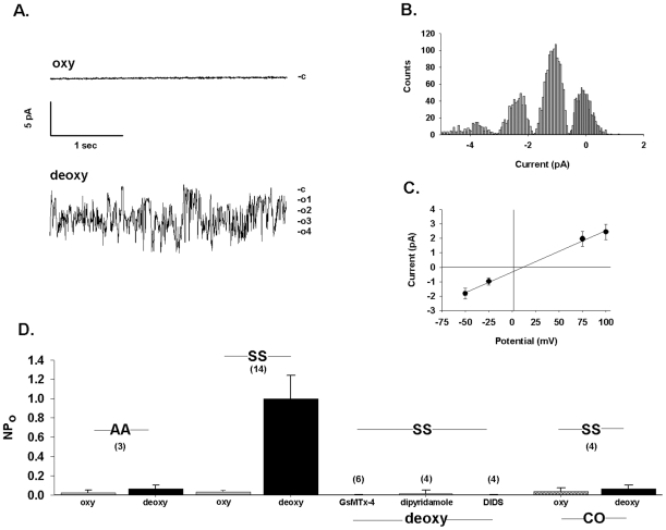
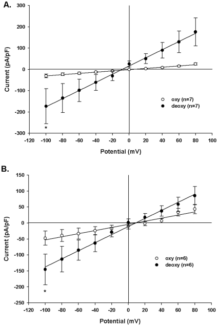
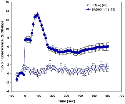
Methodology/principal findings: We now show by cell-attached and nystatin-permeabilized patch clamp recording from sickle erythrocytes of mouse and human that deoxygenation reversibly activates a Ca(2+)- and cation-permeable conductance sensitive to inhibition by Grammastola spatulata mechanotoxin-4 (GsMTx-4; 1 microM), dipyridamole (100 microM), DIDS (100 microM), and carbon monoxide (25 ppm pretreatment). Deoxygenation also elevates sickle erythrocyte [Ca(2+)](i), in a manner similarly inhibited by GsMTx-4 and by carbon monoxide. Normal human and mouse erythrocytes do not exhibit these responses to deoxygenation. Deoxygenation-induced elevation of [Ca(2+)](i) in mouse sickle erythrocytes did not require KCa3.1 activity.

Conclusions/significance: The electrophysiological and fluorimetric data provide compelling evidence in sickle erythrocytes of mouse and human for a deoxygenation-induced, reversible, Ca(2+)-permeable cation conductance blocked by inhibition of HbSS polymerization and by an inhibitor of strctch-activated cation channels. This cation permeability pathway is likely an important source of intracellular Ca(2+) for pathologic activation of KCa3.1 in sickle erythrocytes. Blockade of this pathway represents a novel therapeutic approach for treatment of sickle disease.


Conflict of interest statement

Competing Interests: The authors have declared that no competing interests exist.

Figures

Figure 1
Figure 1. Deoxygenation reversibly activates a conductance in red cells from SAD sickle mice.
A. Representative current trace from an individual cell-attached patch on a SAD sickle mouse erythrocyte before deoxygenation (upper trace, oxy), 1 min post-deoxygenation (middle trace, deoxy), and 4 min post-reoxygenation (lower trace, reoxy); −Vp  = −50 mV. Symmetric pipette and bath solutions contained (in mM) 140 NaCl, 4 KCl, 1CaCl2, 1 MgCl2, 10 Na HEPES, pH 7.4. B. Seal resistance was maintained during deoxygenation in all 9 cells, and during reoxygenation in 6 of the 9 cells (*, p<0.01 vs. oxy; p =  N.S. vs. re-oxy, ANOVA). C. Summary of patches such as in panel A, showing that the increased NPo (product of the number of single channels and the channel open probability) observed after deoxygenation was reversible (without change in seal resistance) upon reoxygenation (*, p<0.02 vs. oxy; p<0.05 vs. reoxy, ANOVA). Values are means ± s.e.m.
Figure 2
Figure 2. Deoxygenation activates conductance and increases [Ca2+]i in red cells from SAD sickle mice.
A. Representative current trace from an individual cell-attached patch on a SAD mouse erythrocyte, recorded first in oxygenated (upper trace, oxy, −Vp = −25 mV) and subsequently in deoxygenated conditions (lower trace, deoxy, −Vp = +75 mV). Symmetrical pipette and bath solutions contained (in mM) 150 Na methanesulfonate, 10 Na EDTA, and 10 Na HEPES, pH 7.4. B. Deoxygenation increased the NPo of 6 cell-attached patches recorded in symmetrical Na methanesulfate (*, p<0.05; n = 6). Substitution of pipette solution Na+ with NMDG blocked the deoxygenation-induced increase in NPo (n = 5). Values are means ± s.e.m, recorded at −Vp = −25 mV. C. Current-voltage relationship in a representative cell-attached patch on a SAD red cell exposed to deoxygenation with symmetrical Na methanesulfonate solutions in pipette and bath. Mean ± s.e.m. for fit of the amplitude histogram. D. Deoxygenation increases [Ca2+]i in SAD red cells but not in WT mouse red cells, in a manner inhibited by 1 µM GsMTx-4 and enhanced by 50 µM vanadate. Values are means ± s.e.m. of Fluo-3 fluorescence increase for (n) red cells from 3 mice studied in 8 experiments (SAD), from 1 mouse studied in 4 experiiments (WT and SAD + GsMTx-4) or from 1 mouse studied in 2 experiments (SAD + vanadate).
Figure 3
Figure 3. Deoxygenation activates conductance and increases [Ca2+]i in red cells from Berkeley sickle mice.
A. Representative current trace from an individual cell-attached patch on a Berkeley sickle mouse erythrocyte. Symmetrical pipette and bath solutions contained (in mM) 150 Na methanesulfonate, 10 Na EDTA, and 10 Na HEPES, pH 7.4. The cell was subjected to deoxygenation at t = 0. −Vp = −25 mV. B. Deoxygenation increased in Berkeley mouse cells (n = 4), but not in C57BL6/J cells (n = 3). Values are means ± s.e.m, recorded at −Vp = −25 mV. C. Current-voltage relationship of channel activity actived by deoxygenation in Berkeley red cells. Mean ± s.e.m. for fit of the amplitude histogram. D. Deoxygenation-increased [Ca2+]i in Berkeley red cells (9 experiments) was prevented by preincubation with 1 µM GsMtx-4 bath preincubation (7 experiments). Values are means ± s.e.m. of Fluo-3 fluorescence increase for (n) red cells from 3 mice.
Figure 4
Figure 4. Deoxygenation elevates [Ca2+]i in human SS red cells but not in human AA red cells.
A. Fluo-3-loaded AA cells exposed to deoxygenation at t = 0 (arrow) did not exhibit increased fluorescence, indicating lack of increase in [Ca2+]i. Values are means ± s.e.m. from AA cells (n = 58) from 2 subjects, as acquired during six experiments. B. Fluo-3-loaded human SS cells responded to deoxygenation with elevation of [Ca2+]i to peak, sustained values within 2–3 min (filled circles, SS cells (n = 152) from 4 subjects, examined in 8 experiments). This increase was blocked by inclusion of 1 µM GsTMx-4 in the bath (open circles, SS cells (n = 117) from 3 subjects, examined in 4 experiments) or by prior treatment with CO as described in Methods (open squares, SS cells (n = 137) from 3 subjects, examined in 5 experiments). See Figure S1 for fluorescence intensities of individual cells of each genotype at single time points of maximal [Ca2+]i elevation.
Figure 5
Figure 5. Deoxygenation activates a Ca2+-permeable conductance in human SS cells.
A. Representative current traces from a cell-attached patch on an individual human SS erythrocyte recorded before (oxy) and after onset of deoxygenation (deoxy). Pipette solution contained (in mM) 100 CaCl2, 10 Na HEPES, pH 7.4. Bath solution contained (in mM) 150 Na methanesulfonate, 10 Na HEPES, pH 7.4. Holding potential was −Vp = −25 mV. Open states at right are derived from the open state histogram (not shown). B. Current-voltage relationship derived from the deoxygenated currents measured in the patch of panel A. Mean ± s.e.m. for fit of the amplitude histogram. C. The low NPo of inward single channel activity of human SS cells recorded in the on-cell configuration with Ca2+ in the pipet is increased by deoxygenation (*, P<0.02). The deoxygenation-induced increase in NPo is prevented by inclusion of 1 µM GsMTx-4 in the pipette. Values are means ± s.e.m. (n = 4–5), recorded at −Vp = −25 mV.
Figure 6
Figure 6. Deoxygenation activates a cation-permeable conductance in human SS cells.
A. Current traces recorded from an individual SS red cell patch of 4 GΩ initial seal resistance before (upper trace, oxy) and 2 min after deoxygenation (lower trace, deoxy). Symmetrical pipette and bath solutions contained (in mM) 150 Na methanesulfonate, 10 Na EDTA, and 10 Na HEPES, pH 7.4. Holding potential was −Vp = +50 mV. Open states are at right. Tight seal recording continued under deoxygenated conditions for 8 min beyond the “deoxy” trace shown. Total patch duration was 14 min 18 sec. B. Amplitude histogram from 5 min recording in deoxygenated conditions showing the presence in the panel A patch of at least three equally spaced conductance levels of 1.2±0.3 pA magnitude (mean ± s.d.), consistent with up to four channels in the patch. Estimates of the mean Gaussian fit in the histogram were made with the Simplex least squares method (pCLAMP). C. Current-voltage relationship from the patch shown in panel A, with chord conductance of 29 pS. Mean ± s.e.m. for fit of the amplitude histogram. D. NPo in AA red cells (leftmost two bars) and in SS red cells recorded at −Vp = −50 mV, first in room air and subsequently in deoxygenated conditions (leftmost 4 bars). NPo was measured in on-cell patches of additional SS cells before (not shown) and after deoxygenation in the presence of pipette solution containing GsMTx-4 (1 µM), dipyridamole (100 µM), or DIDS (100 µM), as indicated. Additional cells pretreated with CO prior to on-cell recording were recorded first in oxygenated and then subsequently in deoxygenated conditions (rightmost two bars). The drugs and the pretreatment with CO prevented deoxygenation-induced activation of conductance. Values are means ± s.e.m. for (n) red cells.
Figure 7
Figure 7. Deoxygenation increases whole cell currents in human and SAD mouse sickle red cells.
A. Capacitance-normalized whole cell currents in nystatin-permeabilized patches on intact SAD mouse red cells recorded first in room air (oxy) and then in deoxygenated conditions (deoxy; *, p = 0.016, Wilcoxon; n = 7). B. Capacitance-normalized currents in nystatin-permeabilized patches on intact human SS cells recorded first in room air (oxy) and then in deoxygenated conditions (deoxy; *, p = 0.031, Wilcoxon; n = 6). In both cell types, symmetric pipette and bath solutions contained 150 mM Na methanesulfonate. Values are means ± s.e.m.
Figure 8
Figure 8. Deoxygenation elevates [Ca2+]i in SAD sickle red cells in the absence of Kcnn4/KCa3.1/IK1 “Gardos channel”.
Fluo-3-loaded SAD red cells were subjected to deoxygenation at t = 0. Whereas red cells with normal mouse hemoglobin that lacked KCa3.1 [IK1(−/−)] showed no change in [Ca2+]i, SAD red cells lacking KCa3.1 [SAD/IK1(−/−)] responded to deoxygenation with a substantial increase in [Ca2+]i that later fell to a sustained value ∼50% of peak levels. Values are means ± s.e.m. for (n) red cells from two mice studied in two experiments.

Similar articles

Cited by

References

    1. Platt OS. Hydroxyurea for the treatment of sickle cell anemia. N Engl J Med. 2008;358:1362–1369. - PubMed
    1. Steinberg MH. Pathophysiologically based drug treatment of sickle cell disease. Trends Pharmacol Sci. 2006;27:204–210. - PubMed
    1. Steinberg MH, Brugnara C. Pathophysiological-based approaches to treatment of sickle cell disease. Annu Rev Med. 2003;54:89–112. - PubMed
    1. Brugnara C, De Franceschi L, Bennekou P, Alper SL, Christophersen P. Novel therapies for prevention of erythrocyte dehydration in sickle cell anemia. Drug News Perspect. 2001;14:208–220. - PubMed
    1. Rust MB, Alper SL, Rudhard Y, Shmukler BE, Vicente R, et al. Disruption of erythroid K-Cl cotransporters alters erythrocyte volume and partially rescues erythrocyte dehydration in SAD mice. J Clin Invest. 2007;117:1708–1717. - PMC - PubMed

Publication types

MeSH terms