. 2010 Jan 15;5(1):e8727.
doi: 10.1371/journal.pone.0008727.

Changes in vascular permeability and expression of different angiogenic factors following anti-angiogenic treatment in rat glioma

Affiliations

Changes in vascular permeability and expression of different angiogenic factors following anti-angiogenic treatment in rat glioma

Meser M Ali et al. PLoS One. .

Abstract

Background: Anti-angiogenic treatments of malignant tumors targeting vascular endothelial growth factor receptors (VEGFR) tyrosine kinase are being used in different early stages of clinical trials. Very recently, VEGFR tyrosine kinase inhibitor (Vetanalib, PTK787) was used in glioma patient in conjunction with chemotherapy and radiotherapy. However, changes in the tumor size, tumor vascular permeability, vascular density, expression of VEGFR2 and other angiogenic factors in response to PTK787 are not well documented. This study was to determine the changes in tumor size, vascular permeability, fractional plasma volume and expression of VEGFR2 in PTK787 treated U-251 glioma rat model by in vivo magnetic resonance imaging (MRI) and single photon emission computed tomography (SPECT). The findings were validated with histochemical and western blot studies.

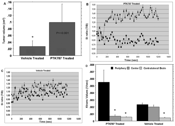
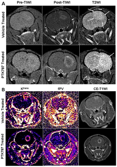
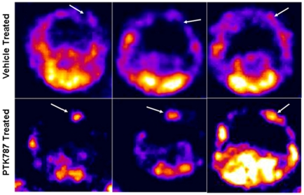
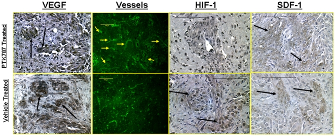
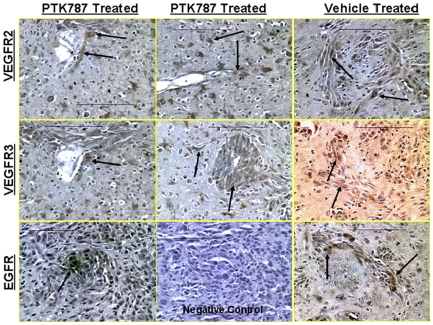
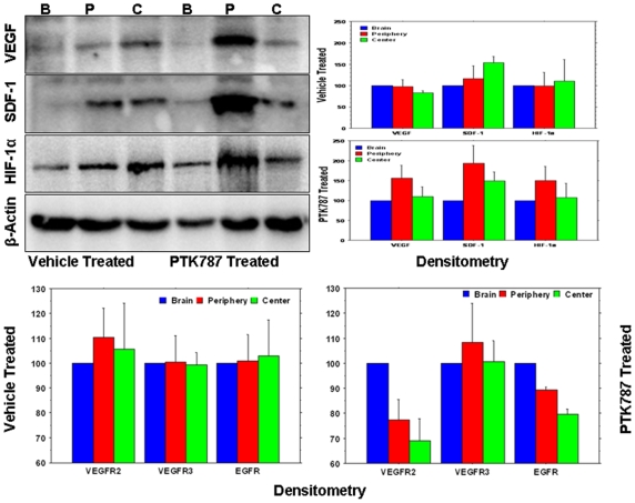
Methodologies and principal findings: Seven days after implantation of U251 glioma cells, animals were treated with either PTK787 or vehicle-only for two weeks, and then tumor size, tumor vascular permeability transfer constant (K(trans)), fractional plasma volume (fPV) and expression of VEGFR2 and other relevant angiogenic factors were assessed by in vivo MRI and SPECT (Tc-99-HYNIC-VEGF), and by immunohistochemistry and western blot analysis. Dynamic contrast-enhanced MRI (DCE-MRI) using a high molecular weight contrast agent albumin-(GdDTPA) showed significantly increased K(trans) at the rim of the treated tumors compared to that of the central part of the treated as well as the untreated (vehicle treated) tumors. Size of the tumors was also increased in the treated group. Expression of VEGFR2 detected by Tc-99m-HYNIC-VEGF SPECT also showed significantly increased activity in the treated tumors. In PTK787-treated tumors, histological staining revealed increase in microvessel density in the close proximity to the tumor border. Western blot analysis indicated increased expression of VEGF, SDF-1, HIF-1alpha, VEGFR2, VEGFR3 and EGFR at the peripheral part of the treated tumors compared to that of central part of the treated tumors. Similar expression patters were not observed in vehicle treated tumors.

Conclusion: These findings indicate that PTK787 treatment induced over expression of VEGF as well as the Flk-1/VEGFR2 receptor tyrosine kinase, especially at the rim of the tumor, as proven by DCE-MRI, SPECT imaging, immunohistochemistry and western blot.


Conflict of interest statement

Competing Interests: The authors have declared that no competing interests exist.

Figures

Figure 1
Figure 1. Tumor volume, signal intensity changes and Ktrans in PTK787 and vehicle treated tumors.
Measurement of tumor volume from the post contrast T1WI show significantly higher tumor volume in rats that received PTK787 (A). Signal intensity (SI) changes at the peripheral and central parts of the tumors in PTK787 (B) and vehicle (C) treated animals show remarkable differences in the SI-change patterns. SI changes at the peripheral part (▴) in PTK787 treated tumor are significantly higher from that of central part (▪) of the PTK787 treated tumors as early as 3 minutes after the injection of contrast agent, which is obviously different from that of vehicle treated tumors. Analysis of Ktrans values (D) shows significantly higher Ktrans at the peripheral part of PTK87 treated tumors, compared to that of the corresponding central part. Vehicle treated tumors did not show any differences in Ktrans values between the peripheral and central parts. Data are expressed as mean ± SEM, n≥3, * p<0.05.
Figure 2
Figure 2. MRI and parametric analysis of PTK787 treated and vehicle treated tumors.
(A) Representative cases from vehicle and PTK787 treated tumors show enhancement following administration of contrast agents (Post T1WI) and changes in T2WI. PTK787 treated tumor show larger tumor with prominent rim enhancement and central hollow compared to that of vehicle treated tumor. T2WI also show larger area of high signal intensity beyond the margin of enhancement (arrows). (B) Representative cases from vehicle and PTK787 treated tumors showing Ktrans, fractional plasma volume (fPV) and corresponding post contrast T1W images. Clearly increased Ktrans and fPV at the periphery of PTK787 treated tumor (calculated Ktrans and fPV maps are adjusted to same scale level for both vehicle treated and PTK787 treated tumors) indicate higher permeability and fPV, which are in agreement with the findings of dilated vessels at the peripheral part of PTK787 treated tumors (see below).
Figure 3
Figure 3. SPECT analysis of in vivo accumulation of Tc-99m-HYNIC-VEGF-c.
VEGF-c (which targets both VEGFR2 and VEGFR3) was tagged with HYNIC chelators and then labeled with Tc-99m-pertechnetate (Tc-99m) and injected intravenously in PTK787 and vehicle treated rats. One hour after injection, SPECT images were obtained using dedicated animal scanner. All PTK787 treated rats showed increased accumulation of Tc-99m-HYNIC-VEGF-c in the tumors (lower panel, arrows) compared to that of vehicle treated tumors (upper panel, arrows).
Figure 4
Figure 4. Immunohistochemistry of PTK787 and vehicle treated tumor showing expression of VEGF, HIF-1α, SDF-1 and vessel morphology.
Expression of vascular endothelial growth factor (VEGF) (dark brown colored) at different parts of the PTK787 treated and vehicle treated tumors. There were no differences observed in the expression of VEGF on immunohistochemistry at different parts of the tumors treated with either PTK787 or vehicle. However, delineation of vessels using FITC tagged tomato lectin indicated higher number of dilated vessels at the tumor periphery in rats that received PTK787 treatment. These dilated vessels may be indicative of increased permeability, fPV and signal intensity changes in PTK787 treated tumors (see Figures 1 and 2 ). Increased permeability may also be due to increased VEGF expression. HIF-1α expression was mostly seen in the central part of the vehicle treated tumors (arrows), however, SDF-1 expressions were observed both in PTK787 and vehicle treated tumors (arrows).
Figure 5
Figure 5. Immunohistochemistry of PTK787 and vehicle treated tumor showing expression of VEGFR2, VEGFR3 and EGFR.
Immunohistochemistry confirmed the findings of SPECT studies, where PTK787 treated tumors showed increased expression of VEGFR2 and VEFGR3 at the peripheral parts of the tumors, especially around the vessels (arrows) compared to that of vehicle treated tumors (right column). Both PTK787 (lower panel, left column) and vehicle treated (lower panel, right column) showed expression of EGFR in the tumors. Lower panel, middle column show no brown cells in negative control slide.
Figure 6
Figure 6. Western blot images and densitometry analysis of western blots.
(A) Expression of different angiogenic factors (left panel) in the vehicle- and PTK787 treated tumors from representative cases at the peripheral (P), central part of the tumors (C) and contralateral brains (B). Note the increased expression of VEGF, SDF-1 and HIF-1α at the peripheral part of PTK787 treated tumors. Right panel shows the densitometry analysis of the blot (normalized to β-actin and contralateral brain). The analysis also confirmed the finding of the blot. Note the patterns of VEGF, SDF-1 and HIF-1α in PTK787 treated tumors which are different from that of vehicle treated tumors. (B) Densitometry analysis of VEGFR2, VEGFR3 and EGFR blot (normalized to β-actin and contralateral brain). Expression of VEGFR2, VEGFR3 and EGFR showed higher normalized values at the peripheral part of the PTK787 treated tumors compared to that of central part and the expression patterns are different in vehicle treated tumors. Please note that PTK787 treated tumors showed lower normalized values of VEGFR2 and EGFR both at the periphery and central parts of the tumors compared to that of corresponding contralateral brain; whereas vehicle treated tumors did not show any changes in the normalized values of VEGFR2 and EGFR compared to that of corresponding contralateral brain. Data are expressed as mean ± SEM, n = 3.

References

    1. Remer S, Murphy ME. The challenges of long-term treatment outcomes in adults with malignant gliomas. Clin J Oncol Nurs. 2004;8:368–376. - PubMed
    1. Dhermain F, Ducreux D, Bidault F, Bruna A, Parker F, et al. [Use of the functional imaging modalities in radiation therapy treatment planning in patients with glioblastoma]. Bull Cancer. 2005;92:333–342. - PubMed
    1. Los M, Roodhart JM, Voest EE. Target practice: lessons from phase III trials with bevacizumab and vatalanib in the treatment of advanced colorectal cancer. Oncologist. 2007;12:443–450. - PubMed
    1. Norden AD, Young GS, Setayesh K, Muzikansky A, Klufas R, et al. Bevacizumab for recurrent malignant gliomas: efficacy, toxicity, and patterns of recurrence. Neurology. 2008;70:779–787. - PubMed
    1. Norden AD, Drappatz J, Wen PY. Novel anti-angiogenic therapies for malignant gliomas. Lancet Neurol. 2008;7:1152–1160. - PubMed

Publication types

Substances