. 2010 Jan 15;5(1):e8729.
doi: 10.1371/journal.pone.0008729.

Dynamic innate immune responses of human bronchial epithelial cells to severe acute respiratory syndrome-associated coronavirus infection

Affiliations

Dynamic innate immune responses of human bronchial epithelial cells to severe acute respiratory syndrome-associated coronavirus infection

Tomoki Yoshikawa et al. PLoS One. .

Abstract

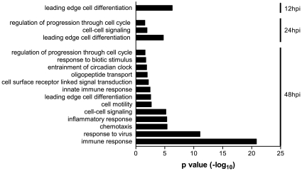
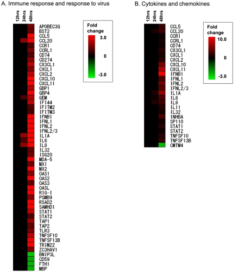
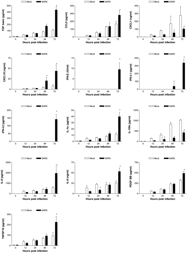
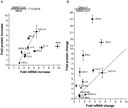
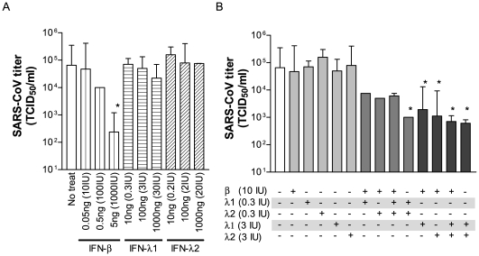
Human lung epithelial cells are likely among the first targets to encounter invading severe acute respiratory syndrome-associated coronavirus (SARS-CoV). Not only can these cells support the growth of SARS-CoV infection, but they are also capable of secreting inflammatory cytokines to initiate and, eventually, aggravate host innate inflammatory responses, causing detrimental immune-mediated pathology within the lungs. Thus, a comprehensive evaluation of the complex epithelial signaling to SARS-CoV is crucial for paving the way to better understand SARS pathogenesis. Based on microarray-based functional genomics, we report here the global gene response of 2B4 cells, a cloned bronchial epithelial cell line derived from Calu-3 cells. Specifically, we found a temporal and spatial activation of nuclear factor (NF)kappaB, activator protein (AP)-1, and interferon regulatory factor (IRF)-3/7 in infected 2B4 cells at 12-, 24-, and 48-hrs post infection (p.i.), resulting in the activation of many antiviral genes, including interferon (IFN)-beta, -lambdas, inflammatory mediators, and many IFN-stimulated genes (ISGs). We also showed, for the first time, that IFN-beta and IFN-lambdas were capable of exerting previously unrecognized, non-redundant, and complementary abilities to limit SARS-CoV replication, even though their expression could not be detected in infected 2B4 bronchial epithelial cells until 48 hrs p.i. Collectively, our results highlight the mechanics of the sequential events of antiviral signaling pathway/s triggered by SARS-CoV in bronchial epithelial cells and identify novel cellular targets for future studies, aiming at advancing strategies against SARS.


Conflict of interest statement

Competing Interests: The authors have declared that no competing interests exist.

Figures

Figure 1
Figure 1. Characteristics of 2B4 cells clonally derived from human bronchial epithelial Calu-3 cells.
Expressions of the viral ACE2 receptor in indicated passages of 2B4 cells and their parental Calu-3 cells were assessed by standard IHC (A) and Western blot analysis (B), whereas the morphological features of polarized 2B4 cells were assessed by TEM (C), as described in Materials and Methods. The images were taken at 6,270 magnifications. The scale bar represents 1 µm. To compare the permissiveness of 2B4 cells to their parental Calu-3 cells, confluent 2B4 cells, at passages #6 and #12, and Calu-3 cells were subjected to SARS-CoV (MOI = 0.1). The growth kinetics of SARS-CoV in culture supernatant and proportion of SARS-CoV-infected 2B4 cells were assessed at indicated time points by the standard Vero E6-based infectivity assay of the resulting cell-free supernatants (D) and infectious center assay (E). Finally, 2B4 cells (passage #6) were infected with SARS-CoV (MOI = 0.1) for 24, 48, and 72 hrs before being fixed with 4% paraformaldehyde for monitoring the morphological changes of infected cells, as visualized by the expression of SARS-CoV NP protein (red) by using the standard IHC (F).
Figure 2
Figure 2. Temporal and overlapping gene expression of 2B4 cells triggered by SARS-CoV infection.
Confluent 2B4 cells grown in T-75 flasks were infected with SARS-CoV at MOI = 0.1 or remained uninfected (as controls). The cells were lysed for extracting total RNAs for the subsequent microarray analysis by using Affymetrix Genechips. After the stringent pairwise comparisons and the statistical analysis, genes whose expressions were significantly altered (e.g., fold-change ≥1.5 and at least 50% greater in magnitude than any change observed between control samples, p<0.05) in SARS-CoV-infected versus uninfected 2B4 cells were selected for the additional analyses, as described in Materials and Methods. Two Venn diagrams were created to reflect the temporal and overlapping expressions of those up-regulated (A) and down-regulated genes (B), respectively.
Figure 3
Figure 3. Temporal and differential activation of transcription factors (TFs) in SARS-CoV-infected 2B4 cells.
Genes whose expressions were significantly altered in SARS-CoV-infected 2B4 cells were subjected to the TRANSFAC database-based analysis of TF activation. Adjusted p values of <0.05 among stringent pairwise comparisons were used for selecting those TFs that were significantly enriched at 12, 24, and 48 hrs p.i., respectively.
Figure 4
Figure 4. Enriched molecular functions of those genes whose expressions in 2B4 cells were significantly altered by SARS-CoV.
Expressions of both up- and down-regulated genes at 12, 24, and/or 48 hrs after SARS-CoV infection were analyzed against the entire human genome gene set. The enriched GO-annotated terms identified for those up-regulated and down-regulated genes are presented in A and B, respectively. The height of an individual bar represents the level of the statistical significance of the enriched GO-annotated term. An adjusted p value of <0.05 was used as the criterion for selecting enriched molecular functions.
Figure 5
Figure 5. Enriched entities of biological processes for those genes that were highly activated in 2B4 cells in response to SARS-CoV infection.
While no enriched GO-annotated biological term could be identified for those genes whose expressions were significantly down-regulated, 13 enriched biological entities were readily revealed for those highly activated genes of SARS-CoV-infected 2B4 cells at 12, 24, and/or 48 hrs p.i.. The height of each individual bar represents the level of statistical significance of the enriched GO-annotated biological process. An adjusted p value of <0.05 was used as the minimal criterion for selecting the enriched GO-annotated biological functions.
Figure 6
Figure 6. SARS-CoV-infected 2B4 cells elicit temporal and transitional expressions of genes relevant to innate immune responses.
The expression of 52 and 26 genes relevant to host viral immune responses (A) and the expression of cytokines and chemokines (B) elicited by SARS-CoV-infected 2B4 cells were hierarchically clustered along with time points after infection to more comprehensively reveal the temporal and transitional nature of host antiviral and inflammatory responses to SARS-CoV infection.
Figure 7
Figure 7. Confirmation of the transcription of pro-inflammatory genes at the translational level.
Cell-free supernatants harvested from mock- and SARS-CoV-infected 2B4 cultures at indicated time points p.i. were assessed for the concentration of various inflammatory mediators by using the human group I 27-plex and group II 8-plex Cytometric Bead Array, ELISA (for IFN-β and –λs), as described in Materials and Methods. *, p<0.05 by two-way ANOVA with Bonferroni's post-hoc test in comparison with the mock-infected group at the same hrs p.i.
Figure 8
Figure 8. Correlation between transcript and protein levels of genes encoding chemokines, cytokines and IFNs.
The relative transcriptional-versus-translational efficacies of selected genes in 2B4 cells were assessed, based on the fold-increase of mRNA and the corresponding protein levels of infected cells over those of mock-infected ones. A strong correlation between transcriptional and translational expressions of genes coding for CCL5/RANTES, CXCL1/GROα, CXCL10/IP-10, IFN-λ2, IL-6, IL-8, and TNFSF10/TRAIL (r2 = 0.8378) was identified in SARS-CoV-infected 2B4 cells. Dotted line indicates regression line (A). The relative efficacy of gene expression among those highly activated in SARS-CoV-infected and DHOV-infected 2B4 cells was compared as described in a subsection of Results. Dotted line (y = x) separates the genes to verify which virus (DOHV; upper part, SARS-CoV; lower part) -infected 2B4 cells effectively translate the genes than the other virus-infected one. It became clear that translational expressions of CCL5/RANTES, CXCL1/GROα, IL-1α, IL-6, and, particularly of IFN-β, IFN-λ1 and –λ2 were efficient in DHOV-infected 2B4 cells. The efficacy of CXCL-10/IP-10, IL-8 and TNFSF10/TRAIL expression was similar between these two differentially infected 2B4 cells (B). Results are shown as mean ± SEM for nine calculated results by division of each triplicated samples in indicated cytokines.
Figure 9
Figure 9. Efficacy of IFN-λs in the host defense against SARS-CoV infection.
Confluent cultures of 2B4 cells were treated with rIFN-β, rIFN-λ1, or rIFN-λ2 at the indicated concentrations alone (A) or in combination (B) for 24 hrs prior to their infection with SARS-CoV (MOI = 0.01). Resulting supernatants were harvested at day 2 after infection for assessing yields of infectious progeny virus by the standard Vero E6-based infectivity assay. *, p<0.05 by a two-way ANOVA with Bonferroni's post-hoc test in comparison with IFN-untreated cultures. Data shown were representative of two independent experiments.

References

    1. Peiris JSM, Lai ST, Poon LLM, Guan Y, Yam LYC, et al. Coronavirus as a possible cause of severe acute respiratory syndrome. Lancet. 2003;361:1319–1325. - PMC - PubMed
    1. Ksiazek TG, Erdman D, Goldsmith CS, Zaki SR, Peret T, et al. A novel coronavirus associated with severe acute respiratory syndrome. The New England Journal of Medicine. 2003;348:1953–1966. - PubMed
    1. Rota PA, Oberste MS, Monroe SS, Nix WA, Campagnoli R, et al. Characterization of a novel coronavirus associated with severe acute respiratory syndrome. Science. 2003;300:1394–1399. - PubMed
    1. Drosten C, Gunther S, Preiser W, van der Werf S, Brodt HR, et al. Identification of a novel coronavirus in patients with severe acute respiratory syndrome. N Engl J Med. 2003;348:1967–1976. - PubMed
    1. Chen PC, Hsiao CH. Re: To KF, Tong JH, Chan PK, et al. Tissue and cellular tropism of the coronavirus associated with severe acute respiratory syndrome: an in-situ hybridization study of fatal cases. J Pathol 2004. 2004;202:157–163. J Pathol 203: 729–730; author reply 730–721. - PMC - PubMed

Publication types

MeSH terms

Substances