Review
. 2010 Jul;460(2):223-37.
doi: 10.1007/s00424-009-0761-0. Epub 2009 Nov 29.

Cardiac sodium channelopathies

Affiliations
Review

Cardiac sodium channelopathies

Ahmad S Amin et al. Pflugers Arch. 2010 Jul.

Abstract

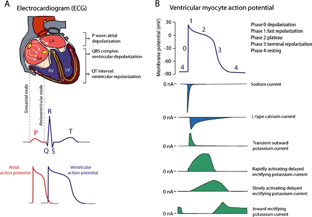
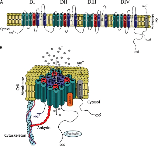
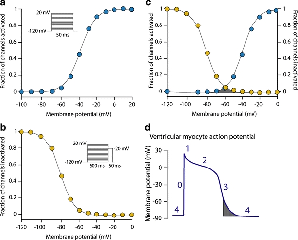
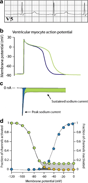
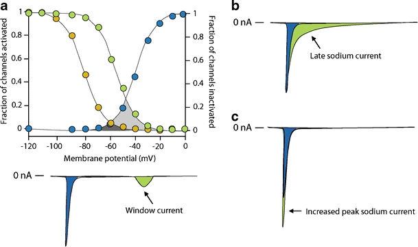
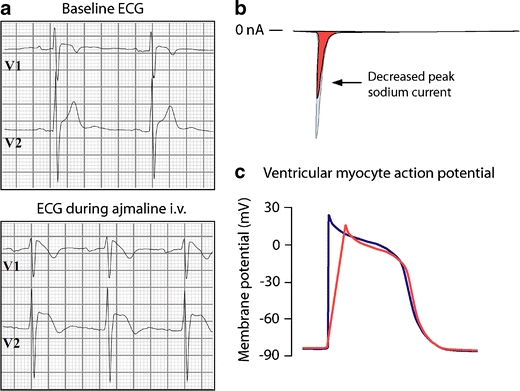
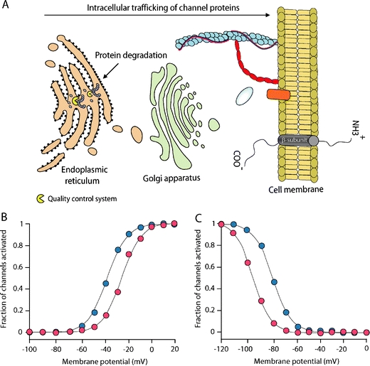
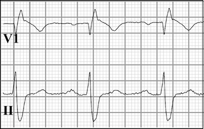
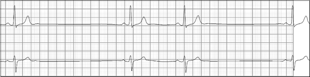
Cardiac sodium channel are protein complexes that are expressed in the sarcolemma of cardiomyocytes to carry a large inward depolarizing current (INa) during phase 0 of the cardiac action potential. The importance of INa for normal cardiac electrical activity is reflected by the high incidence of arrhythmias in cardiac sodium channelopathies, i.e., arrhythmogenic diseases in patients with mutations in SCN5A, the gene responsible for the pore-forming ion-conducting alpha-subunit, or in genes that encode the ancillary beta-subunits or regulatory proteins of the cardiac sodium channel. While clinical and genetic studies have laid the foundation for our understanding of cardiac sodium channelopathies by establishing links between arrhythmogenic diseases and mutations in genes that encode various subunits of the cardiac sodium channel, biophysical studies (particularly in heterologous expression systems and transgenic mouse models) have provided insights into the mechanisms by which INa dysfunction causes disease in such channelopathies. It is now recognized that mutations that increase INa delay cardiac repolarization, prolong action potential duration, and cause long QT syndrome, while mutations that reduce INa decrease cardiac excitability, reduce electrical conduction velocity, and induce Brugada syndrome, progressive cardiac conduction disease, sick sinus syndrome, or combinations thereof. Recently, mutation-induced INa dysfunction was also linked to dilated cardiomyopathy, atrial fibrillation, and sudden infant death syndrome. This review describes the structure and function of the cardiac sodium channel and its various subunits, summarizes major cardiac sodium channelopathies and the current knowledge concerning their genetic background and underlying molecular mechanisms, and discusses recent advances in the discovery of mutation-specific therapies in the management of these channelopathies.


Figures

Fig. 1
Fig. 1
The cardiac electrical activity and cardiac ion currents. a The electrical activity of the heart is represented on the surface electrocardiogram (ECG), and results from coordinated action potential generation in individual cardiomyocytes. The electrical activity starts by the spontaneous generation of action potentials in pacemaker cells in the sinoatrial node. Propagation of these action potentials creates an excitation wave through the atria, leading to atrial depolarization. After traveling through the atrioventricular node, the excitation wave reaches the ventricles, and leads to ventricular depolarization. b The cardiac action potential is generated by transmembrane inwardly and outwardly directed ion currents. The inward (depolarizing) sodium and calcium currents are pointed downwards and colored blue. The outward (repolarizing) potassium currents are pointed upwards and colored green
Fig. 2
Fig. 2
Molecular structure of the cardiac sodium channel. a Cartoon of the α-subunit (Nav1.5) and the β-subunit of the cardiac sodium channel. Nav1.5 consists of four domains (DIDIV), each containing six transmembrane segments (S1–S6); S4 segments are positively charged and act as voltage sensors. The β-subunit consists of one single transmembrane segment. b The four domains of Nav1.5 fold around an ion-conducting pore, which is lined by the loops between the S5 and S6 segments. The expression and function of Nav1.5 is regulated by β-subunits and several directly or indirectly interacting regulatory proteins
Fig. 3
Fig. 3
Voltage-dependent activation and inactivation of the cardiac sodium channel. a Using the patch-clamp technique, the membrane potential dependence of activation is studied by applying 50 ms depolarizing voltage steps from a holding potential of −120 mV (inset). The activation curve is obtained by dividing the amplitude of the resulting sodium current at each voltage step by the maximum peak sodium current amplitude, and plotting versus the corresponding voltage. b The membrane potential dependence of inactivation is studied by applying 500 ms depolarizing voltage steps from a holding potential of −120 mV to inactivate the channels (prepulse). Next, the fraction of channels that is not inactivated by the prepulse is measured by applying a voltage step to 20 mV (test pulse). The inactivation curve is obtained by dividing the amplitude of sodium current at each test pulse by the maximum peak sodium current amplitude, and plotting versus the corresponding prepulse voltage. c The window current (gray area) arises when the sarcolemma reaches a potential that is depolarized sufficiently to reactivate some channels, but not enough to cause complete inactivation. d The voltage range for the window current is normally narrow and achieved during phase 3 of the ventricular action potential
Fig. 4
Fig. 4
Long QT syndrome type 3. a Prolonged QT intervals on the surface ECG of an individual with LQT-3. b QT interval prolongation results from delayed repolarization of ventricular action potentials. c Delayed repolarization in LQT-3 is often due to the presence of abnormal sustained non-inactivating sodium current (green area). d Sustained current results from incomplete inactivation of the sodium channels (green circles)
Fig. 5
Fig. 5
Alternative mechanisms of sodium channel gain-of-function in long QT syndrome type 3. a Increased window current due to delayed inactivation of cardiac sodium channels (green circles). Increased windows current is carried at potentials corresponding to phases 2 and 3 of the ventricular action potential (green area), remote from the peak sodium current during phase 0 (blue area). b Slower inactivation creates a late sodium current (green area). c Increased peak sodium current
Fig. 6
Fig. 6
Brugada syndrome. a Coved-type ST segment elevation in the right-precordial ECG leads V1 and V2 after intravenous (i.v.) administration of sodium channel blocking drug ajmaline in an individual with BrS. b BrS-linked SCN5A mutations often lead to peak sodium current reduction. c Reduced peak sodium current decreases the upstroke velocity of action potential phase 0, which slows cardiac electrical conduction
Fig. 7
Fig. 7
Mechanisms of sodium channel loss-of-function in Brugada syndrome, progressive cardiac conduction disease, and sick sinus syndrome. a Decreased sarcolemmal expression of Nav1.5 due to premature degradation of mutant channel proteins by the quality control system in the endoplasmic reticulum. b Delayed activation of cardiac sodium channels (red circles). c Earlier inactivation of cardiac sodium channels (red circles)
Fig. 8
Fig. 8
Prolonged conduction parameters (P wave, PR and QRS intervals), and right bundle branch block in an individual with progressive cardiac conduction disease
Fig. 9
Fig. 9
Sinus arrest in an individual with sick sinus syndrome. The patient also suffered from sinus bradycardia

Similar articles

Cited by

References

    1. Abriel H, Kass RS. Regulation of the voltage-gated cardiac sodium channel Nav1.5 by interacting proteins. Trends Cardiovasc Med. 2005;15:35–40. doi: 10.1016/j.tcm.2005.01.001. - DOI - PubMed
    1. Albert CM, Nam EG, Rimm EB, Jin HW, Hajjar RJ, Hunter DJ, MacRae CA, Ellinor PT. Cardiac sodium channel gene variants and sudden cardiac death in women. Circulation. 2008;117:16–23. doi: 10.1161/CIRCULATIONAHA.107.736330. - DOI - PubMed
    1. Amin AS, de Groot EAA, Ruijter JM, Wilde AA, Tan HL. Exercise-induced ECG changes in Brugada syndrome. Circ Arrhythmia Electrophysiol. 2009;2:531–539. doi: 10.1161/CIRCEP.109.862441. - DOI - PubMed
    1. Amin AS, Meregalli PG, Bardai A, Wilde AA, Tan HL. Fever increases the risk for cardiac arrest in the Brugada syndrome. Ann Intern Med. 2008;149:216–218. - PubMed
    1. Amin AS, Tan HL, Wilde AAM. Cardiac ion channels in health and disease. Heart Rhythm. 2009 - PubMed

Publication types

MeSH terms