. 2010 Apr;67(8):1353-69.
doi: 10.1007/s00018-010-0257-2. Epub 2010 Jan 21.

Cell-specific and lamin-dependent targeting of novel transmembrane proteins in the nuclear envelope

Affiliations

Cell-specific and lamin-dependent targeting of novel transmembrane proteins in the nuclear envelope

Poonam Malik et al. Cell Mol Life Sci. 2010 Apr.

Abstract

Nuclear envelope complexity is expanding with respect to identification of protein components. Here we test the validity of proteomics results that identified 67 novel predicted nuclear envelope transmembrane proteins (NETs) from liver by directly comparing 30 as tagged fusions using targeting assays. This confirmed 21 as NETs, but 4 only targeted in certain cell types, underscoring the complexity of interactions that tether NETs to the nuclear envelope. Four NETs accumulated at the nuclear rim in normal fibroblasts but not in fibroblasts lacking lamin A, suggesting involvement of lamin A in tethering them in the nucleus. However, intriguingly, for the NETs tested alternative mechanisms for nuclear envelope retention could be found in Jurkat cells that normally lack lamin A. This study expands by a factor of three the number of liver NETs analyzed, bringing the total confirmed to 31, and shows that several have multiple mechanisms for nuclear envelope retention.


Figures
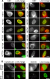
Fig. 1
Fig. 1
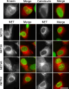
NE localization and detergent resistance of novel NETs. (a) NETs fused to mRFP at their carboxy-termini (except NET25 fused at its amino-terminus to an HA epitope tag) were transiently expressed in HT1080 cells also expressing lamin A-GFP. Cells were pre-extracted with Triton X-100 prior to fixation to remove membranes and soluble proteins, which typically also distorts morphology. NETs alone (left) and the merge between the NET (red) and lamin A (green) are shown. In some panels adjacent untransfected cells are shown, confirming that NE signal is not due to bleedthrough from the lamin channel. Like the emerin control (top), all NETs shown resisted detergent pre-extraction. Such resistance typically indicates association with the lamin polymer. Scale bars 7.5 μm. (b) Controls showing that the ER was fully removed by the detergent pre-extraction. Left calreticulin fused to GFP (but colored red) was overexpressed in cells either directly fixed or pre-extracted with detergent. No colocalization with lamins (green) was observed, and no calreticulin remained after extraction, though lamins did remain. Both direct fixed and pre-extracted images were exposed for 2 s. Right untransfected cells were similarly treated, then stained with the ER lipid dye DiOC6. Endogenous ER staining did not exhibit notable accumulation at the NE and was completely removed by the pre-extraction. Both images were exposed for 500 ms. Scale bars 20 μm
Fig. 2
Fig. 2
NETs that did not resist detergent pre-extraction re-tested for NE accumulation in directly fixed cells. HA-tagged NET62 and mRFP-tagged NETs 20, 38, and 46 were transiently transfected into HT1080 cells expressing lamin A-GFP. Tagged emerin and calreticulin were separately overexpressed as controls. The NET alone (black and white) and the merge between the NET (red) and lamin A (green) are shown. The new NETs yielded clear nuclear rims against the high cytoplasmic accumulation, colocalizing with lamins (yellow merge) similarly to emerin (top left). This staining pattern clearly differs from the ER localization of calreticulin (top right). Scale bars 20 μm
Fig. 3
Fig. 3
Relative amounts of NETs in the NE vs ER. NEs and microsomal membranes representing ER were prepared, and an equal amount of total protein for each fraction loaded on SDS-PAGE based upon measurements with Bradford assay. The proteins were transferred to nitrocellulose membranes, reacted with NET antibodies and fluorescent secondary antibodies, and fluorescent signals quantified using a LI-COR Odyssey. The averaged results of three separate Western blots are plotted showing the percentage of the combined signal coming from NE and microsome lanes. Most NETs were principally in the NE fraction, but NET34 was principally in the microsome fraction
Fig. 4
Fig. 4
Antibody staining in liver tissue sections. Cryosections of rat liver were stained with various NET antibodies and imaged on a confocal microscope. Nuclear rim staining could be observed in multiple cells in all fields, though for NET29 and NET30 not all cells in a given field yielded nuclear rims (cells indicated by arrows, as determined by comparing DAPI staining for mid-sectioned nuclei). Some background staining is observed with all NETs in the cytoplasm: this was slightly diminished with use of an affinity-purified antibody for NET30 (NET30AP), but may also indicate multiple cellular localizations for NETs. Scale bars 10 μm
Fig. 5
Fig. 5
INM versus ONM targeting. NETs were imaged using high-resolution systems for localization in relation to the two sides of the NPC using Nup153 from the nuclear basket and Nup358 from the cytoplasmic filaments. (a) Schematic of expected patterns indicating INM or ONM localization. If a protein is in the INM, the NET and Nup153 signals should occur in the same plane, and the NET signal should appear internal to the Nup358 signal. If the protein is in the ONM, the Nup153 signal should be internal to the NET signal and the Nup358 signal should occur in the same plane as the NET signal. (b) Images using structured illumination show both characterized (LAP2ß) and many novel NETs in the same plane of the inner nuclear membrane with Nup153 and internal to Nup358. Only NET23 and controls Sec61ß and calreticulin yielded the pattern expected for ONM residence. (c) High-resolution deconvolved Deltavision images also can distinguish inner from outer nuclear membranes with Nup153 shown in red and Nup358 shown in green. (d) Many additional novel NETs appeared in the INM using again LAP2ß as a control and NET55 that had been separately tested with the OMX system. In contrast NETs 4, 24, and 31 together with the calreticulin control yielded ONM targeting. Scale bars for b-d 5 μm. (e) Immunogold-EM confirms the validity of OMX and Deltavision results as 5-nm gold particles recognizing GFP antibodies for expressed NET51 and NET55 proteins appeared in the INM, similarly to controls. C and N denote cytoplasmic and nucleoplasmic sides where NPCs are inserted in the membrane. Bars 100 nm
Fig. 6
Fig. 6
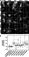
Some NETs only accumulate at the NE in certain cell types. (a) NETs that failed to target to the NE in HT1080 (human fibrosarcoma) cells were re-tested in other cell lines derived from different tissues: HepG2 (human liver tumor), 293T (human embryonic kidney), C2C12 (mouse skeletal muscle), and 3T3-L1 (mouse pre-adipocyte). HA-tagged NET32 and mRFP-tagged NETs 11, 13, 45, 59, and emerin were transiently transfected into the different cells. To ensure that rim accumulation was not due to bleedthrough or cross-reactivity with NE markers, cells were not co-transfected with or stained for other NE proteins. Arrows mark cells in which different NETs yielded discernible rim staining by having a strong distinct rim as opposed to one that could be accounted by ER condensed against the nucleus. Scale bars 20 μm. (b) To further validate NE targeting, the relative pixel intensities in ER and NE were quantified compared to ER controls. Pixel intensity was measured at a point in the nuclear rim (based on DAPI staining) and at a point approximately 2 μm distant into the ER, and the NE/ER ratio was calculated. Eight such measurements were taken from each NET from 5 different cells, and Tukey’s boxplots [45] for the 40 ratios for each NET in each cell line are shown with the median (central line), two quartiles above and below (box) and third quartile (error bars) shown. We compared each sample to each control (DiOC6 or calreticulin expressed in HT1080 cells) with the null hypothesis that ‘control to sample differences are by random chance.’ After analysis we reject the null hypothesis for each sample at P < 0.001 by Mann-Whitney (Wilcoxon) U test (Table 2). The ER dye DiOC6 and calreticulin-GFP both were very similar in intensity between the ER and NE, yielding a ratio of ~1. Though NET ratios tended to be in the 1.3–1.5 range, they were highly statistically significant, even for NET11 in C2C12 cells where a strong rim was not visually evident
Fig. 7
Fig. 7
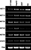
Expression profiles of NETs only sometimes correlate with their targeting. mRNAs were prepared from the human cell lines used in Fig. 6. RD myoblast cells (human) were used to represent muscle because C2C12 myoblast cells are derived from mouse. RT-PCR reactions were performed to determine the relative NET levels in the cell lines, using peptidylprolyl isomerase A (Ppia) as a loading control. Each was repeated at least three times, and representative gels are shown
Fig. 8
Fig. 8
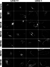
NETs that mistarget in cells lacking lamin A/C. Mouse embryonic fibroblasts extracted from a wild-type mouse (Lmna +/+) or from a matched LMNA knockout mouse (Lmna −/−) [29] were transfected with NET fusion constructs. At 30 h post-transfection, cells were directly fixed with formaldehyde and processed for immunofluorescence microscopy. Upper panels above the break show emerin and NETs that produced a distinctive rim in the Lmna +/+ cells, but did not in the Lmna −/− cells. Other NETs tested yielded no striking or reproducible differences in presence or absence of lamin A/C (only NET51 and NET55 are shown in bottom panels). Deconvolved images are shown. Scale bars 20 μm
Fig. 9
Fig. 9
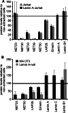
Indirect assay for lamin A interactions. If a NET that resists detergent extraction depends on lamin A for its NE retention either through direct or indirect binding, then it would be expected to be more resistant to detergent extraction in cells that express lamin A. Relative NET and lamin protein levels were quantified between lamin A expressing and not expressing cells from lysates run on Western blots using anti-NET and anti-lamin antibodies. The graphs show the levels of the protein left in cells extracted with 1% Triton X-100 as a percentage of the levels measured in unextracted cells. The numbers used for NETs were normalized according to the amount of lamin B1 remaining after extraction, which is shown here as absolute values, and three separate experiments were averaged to generate standard deviations. (a) Jurkat cells are suspension cells derived from a T-cell lymphoma that do not normally express lamin A. To test if adding lamin A to these cells would affect NET resistance to detergent extraction, Jurkats stably expressing lamin A-GFP were generated. Several NETs that resisted detergent extraction in the HT1080 fibroblasts grown on coverslips did not resist extraction in the Jurkat cells whether or not lamin A was present. Those that did resist showed no difference between the lamin A expressing and non-expressing cells. (b) Lmna −/− mouse fibroblasts (216−/−; [29]) and control mouse fibroblasts (NIH3T3) were also compared. In the fibroblast system emerin, LAP2ß, and NET33 differed in their extractibility between the lamin A null cells and the lamin A-expressing cells

Similar articles

Cited by

References

    1. Worman HJ, Bonne G. “Laminopathies”: a wide spectrum of human diseases. Exp Cell Res. 2007;313:2121–2133. doi: 10.1016/j.yexcr.2007.03.028. - DOI - PMC - PubMed
    1. Mattout A, Dechat T, Adam SA, Goldman RD, Gruenbaum Y. Nuclear lamins, diseases and aging. Curr Opin Cell Biol. 2006;18:335–341. doi: 10.1016/j.ceb.2006.03.007. - DOI - PubMed
    1. Bonne G, Di Barletta MR, Varnous S, Becane HM, Hammouda EH, Merlini L, Muntoni F, Greenberg CR, Gary F, Urtizberea JA, Duboc D, Fardeau M, Toniolo D, Schwartz K. Mutations in the gene encoding lamin A/C cause autosomal dominant Emery-Dreifuss muscular dystrophy. Nat Genet. 1999;21:285–288. doi: 10.1038/6799. - DOI - PubMed
    1. Bione S, Maestrini E, Rivella S, Mancini M, Regis S, Romeo G, Toniolo D. Identification of a novel X-linked gene responsible for Emery-Dreifuss muscular dystrophy. Nat Genet. 1994;8:323–327. doi: 10.1038/ng1294-323. - DOI - PubMed
    1. Clements L, Manilal S, Love DR, Morris GE. Direct interaction between emerin and lamin A. Biochem Biophys Res Commun. 2000;267:709–714. doi: 10.1006/bbrc.1999.2023. - DOI - PubMed

Publication types

MeSH terms