. 2010 May;119(5):579-89.
doi: 10.1007/s00401-009-0635-8. Epub 2010 Jan 21.

Quantification of myelin loss in frontal lobe white matter in vascular dementia, Alzheimer's disease, and dementia with Lewy bodies

Affiliations

Quantification of myelin loss in frontal lobe white matter in vascular dementia, Alzheimer's disease, and dementia with Lewy bodies

Masafumi Ihara et al. Acta Neuropathol. 2010 May.

Abstract

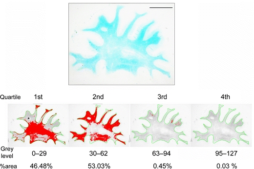
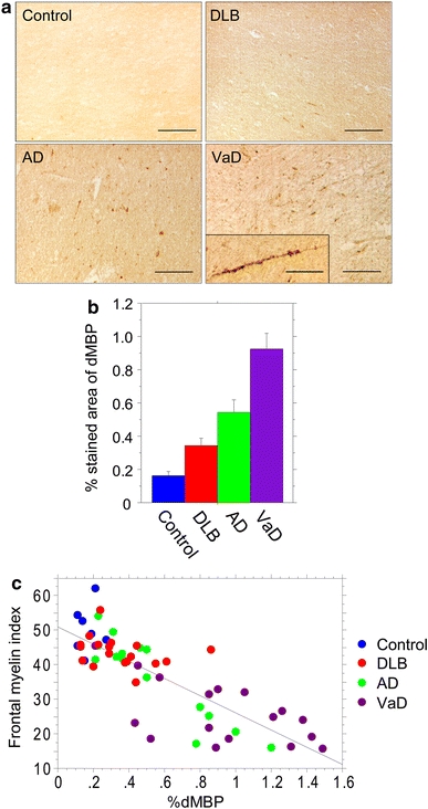
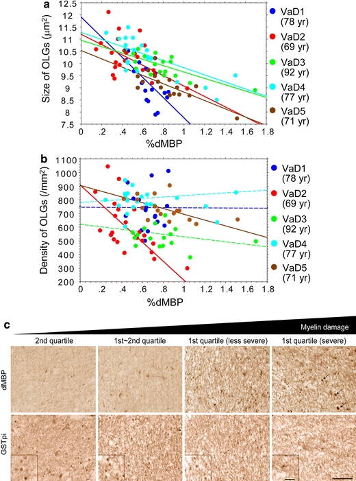
The aim of this study was to characterize myelin loss as one of the features of white matter abnormalities across three common dementing disorders. We evaluated post-mortem brain tissue from frontal and temporal lobes from 20 vascular dementia (VaD), 19 Alzheimer's disease (AD) and 31 dementia with Lewy bodies (DLB) cases and 12 comparable age controls. Images of sections stained with conventional luxol fast blue were analysed to estimate myelin attenuation by optical density. Serial adjacent sections were then immunostained for degraded myelin basic protein (dMBP) and the mean percentage area containing dMBP (%dMBP) was determined as an indicator of myelin degeneration. We further assessed the relationship between dMBP and glutathione S-transferase (a marker of mature oligodendrocytes) immunoreactivities. Pathological diagnosis significantly affected the frontal but not temporal lobe myelin attenuation: myelin density was most reduced in VaD compared to AD and DLB, which still significantly exhibited lower myelin density compared to ageing controls. Consistent with this, the degree of myelin loss was correlated with greater %dMBP, with the highest %dMBP in VaD compared to the other groups. The %dMBP was inversely correlated with the mean size of oligodendrocytes in VaD, whereas it was positively correlated with their density in AD. A two-tier regression model analysis confirmed that the type of disorder (VaD or AD) determines the relationship between %dMBP and the size or density of oligodendrocytes across the cases. Our findings, attested by the use of three markers, suggest that myelin loss may evolve in parallel with shrunken oligodendrocytes in VaD but their increased density in AD, highlighting partially different mechanisms are associated with myelin degeneration, which could originate from hypoxic-ischaemic damage to oligodendrocytes in VaD whereas secondary to axonal degeneration in AD.


Figures

Fig. 1
Fig. 1
Quartile analysis of LFB-stained section from the frontal lobe of a VaD patient (74 years). The WM was outlined in green and the grey levels within the WM were divided into four quartiles. The area percentage (%area) for each quartile was calculated (labelled in red). Myelin index was then calculated for each case and control (see “Methods”) (scale bar 1 cm)
Fig. 2
Fig. 2
Boxplot analysis for frontal myelin indices from LFB-stained sections in cases with microscopically evaluated normal WM (n = 3) and in those with WM changes categorized as mild (n = 18), moderate (n = 6), and severe (n = 8) in H&E. The line within the box indicates the median value. Error bars are drawn at the 5% and the 95% confidence intervals
Fig. 3
Fig. 3
Myelin loss in the frontal lobes in dementing disorders as assessed by LFB staining. a Representative images of LFB staining in cases of control (89 years), AD (87 years), DLB (77 years), and VaD (78 years) (scale bars 1 cm). b The %area for the first quartile in the four groups (Table 1). Differences were significant between any two groups except for that between controls and DLB; controls versus DLB, P = 0.0537; controls versus AD, P = 0.0032; controls versus VaD, P < 0.0001; DLB versus AD, P = 0.0464; DLB versus VaD, P < 0.0001; AD versus VaD, P = 0.0045. c The frontal myelin index in the four groups. Differences were significant between any two groups: controls versus DLB, P = 0.0027; controls versus AD, P = 0.0005; controls versus VaD, P < 0.0001; DLB versus AD, P = 0.0473; DLB versus VaD, P < 0.0001; AD versus VaD, P = 0.0060. Vertical bars represent mean ± SE
Fig. 4
Fig. 4
Myelin damage in the frontal lobes in dementing disorders as assessed by novel dMBP labelling. a Representative images of dMBP staining in cases of control (89 years), AD (87 years), DLB (77 years), and VaD (78 years) (scale bars 100 μm). Inset in VaD shows degenerative dMBP-positive myelin sheaths along the axon (scale bar 50 μm). b %dMBP in the four groups. Vertical bars represent mean ± SE. Differences were significant between any two groups: controls versus DLB, P = 0.0160; controls versus AD, P = 0.0029; controls versus VaD, P < 0.0001; DLB versus AD, P = 0.0331; DLB versus VaD, P < 0.0001; AD versus VaD, P = 0.0044. c An inverse correlation between the %dMBP and the myelin index in the 59 cases combined (r = −0.823, P < 0.0001)
Fig. 5
Fig. 5
Correlation analysis between %dMBP and density/size of oligodendrocytes in the frontal lobe of VaD cases. a The %dMBP was inversely correlated with the size of oligodendrocytes (OLGs) with statistical significance (blue points, r = −0.70; red points, r = −0.48; green points, r = −0.72; light-blue points, r = −0.76; brown points, r = −0.79). Each point represents a ROI (n = 20–22) within WM per Fig. 3a. b The %dMBP inversely correlated with the density of oligodendrocytes in two VaD cases with statistical significance (red points, r = −0.64; brown points, r = −0.54) but not in the others. c Images for dMBP and GSTpi in two adjacent sections in a 78-year-old patient with VaD, showing that increased myelin damage (from left to right panels) is accompanied by shrunken oligodendrocytes. The infarcted areas with dMBP-positive macrophages (arrows) as shown in the rightmost panels were excluded from the dMBP analysis. Magnified images of GSTpi-positive oligodendrocytes are shown in insets (scale bars 50 μm)
Fig. 6
Fig. 6
Correlation analysis between %dMBP and density/size of oligodendrocytes in the frontal lobe of AD cases. a The %dMBP did not significantly correlate with the size of oligodendrocytes (OLGs) in two AD cases but it did in one AD case (r = 0.56). Each point represents a ROI (n = 20–22) within WM per Fig. 3a. b The %dMBP significantly correlated with the density of oligodendrocytes (blue points, r = 0.84; red points, r = 0.68; green points, r = 0.82; light-blue points, r = 0.85; brown points, r = 0.87). c Images for dMBP and GSTpi in two adjacent sections in an 87-year-old patient with AD, showing that increased myelin damage (from left to right panels) is accompanied by increased density of oligodendrocytes (scale bars 50 μm)

Similar articles

Cited by

References

    1. Akiguchi I, Tomimoto H, Suenaga T, Wakita H, Budka H. Alterations in glia and axons in the brains of Binswanger’s disease patients. Stroke. 1997;28:1423–1429. - PubMed
    1. Akiguchi I, Tomimoto H, Wakita H, et al. Topographical and cytopathological lesion analysis of the white matter in Binswanger’s disease brains. Acta Neuropathol. 2004;107:563–570. doi: 10.1007/s00401-004-0850-2. - DOI - PubMed
    1. Alafuzoff I, Thal DR, Arzberger T, et al. Assessment of beta-amyloid deposits in human brain: a study of the BrainNet Europe Consortium. Acta Neuropathol. 2009;117:309–320. doi: 10.1007/s00401-009-0485-4. - DOI - PMC - PubMed
    1. Bannykh SI, Stolt CC, Kim J, Perry A, Wegner M. Oligodendroglial-specific transcriptional factor SOX10 is ubiquitously expressed in human gliomas. J Neurooncol. 2006;76:115–127. doi: 10.1007/s11060-005-5533-x. - DOI - PubMed
    1. Barber R, Scheltens P, Gholkar A, et al. White matter lesions on magnetic resonance imaging in dementia with Lewy bodies, Alzheimer’s disease, vascular dementia, and normal aging. J Neurol Neurosurg Psychiatry. 1999;67:66–72. doi: 10.1136/jnnp.67.1.66. - DOI - PMC - PubMed

Publication types

MeSH terms

Substances