

BDNF+/- mice exhibit deficits in oligodendrocyte lineage cells of the basal forebrain
- PMID: 20091777
- PMCID: PMC2851835
- DOI: 10.1002/glia.20969
BDNF+/- mice exhibit deficits in oligodendrocyte lineage cells of the basal forebrain
Abstract
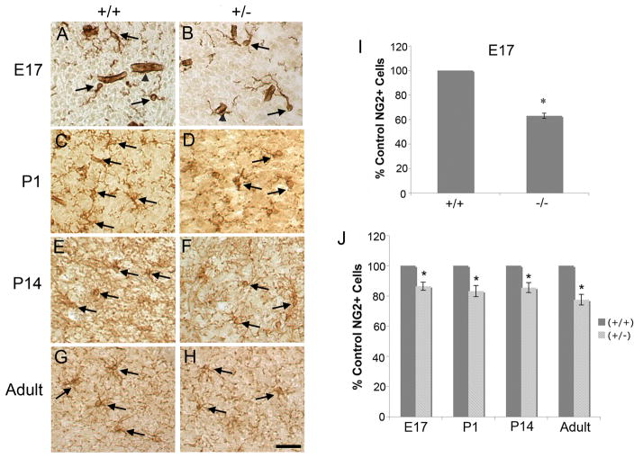
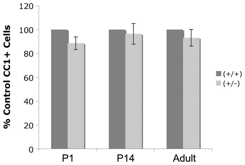
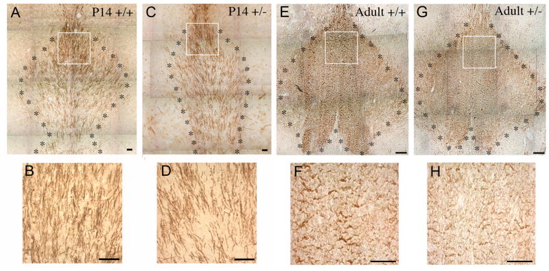
Previous work indicated that brain-derived neurotrophic factor (BDNF), through the trkB receptor, increases DNA synthesis in oligodendrocyte (OLG) progenitor cells (OPCs) and differentiation of postmitotic OLGs of the basal forebrain (BF). In the present studies, BDNF knockout animals were used to investigate BDNF's effects on OLG lineage cells (OLCs) in vivo. OLCs of the BF were found to express the trkB receptor, suggesting they are responsive to BDNF. Immunohistochemistry using NG2 and CC1 antibodies was utilized to examine the numbers of NG2+ OPCs and CC1+ postmitotic BF OLGs. At embryonic day 17 (E17), BDNF-/- animals display reduced NG2+ cells. This reduction was also observed in BDNF+/- mice at E17 and at postnatal day 1 (P1), P14, and adult stage, suggesting that BDNF plays a role in OPC development. BDNF+/- mice do not exhibit deficits in numbers of CC1+ OLGs. However, myelin basic protein, myelin associated glycoprotein, and proteolipid protein are reduced in BDNF+/- mice, suggesting that BDNF plays a role in differentiation. These data indicate that progenitor cells and myelin proteins may be affected in vivo by a decrease in BDNF.
Figures


Similar articles
-
Levels of BDNF impact oligodendrocyte lineage cells following a cuprizone lesion.J Neurosci. 2011 Oct 5;31(40):14182-90. doi: 10.1523/JNEUROSCI.6595-10.2011. J Neurosci. 2011. PMID: 21976503 Free PMC article.
-
Oligodendroglial expression of TrkB independently regulates myelination and progenitor cell proliferation.J Neurosci. 2013 Mar 13;33(11):4947-57. doi: 10.1523/JNEUROSCI.3990-12.2013. J Neurosci. 2013. PMID: 23486965 Free PMC article.
-
Brain-derived neurotrophic factor deficiency restricts proliferation of oligodendrocyte progenitors following cuprizone-induced demyelination.ASN Neuro. 2015 Jan 13;7(1):1759091414566878. doi: 10.1177/1759091414566878. Print 2015 Jan-Feb. ASN Neuro. 2015. PMID: 25586993 Free PMC article.
-
NG2-expressing cells as oligodendrocyte progenitors in the normal and demyelinated adult central nervous system.J Anat. 2005 Dec;207(6):707-16. doi: 10.1111/j.1469-7580.2005.00454.x. J Anat. 2005. PMID: 16367798 Free PMC article. Review.
-
Cytology and lineage of NG2-positive glia.J Neurocytol. 2002 Jul-Aug;31(6-7):457-67. doi: 10.1023/a:1025735513560. J Neurocytol. 2002. PMID: 14501216 Review.
Cited by
-
Reversal of the Detrimental Effects of Post-Stroke Social Isolation by Pair-Housing is Mediated by Activation of BDNF-MAPK/ERK in Aged Mice.Sci Rep. 2016 Apr 29;6:25176. doi: 10.1038/srep25176. Sci Rep. 2016. PMID: 27125783 Free PMC article.
-
Presentation and integration of multiple signals that modulate oligodendrocyte lineage progression and myelination.Front Cell Neurosci. 2022 Nov 14;16:1041853. doi: 10.3389/fncel.2022.1041853. eCollection 2022. Front Cell Neurosci. 2022. PMID: 36451655 Free PMC article. Review.
-
Cortical spreading depolarization stimulates gliogenesis in the rat entorhinal cortex.J Cereb Blood Flow Metab. 2015 Mar 31;35(4):576-82. doi: 10.1038/jcbfm.2014.232. J Cereb Blood Flow Metab. 2015. PMID: 25515215 Free PMC article.
-
Microenvironmental interactions of oligodendroglial cells.Dev Cell. 2021 Jul 12;56(13):1821-1832. doi: 10.1016/j.devcel.2021.06.006. Epub 2021 Jun 29. Dev Cell. 2021. PMID: 34192527 Free PMC article. Review.
-
BDNF-TrkB Signaling Pathway in Spinal Cord Injury: Insights and Implications.Mol Neurobiol. 2025 Feb;62(2):1904-1944. doi: 10.1007/s12035-024-04381-4. Epub 2024 Jul 24. Mol Neurobiol. 2025. PMID: 39046702 Review.
References
-
- Abercrombie M. Estimation of nuclear population from microtome sections. Anat Rec. 1946;94:239–247. - PubMed
-
- Alderson RF, Alterman AL, Barde YA, Lindsay RM. Brain-derived neurotrophic factor increases survival and differentiated functions of rat septal cholinergic neurons in culture. Neuron. 1990;5(3):297–306. - PubMed
-
- Auld DS, Kornecook TJ, Bastianetto S, Quirion R. Alzheimer’s disease and the basal forebrain cholinergic system: relations to beta-amyloid peptides, cognition, and treatment strategies. Prog Neurobiol. 2002;68(3):209–45. - PubMed
-
- Azoulay D, Vachapova V, Shihman B, Miler A, Karni A. Lower brain-derived neurotrophic factor in serum of relapsing remitting MS: reversal by glatiramer acetate. J Neuroimmunol. 2005;167(1–2):215–8. - PubMed
Publication types
MeSH terms
Substances
Grants and funding
LinkOut - more resources
Full Text Sources
Other Literature Sources
Medical
Molecular Biology Databases
Research Materials
Miscellaneous
