. 2010 Jan;31(1):134-47.
doi: 10.1111/j.1460-9568.2009.07042.x. Epub 2009 Dec 23.

Qualitatively different modes of perirhinal-hippocampal engagement when rats explore novel vs. familiar objects as revealed by c-Fos imaging

Affiliations

Qualitatively different modes of perirhinal-hippocampal engagement when rats explore novel vs. familiar objects as revealed by c-Fos imaging

Mathieu M Albasser et al. Eur J Neurosci. 2010 Jan.

Abstract

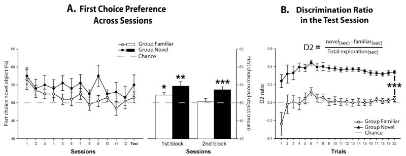
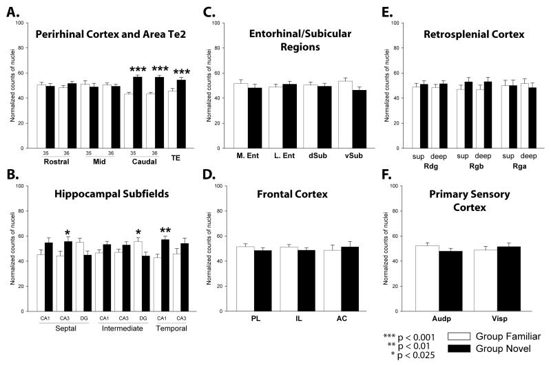
Expression of the immediate-early gene c-fos was used to test for different patterns of temporal lobe interactions when rats explore either novel or familiar objects. A new behavioural test of recognition memory was first devised to generate robust levels of novelty discrimination and to provide a matched control condition using familiar objects. Increased c-Fos activity was found in caudal but not rostral portions of the perirhinal cortex (areas 35/36) and in area Te2 in rats showing object recognition, i.e. preferential exploration of novel vs. familiar objects. The findings are presented at a higher anatomical resolution than previous studies of immediate-early gene expression and object novelty and, crucially, provide the first analyses when animals are actively discriminating the novel objects. Novel vs. familiar object comparisons also revealed altered c-Fos patterns in hippocampal subfields, with relative increases in CA3 and CA1 and decreases in the dentate gyrus. These hippocampal changes match those previously reported for the automatic coding of object-spatial associations. Additional analyses of the c-Fos data using structural equation modelling indicated the presence of pathways starting in the caudal perirhinal cortex that display a direction of effects from the entorhinal cortex to the CA1 field (temporo-ammonic) when presented with familiar objects, but switch to the engagement of the direct entorhinal cortex pathway to the dentate gyrus (perforant) with novel object discrimination. This entorhinal switch provides a potential route by which the rhinal cortex can moderate hippocampal processing, with a dynamic change from temporo-ammonic (familiar stimuli) to perforant pathway (novel stimuli) influences.


Figures

Figure 1
Figure 1
A. Schematic of the Bow-tie maze, with dimensions in centimetres. Food wells shown in gray. B. General procedure showing the presentation order of the objects. All objects are rewarded (+). Arrow shows rats movements. Group Novel: Black print represents the novel objects and gray print the familiar objects. C. Spatial placement of a now familiar object compared to its previous trial location. Two possible configurations are possible: same (left) or displaced (right).
Figure 2
Figure 2
Specific procedure for Group Novel (left) and Group Familiar (right) during the penultimate session (top, session 12) and the final session (bottom, Test session). The letters α, β, γ represent a novel set of objects, whereas objects A, B, C are only new for Group Novel.
Figure 3
Figure 3
Coronal sections indicating regions of interest: anterior cingulate cortex (AC), areas 35 and 36 for the rostral, mid and caudal perirhinal (Prh) cortex, area Te2, primary auditory cortex (Audp), CA1, CA3, dentate gyrus (DG) for the septal, intermediate and temporal hippocampus, lateral entorhinal cortex (lEnt), medial entorhinal cortex (mEnt), infralimbic cortex (IL), prelimbic cortex (PL), retrosplenial dysgranular cortex (Rdg), retrosplenial granular a (Rga), retrosplenial granular b (Rgb), dorsal subiculum (d Sub), ventral subiculum (v Sub), primary visual cortex (Visp). The numbers refer to the distance (mm) from bregma according to the atlas of Paxinos and Watson (2005).
Figure 4
Figure 4
A. First choice preference of novel objects. The line plot graph shows the scores during the 13 sessions (left). The bar chart represents mean scores over the first set of six sessions (1st block) and the subsequent set of seven sessions (2nd block). Data are shown as mean ± SEM. A score significantly above 50 (one-sample t test) indicates first choice preference for the novel object. * p < 0.05, ** p < 0.01, *** p 0< 0.001 (right). B. Discrimination ratio D2 = [(novel(sec) -familiar(sec)) / total exploration(sec)] for final session. The D2 score is re-calculated after each trial as data from successive trials are accumulated. A score of zero reflects a failure to discriminate novel from familiar. Data shown are mean ± SEM. Group differences: *** p < 0.001.
Figure 5
Figure 5
c-Fos levels following familiar (white) or novel (black) object exposure. A. Perirhinal cortex and Area Te2: areas 35 and 36 of the rostral, mid and caudal perirhinal cortex and area Te2. B. Hippocampal Subfields: dentate gyrus (DG), CA3, CA1, for septal, intermediate and temporal hippocampus. C. Entorhinal/Subicular Regions: lateral entorhinal (lEnt) cortex, medial entorhinal (mEnt) cortex, dorsal subiculum (d Sub) and ventral subiculum (v Sub). D. Frontal Cortex: prelimbic (PL), infralimbic (IL) and anterior cingulate (AC) cortices. E. Retrosplenial Cortex: superficial and deep layers of the dysgranular (Rdg) and granular (Rgb and Rga) retrosplenial cortex. F. Primary Sensory Cortex: primary auditory cortex (Audp), primary visual cortex (Vis). Normalized counts of c-Fos-positive cells are presented as mean ± SEM. Significance of group differences: * p < 0.025; ** p < 0.01; *** p < 0.001.
Figure 6
Figure 6
Photomicrographs comparing c-Fos levels between Group Novel (left-hand side) and Group Familiar (right-hand side). A. Caudal perirhinal cortex (the numbers refer to areas 35 and 36). B. Septal hippocampus. C. Primary visual cortex. The photomicrographs are taken from behaviourally-matched pairs of animals where tissue was processed simultaneously to reduce variation. The brightfield photomicrographs of coronal sections show the comparable c-Fos levels in the primary auditory cortex, which contrasts, with the specific increase of c-Fos positive cells following novelty in the caudal perirhinal cortex. Scale bar, 100 μm.
Figure 7
Figure 7
Temporal lobe interactions derived from structural equation modelling. Notice the engagement of the tri-synaptic circuit induced by novelty. A. Optimal Group Novel model, B. Consequence of trying to fit Group Familiar data into the optimal model of Group Novel (used for stacked comparison between groups) and C. Optimal Group Familiar model. The strength of the causal influences is depicted in the diagrams in black. The squared multiple correlations (R 2) are in grey. Sites depicted: area Te2, dentate gyrus (DG), CA1, CA3, lateral entorhinal cortex (lEnt), caudal perirhinal cortex (caudPrh). *p < 0.025; ***p< 0.001.

Similar articles

Cited by

References

    1. Aggleton JP. One-trial object recognition by rats. Q J Exp Psychol. 1985;37B:279–294.
    1. Aggleton JP, Brown MW. Contrasting hippocampal and perirhinal cortex function using immediate early gene imaging. Q J Exp Psychol B. 2005;58:218–233. - PubMed
    1. Albasser MM, Davies M, Futter JE, Aggleton JP. Magnitude of the object recognition deficit associated with perirhinal cortex damage in rats: Effects of varying the lesion extent and the duration of the sample period. Behav Neurosci. 2009;123:115–124. - PubMed
    1. Albasser MM, Poirier GL, Warburton EC, Aggleton JP. Hippocampal lesions halve immediate-early gene protein counts in retrosplenial cortex: distal dysfunctions in a spatial memory system. Eur J Neurosci. 2007;26:1254–1266. - PubMed
    1. Bast T. Toward an integrative perspective on hippocampal function: from the rapid encoding of experience to adaptive behavior. Rev Neurosci. 2007;18:253–281. - PubMed

Publication types

Substances