

miR-21: an oncomir on strike in prostate cancer
- PMID: 20092645
- PMCID: PMC2823650
- DOI: 10.1186/1476-4598-9-12
miR-21: an oncomir on strike in prostate cancer
Abstract
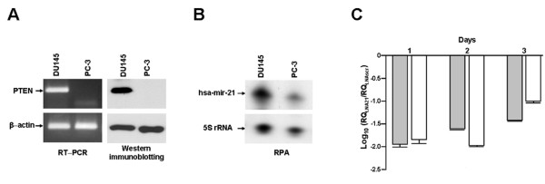
Background: Aberrant expression of microRNAs, small non-coding RNA molecules that post-transcriptionally repress gene expression, seems to be causatively linked to the pathogenesis of cancer. In this context, miR-21 was found to be overexpressed in different human cancers (e.g. glioblastoma, breast cancer). In addition, it is thought to be endowed with oncogenic properties due to its ability to negatively modulate the expression of tumor-suppressor genes (e.g. PTEN) and to cause the reversion of malignant phenotype when knocked- down in several tumor models. On the basis of these findings, miR-21 has been proposed as a widely exploitable cancer-related target. However, scanty information is available concerning the relevance of miR-21 for prostate cancer. In the present study, we investigated the role of miR-21 and its potential as a therapeutic target in two prostate cancer cell lines, characterized by different miR-21 expression levels and PTEN gene status.
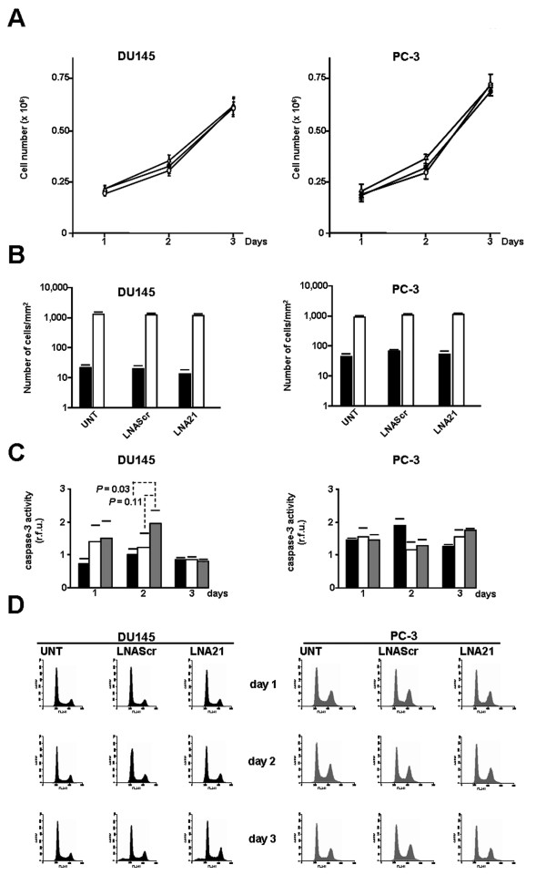
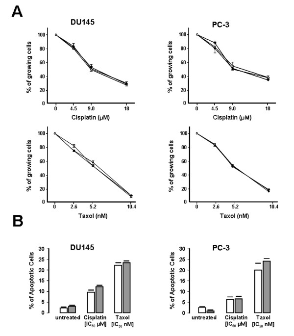
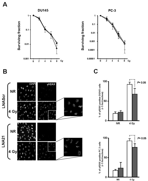
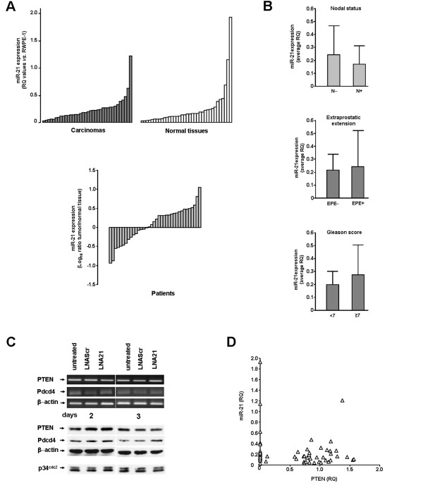
Results: We provide evidence that miR-21 knockdown in prostate cancer cells is not sufficient per se i) to affect the proliferative and invasive potential or the chemo- and radiosensitivity profiles or ii) to modulate the expression of the tumor-suppressors PTEN and Pdcd4, which in other tumor types were found to be regulated by miR-21. We also show that miR-21 is not differently expressed in carcinomas and matched normal tissues obtained from 36 untreated prostate cancer patients subjected to radical prostatectomy.
Conclusions: Overall, our data suggest that miR-21 is not a central player in the onset of prostate cancer and that its single hitting is not a valuable therapeutic strategy in the disease. This supports the notion that the oncogenic properties of miR-21 could be cell and tissue dependent and that the potential role of a given miRNA as a therapeutic target should be contextualized with respect to the disease.
Figures
Similar articles
-
Upregulation of miR-153 promotes cell proliferation via downregulation of the PTEN tumor suppressor gene in human prostate cancer.Prostate. 2013 May;73(6):596-604. doi: 10.1002/pros.22600. Epub 2012 Oct 11. Prostate. 2013. PMID: 23060044
-
miR-410-3p promotes prostate cancer progression via regulating PTEN/AKT/mTOR signaling pathway.Biochem Biophys Res Commun. 2018 Sep 18;503(4):2459-2465. doi: 10.1016/j.bbrc.2018.06.176. Epub 2018 Jul 6. Biochem Biophys Res Commun. 2018. PMID: 29969630
-
Long noncoding RNA GAS5 modulates α-Solanine-induced radiosensitivity by negatively regulating miR-18a in human prostate cancer cells.Biomed Pharmacother. 2019 Apr;112:108656. doi: 10.1016/j.biopha.2019.108656. Epub 2019 Mar 1. Biomed Pharmacother. 2019. PMID: 30970507
-
Role of miRNA let-7 and its major targets in prostate cancer.Biomed Res Int. 2014;2014:376326. doi: 10.1155/2014/376326. Epub 2014 Sep 3. Biomed Res Int. 2014. PMID: 25276782 Free PMC article. Review.
-
An Immunocompetent Environment Unravels the Proto-Oncogenic Role of miR-22.Cancers (Basel). 2022 Dec 19;14(24):6255. doi: 10.3390/cancers14246255. Cancers (Basel). 2022. PMID: 36551740 Free PMC article. Review.
Cited by
-
MicroRNAs as Epigenetic Determinants of Treatment Response and Potential Therapeutic Targets in Prostate Cancer.Cancers (Basel). 2021 May 14;13(10):2380. doi: 10.3390/cancers13102380. Cancers (Basel). 2021. PMID: 34069147 Free PMC article. Review.
-
Urine miR-21-5p and miR-200c-3p as potential non-invasive biomarkers in patients with prostate cancer.Turk J Urol. 2019 Nov 29;46(1):26-30. doi: 10.5152/tud.2019.19163. Print 2020 Jan. Turk J Urol. 2019. PMID: 31905122 Free PMC article.
-
The Effect of Gene Editing by CRISPR-Cas9 of miR-21 and the Indirect Target MMP9 in Metastatic Prostate Cancer.Int J Mol Sci. 2023 Oct 3;24(19):14847. doi: 10.3390/ijms241914847. Int J Mol Sci. 2023. PMID: 37834295 Free PMC article.
-
Emerging biomarkers in the diagnosis of prostate cancer.Pharmgenomics Pers Med. 2018 May 16;11:83-94. doi: 10.2147/PGPM.S136026. eCollection 2018. Pharmgenomics Pers Med. 2018. PMID: 29844697 Free PMC article. Review.
-
Simultaneous subset tracing and miRNA profiling of tumor-derived exosomes via dual-surface-protein orthogonal barcoding.Sci Adv. 2023 Oct 6;9(40):eadi1556. doi: 10.1126/sciadv.adi1556. Epub 2023 Oct 4. Sci Adv. 2023. PMID: 37792944 Free PMC article.
References
-
- Mirnezami AH, Pickard K, Zhang L, Primrose JN, Packham G. MicroRNAs: key players in carcinogenesis and novel therapeutic targets. Eur J Surg Oncol. 2009;35:339–347. - PubMed
Publication types
MeSH terms
Substances
LinkOut - more resources
Full Text Sources
Medical
Research Materials
