

Distribution of airway narrowing responses across generations and at branching points, assessed in vitro by anatomical optical coherence tomography
- PMID: 20092657
- PMCID: PMC2824705
- DOI: 10.1186/1465-9921-11-9
Distribution of airway narrowing responses across generations and at branching points, assessed in vitro by anatomical optical coherence tomography
Abstract
Background: Previous histological and imaging studies have shown the presence of variability in the degree of bronchoconstriction of airways sampled at different locations in the lung (i.e., heterogeneity). Heterogeneity can occur at different airway generations and at branching points in the bronchial tree. Whilst heterogeneity has been detected by previous experimental approaches, its spatial relationship either within or between airways is unknown.
Methods: In this study, distribution of airway narrowing responses across a portion of the porcine bronchial tree was determined in vitro. The portion comprised contiguous airways spanning bronchial generations (#3-11), including the associated side branches. We used a recent optical imaging technique, anatomical optical coherence tomography, to image the bronchial tree in three dimensions. Bronchoconstriction was produced by carbachol administered to either the adventitial or luminal surface of the airway. Luminal cross sectional area was measured before and at different time points after constriction to carbachol and airway narrowing calculated from the percent decrease in luminal cross sectional area.
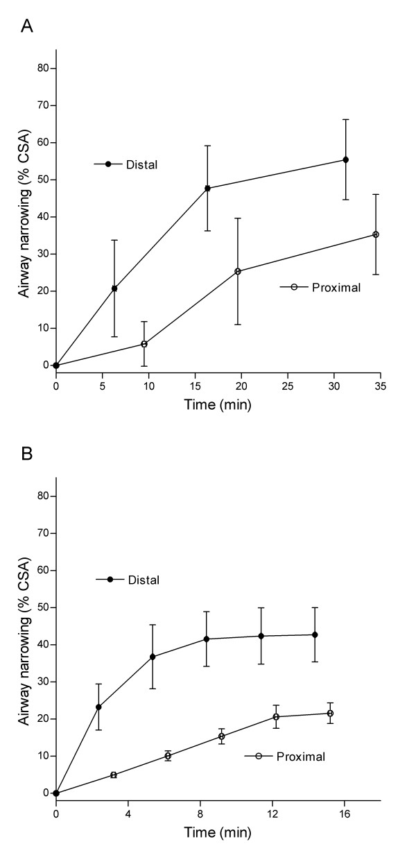
Results: When administered to the adventitial surface, the degree of airway narrowing was progressively increased from proximal to distal generations (r = 0.80 to 0.98, P < 0.05 to 0.001). This 'serial heterogeneity' was also apparent when carbachol was administered via the lumen, though it was less pronounced. In contrast, airway narrowing was not different at side branches, and was uniform both in the parent and daughter airways.
Conclusions: Our findings demonstrate that the bronchial tree expresses intrinsic serial heterogeneity, such that narrowing increases from proximal to distal airways, a relationship that is influenced by the route of drug administration but not by structural variations accompanying branching sites.
Figures


Similar articles
-
Airway narrowing in porcine bronchi with and without lung parenchyma.Eur Respir J. 2005 Nov;26(5):804-11. doi: 10.1183/09031936.05.00065405. Eur Respir J. 2005. PMID: 16264040
-
Relationship of airway narrowing, compliance, and cartilage in isolated bronchial segments.J Appl Physiol (1985). 2002 Mar;92(3):1119-24. doi: 10.1152/japplphysiol.00662.2001. J Appl Physiol (1985). 2002. PMID: 11842048
-
In vivo real-time imaging of airway dynamics during bronchial challenge test.Lasers Surg Med. 2015 Mar;47(3):252-6. doi: 10.1002/lsm.22345. Epub 2015 Mar 13. Lasers Surg Med. 2015. PMID: 25779778
-
Airway wall liquid. Sources and role as an amplifier of bronchoconstriction.Chest. 1995 Mar;107(3 Suppl):105S-110S. doi: 10.1378/chest.107.3_supplement.105s. Chest. 1995. PMID: 7874985 Review.
-
Afferent neural pathways in cough and reflex bronchoconstriction.J Appl Physiol (1985). 1988 Sep;65(3):1007-23. doi: 10.1152/jappl.1988.65.3.1007. J Appl Physiol (1985). 1988. PMID: 3053580 Review.
Cited by
-
Patient-Specific Airway Wall Remodeling in Chronic Lung Disease.Ann Biomed Eng. 2015 Oct;43(10):2538-51. doi: 10.1007/s10439-015-1306-7. Epub 2015 Mar 28. Ann Biomed Eng. 2015. PMID: 25821112 Free PMC article.
-
Examining lung mechanical strains as influenced by breathing volumes and rates using experimental digital image correlation.Respir Res. 2022 Apr 11;23(1):92. doi: 10.1186/s12931-022-01999-7. Respir Res. 2022. PMID: 35410291 Free PMC article.
-
Future of Lung Transplantation: Xenotransplantation and Bioengineering Lungs.Clin Chest Med. 2023 Mar;44(1):201-214. doi: 10.1016/j.ccm.2022.11.003. Clin Chest Med. 2023. PMID: 36774165 Free PMC article. Review.
-
In Vivo Characterization of the Swine Airway Morphometry and Motion Based on Computed Tomographic Imaging During Respiration.J Biomech Eng. 2020 Dec 1;142(12):121009. doi: 10.1115/1.4047550. J Biomech Eng. 2020. PMID: 34043756 Free PMC article.
-
Volumetric optical frequency domain imaging of pulmonary pathology with precise correlation to histopathology.Chest. 2013 Jan;143(1):64-74. doi: 10.1378/chest.11-2797. Chest. 2013. PMID: 22459781 Free PMC article.
References
-
- Amirav I, Kramer SS, Grunstein MM, Hoffman EA. Assessment of methacholine-induced airway constriction by ultrafast high-resolution computed tomography. J Appl Physiol. 1993;75(5):2239–2250. - PubMed
-
- Brown RH, Herold CJ, Hirshman CA, Zerhouni EA, Mitzner W. In vivo measurements of airway reactivity using high-resolution computed tomography. Am Rev Respir Dis. 1991;144(1):208–212. - PubMed
-
- Brown RH, Herold CJ, Hirshman CA, Zerhouni EA, Mitzner W. Individual airway constrictor response heterogeneity to histamine assessed by high-resolution computed tomography. J Appl Physiol. 1993;74(6):2615–2620. - PubMed
Publication types
MeSH terms
LinkOut - more resources
Full Text Sources
