

PET/NIRF/MRI triple functional iron oxide nanoparticles
- PMID: 20092887
- PMCID: PMC2838491
- DOI: 10.1016/j.biomaterials.2010.01.010
PET/NIRF/MRI triple functional iron oxide nanoparticles
Abstract
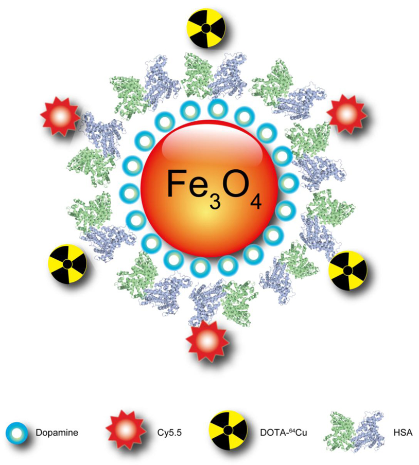
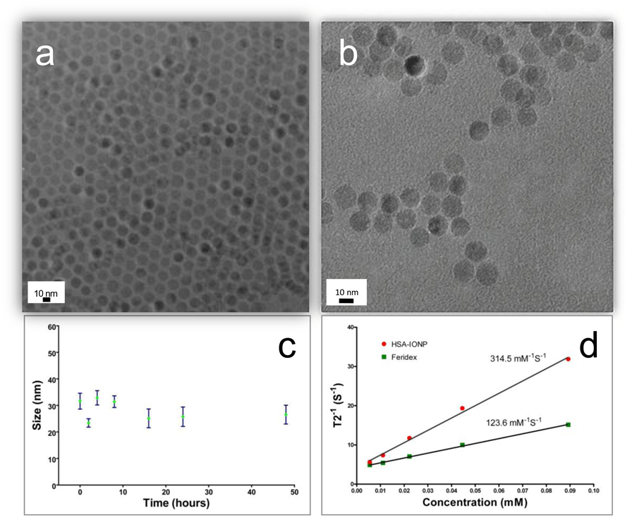
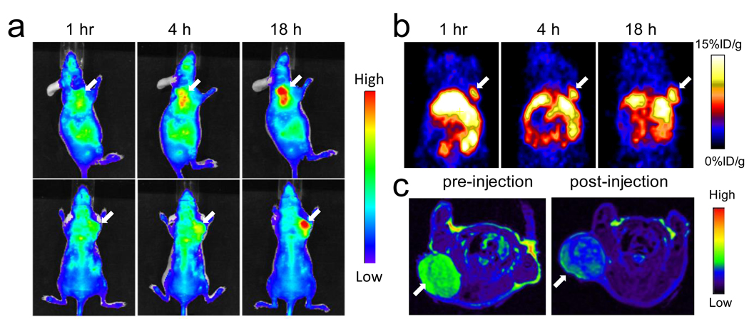
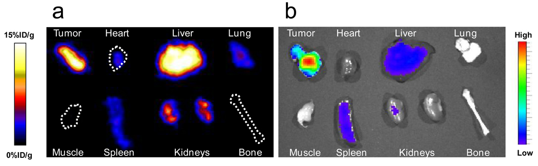
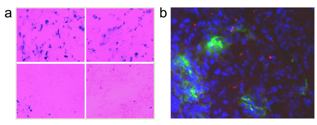
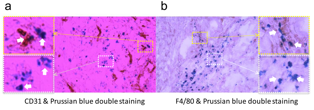
Engineered nanoparticles with theranostic functions have attracted a lot of attention for their potential role in the dawning era of personalized medicine. Iron oxide nanoparticles (IONPs), with their advantages of being non-toxic, biodegradable and inexpensive, are candidate platforms for the buildup of theranostic nanostructures; however, progress in using them has been limited largely due to inefficient drug loading and delivery. In the current study, we utilized dopamine to modify the surface of IONPs, yielding nanoconjugates that can be easily encapsulated into human serum albumin (HSA) matrices (clinically utilized drug carriers). This nanosystem is well-suited for dual encapsulation of IONPs and drug molecules, because the encapsulation is achieved in a way that is similar to common drug loading. To assess the biophysical characteristics of this novel nanosystem, the HSA coated IONPs (HSA-IONPs) were dually labeled with (64)Cu-DOTA and Cy5.5, and tested in a subcutaneous U87MG xenograft mouse model. In vivo positron emission tomography (PET)/near-infrared fluorescence (NIRF)/magnetic resonance imaging (MRI) tri-modality imaging, and ex vivo analyses and histological examinations were carefully conducted to investigate the in vivo behavior of the nanostructures. With the compact HSA coating, the HSA-IONPs manifested a prolonged circulation half-life; more impressively, they showed massive accumulation in lesions, high extravasation rate, and low uptake of the particles by macrophages at the tumor area.
Published by Elsevier Ltd.
Figures
Comment in
-
Multimodal imaging for cancer detection.Nanomedicine (Lond). 2010 Jul;5(5):687-91. doi: 10.2217/nnm.10.47. Nanomedicine (Lond). 2010. PMID: 20662640 No abstract available.
Similar articles
-
A dual-labeled knottin peptide for PET and near-infrared fluorescence imaging of integrin expression in living subjects.Bioconjug Chem. 2010 Mar 17;21(3):436-44. doi: 10.1021/bc9003102. Epub 2010 Feb 4. Bioconjug Chem. 2010. PMID: 20131753 Free PMC article.
-
PET/MRI dual-modality tumor imaging using arginine-glycine-aspartic (RGD)-conjugated radiolabeled iron oxide nanoparticles.J Nucl Med. 2008 Aug;49(8):1371-9. doi: 10.2967/jnumed.108.051243. Epub 2008 Jul 16. J Nucl Med. 2008. PMID: 18632815
-
Dual-function probe for PET and near-infrared fluorescence imaging of tumor vasculature.J Nucl Med. 2007 Nov;48(11):1862-70. doi: 10.2967/jnumed.107.043216. Epub 2007 Oct 17. J Nucl Med. 2007. PMID: 17942800
-
[Development of Molecular Probes Based on Iron Oxide Nanoparticles for in Vivo Magnetic Resonance/Photoacoustic Dual Imaging of Target Molecules in Tumors].Yakugaku Zasshi. 2017;137(1):55-60. doi: 10.1248/yakushi.16-00228. Yakugaku Zasshi. 2017. PMID: 28049896 Review. Japanese.
-
Shape-, size- and structure-controlled synthesis and biocompatibility of iron oxide nanoparticles for magnetic theranostics.Theranostics. 2018 May 11;8(12):3284-3307. doi: 10.7150/thno.25220. eCollection 2018. Theranostics. 2018. PMID: 29930730 Free PMC article. Review.
Cited by
-
Multifunctional nanoparticles for drug delivery and molecular imaging.Annu Rev Biomed Eng. 2013;15:253-82. doi: 10.1146/annurev-bioeng-071812-152409. Epub 2013 Apr 29. Annu Rev Biomed Eng. 2013. PMID: 23642243 Free PMC article. Review.
-
Recent Advances in Smart Biomaterials for the Detection and Treatment of Autoimmune Diseases.Adv Funct Mater. 2020 Sep 10;30(37):1909556. doi: 10.1002/adfm.201909556. Epub 2020 Mar 13. Adv Funct Mater. 2020. PMID: 33071713 Free PMC article.
-
Albumin incorporation into recognising layer of HER2-specific magnetic nanoparticles as a tool for optimal targeting of the acidic tumor microenvironment.Heliyon. 2024 Jul 6;10(14):e34211. doi: 10.1016/j.heliyon.2024.e34211. eCollection 2024 Jul 30. Heliyon. 2024. PMID: 39100472 Free PMC article.
-
Radiolabeled nanoparticles for multimodality tumor imaging.Theranostics. 2014 Jan 24;4(3):290-306. doi: 10.7150/thno.7341. eCollection 2014. Theranostics. 2014. PMID: 24505237 Free PMC article. Review.
-
Radiolabeled polyoxometalate clusters: Kidney dysfunction evaluation and tumor diagnosis by positron emission tomography imaging.Biomaterials. 2018 Jul;171:144-152. doi: 10.1016/j.biomaterials.2018.04.019. Epub 2018 Apr 12. Biomaterials. 2018. PMID: 29689411 Free PMC article.
References
-
- Jun YW, Huh YM, Choi JS, Lee JH, Song HT, Kim S, et al. Nanoscale size effect of magnetic nanocrystals and their utilization for cancer diagnosis via magnetic resonance imaging. J Am Chem Soc. 2005;127:5732–5733. - PubMed
-
- Cai W, Chen X. Nanoplatforms for targeted molecular imaging in living subjects. Small. 2007;3:1840–1854. - PubMed
Publication types
MeSH terms
Substances
Grants and funding
LinkOut - more resources
Full Text Sources
Other Literature Sources
Medical
