. 2010 Nov;21(11):1069-75.
doi: 10.1016/j.jnutbio.2009.09.003. Epub 2010 Jan 25.

Gestational zinc deficiency affects the regulation of transcription factors AP-1, NF-κB and NFAT in fetal brain

Affiliations

Gestational zinc deficiency affects the regulation of transcription factors AP-1, NF-κB and NFAT in fetal brain

Lucila Aimo et al. J Nutr Biochem. 2010 Nov.

Abstract

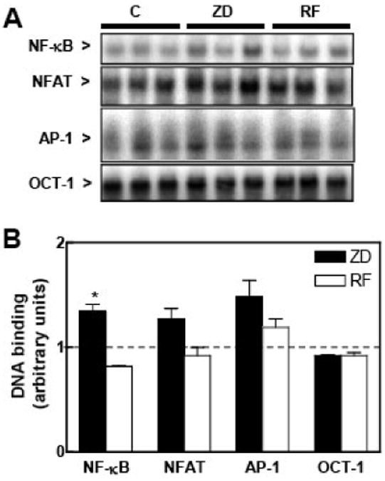
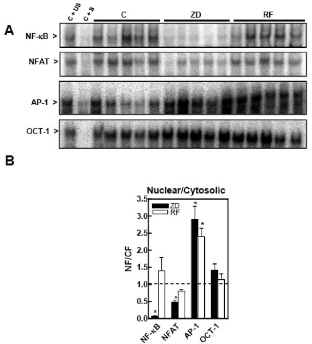
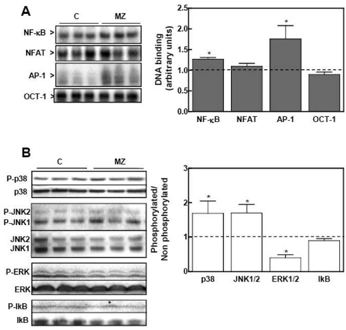
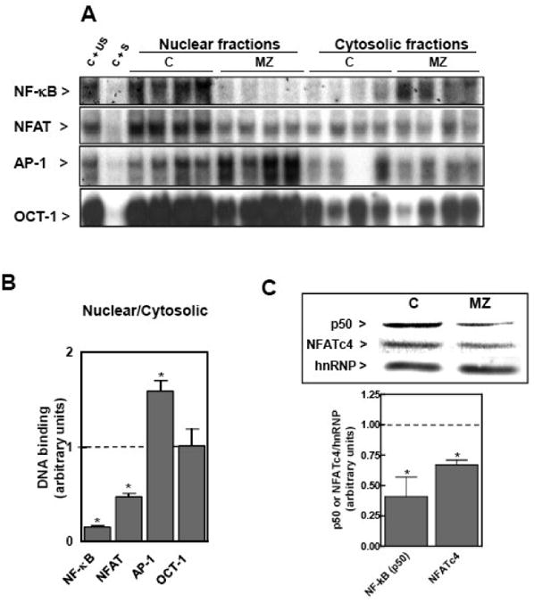
Transcription factors AP-1, nuclear factor κB (NF-κB) and NFAT are central to brain development by regulating the expression of genes that modulate cell proliferation, differentiation, apoptosis and synaptic plasticity. This work investigated the consequences of feeding zinc-deficient and marginal zinc diets to rat dams during gestation on the modulation of AP-1, NF-κB and NFAT in fetal brain. Sprague-Dawley rats were fed from gestation day (GD) 0 a control diet ad libitum (25 μg zinc/g diet, C), a zinc-deficient diet ad libitum (0.5 μg zinc/g diet, ZD), the control diet in the amounts eaten by the ZD rats (restrict fed, RF) or a diet containing a marginal zinc concentration ad libitum (10 μg zinc/g diet, MZD) until GD 19. AP-1-DNA binding was higher (50-190%) in nuclear fraction isolated from ZD, RF and MZD fetal brains compared to controls. In MZD fetal brain, high levels of activation of the upstream mitogen-activated protein kinases JNK and p38 and low levels of ERK phosphorylation were observed. Total levels of NF-κB and NFAT activation were higher or similar in the ZD and MZD groups than in controls, respectively. However, NF-κB- and NFAT-DNA binding in nuclear fractions was markedly lower in ZD and MZD fetal brain than in controls (50-80%). The latter could be related to zinc deficiency-associated alterations of the cytoskeleton, which is required for NF-κB and NFAT nuclear transport. In summary, suboptimal zinc nutrition during gestation could cause long-term effects on brain function, partially through a deregulation of transcription factors AP-1, NF-κB and NFAT.


Figures

Figure 1
Figure 1. A severe gestational zinc deficiency affects NF-κB, NFAT and AP-1 modulation in rat fetal brain total fractions
At GD 0, dams were fed control diets ad libitum (C) or at a restricted level (RF), or a zinc deficient (ZD) diet until GD 19. Total tissue fractions were prepared from fetal brains as described in the Materials and Methods section. A- EMSA for NF-κB, NFAT, AP-1 and OCT-1 in total fractions. B- After the EMSA assays, bands were quantitated, normalized to control levels (dotted line) and results for ZD (full bars) and RF (empty bars) groups are shown as means ± SEM of 4-6 animals per group. *Significantly different compared to the C and RF groups (p < 0.05, one way ANOVA test).
Figure 2
Figure 2. A severe gestational zinc deficiency affects the DNA binding of NF-κB, NFAT and AP-1 in nuclear and cytosolic fractions isolated from rat fetal brain
At GD 0, dams were fed control diets ad libitum (C) or at a restricted level (RF), or a zinc deficient (ZD) diet until GD 19. Nuclear and cytosolic fractions were prepared from fetal brains as described in the Materials and Methods section. A- EMSA for NF-κB, NFAT, AP-1 and OCT-1 in nuclear fractions. To determine the specificity of each transcription factor-DNA complex, the control nuclear fraction (C) was incubated in the presence of 100-fold molar excess of unlabeled oligonucleotide containing the consensus sequence for either the specific (C + S) or an unspecific (C + US) transcription factor before the binding assay. B- After the EMSA assays, bands were quantitated and the ratio nuclear/ cytosolic DNA binding was calculated. Results for ZD (full bars) and RF (empty bars) groups are normalized to control levels (dotted line) and are shown as means ± SEM of 5-6 animals per group. *Significantly different compared to the C group (p < 0.05, one way ANOVA test).
Figure 3
Figure 3. A marginal zinc nutrition during gestation affects NF-κB, NFAT, AP-1 transcription factors, IκB and MAPKs in fetal brain total fractions
At GD 0, dams were fed ad libitum a control (C) or a marginal zinc (MZD) diet until GD 19. Total fetal brain tissue fractions were prepared as described in the Materials and Methods section. A- Transcription factor-DNA binding. Left panel: EMSA for NF-κB, NFAT, AP-1 and OCT-1 in total tissue fractions. Right panel: after the EMSA assays, bands were quantitated, normalized to control levels (dotted line) and results for the MZD group (gray bars) are shown as means ± SEM of 4-6 animals per group. *Significantly different compared to the C group (p < 0.05, one way ANOVA test). B- Immunoblots for IκB and MAPKs. Left panel: Western blots for phosphorylated JNK1/2 (p-JNK1/2), phosphorylated-p38 (p-p38), phosphorylated ERK1/2 (p-ERK1/2), phosphorylated IκB (p-IκBα) and their non- phosphorylated forms. Right panel: After quantitation, results were expressed as the ratio phosphorylated/non-phosphorylated protein. Results for the MZD group were normalized to control values (dotted line) and are shown as means ± SEM of 6 animals per group. *Significantly different compared to the C group (p < 0.05, one way ANOVA test).
Figure 4
Figure 4. A marginal zinc nutrition during gestation affects the DNA binding of NF-κB, NFAT and AP-1 in fetal brain nuclear and cytosolic fractions
At GD 0, dams were fed ad libitum a control (C) o or a marginal zinc (MZD) diet until GD 19. Nuclear and cytosolic fractions were prepared from fetal brains as described in the Materials and Methods section. A- EMSA for NF-κB, NFAT, AP-1 and OCT-1 in nuclear fractions. To determine the specificity of each transcription factor-DNA complex, the control nuclear fraction (C) was incubated in the presence of 100-fold molar excess of unlabeled oligonucleotide containing the consensus sequence for either the specific (C + S) or an unspecific (C + US) transcription factor before the binding assay. B- After the EMSA assays, bands were quantitated and the ratio nuclear/cytosolic DNA binding was calculated. Results for the MZD group were normalized to control values (dotted line) and are shown as means ± SEM of 6 animals per group. *Significantly different compared to the C group (p < 0.05, one way ANOVA test). C- Western blots for NFATc4, p50 and hnRNP in nuclear fractions. After quantitation, results were expressed by the hnRNP content as loading control. Results for the MZD group were normalized to control values (dotted line) and are shown as means ± SEM of 3 animals per group. *Significantly different compared to the C group (p < 0.05, one way ANOVA test).

References

    1. Scheplyagina LA. Impact of the mother's zinc deficiency on the woman's and newborn's health status. J Trace Elem Med Biol. 2005;19:29–35. - PubMed
    1. Briefel RR, Bialostosky K, Kennedy-Stephenson J, McDowell MA, Ervin RB, Wright JD. Zinc intake of the U.S. population: findings from the third National Health and Nutrition Examination Survey, 1988-1994. J Nutr. 2000 May;130:1367S–73S. - PubMed
    1. Wada L, King JC. Trace element nutrition during pregnancy. Clin Obstet Gynecol. 1994 Sep;37:574–86. - PubMed
    1. Walsh CT, Sandstead HH, Prasad AS, Newberne PM, Fraker PJ. Zinc: health effects and research priorities for the 1990s. Environ Health Perspect. 1994 Jun;102(2):5–46. - PMC - PubMed
    1. Caulfield LE, Zavaleta N, Shankar AH, Merialdi M. Potential contribution of maternal zinc supplementation during pregnancy to maternal and child survival. Am J Clin Nutr. 1998 Aug;68:499S–508S. - PubMed

Publication types

MeSH terms