. 2010 May;9(5):791-803.
doi: 10.1074/mcp.M900516-MCP200. Epub 2010 Jan 21.

Post-translational modifications of integral membrane proteins resolved by top-down Fourier transform mass spectrometry with collisionally activated dissociation

Affiliations

Post-translational modifications of integral membrane proteins resolved by top-down Fourier transform mass spectrometry with collisionally activated dissociation

Christopher M Ryan et al. Mol Cell Proteomics. 2010 May.

Abstract

Integral membrane proteins remain a challenge to proteomics because they contain domains with physicochemical properties poorly suited to today's bottom-up protocols. These transmembrane regions may potentially contain post-translational modifications of functional significance, and thus development of protocols for improved coverage in these domains is important. One way to achieve this goal is by using top-down mass spectrometry whereby the intact protein is subjected to mass spectrometry and dissociation. Here we describe top-down high resolution Fourier transform mass spectrometry with collisionally activated dissociation to study post-translationally modified integral membrane proteins with polyhelix bundle and transmembrane porin motifs and molecular masses up to 35 kDa. On-line LC-MS analysis of the bacteriorhodopsin holoprotein yielded b- and y-ions that covered the full sequence of the protein and cleaved 79 of 247 peptide bonds (32%). The experiment proved that the mature sequence consists of residues 14-261, confirming N-terminal propeptide cleavage and conversion of N-terminal Gln-14 to pyrrolidone carboxylic acid (-17.02 Da) and C-terminal removal of Asp-262. Collisionally activated dissociation fragments localized the N(6)-(retinylidene) modification (266.20 Da) between residues 225-248 at Lys-229, the sole available amine in this stretch. Off-line nanospray of all eight subunits of the cytochrome b(6)f complex from the cyanobacterium Nostoc PCC 7120 defined various post-translational modifications, including covalently attached c-hemes (615.17 Da) on cytochromes f and b. Analysis of murine mitochondrial voltage-dependent anion channel established the amenability of the transmembrane beta-barrel to top-down MS and localized a modification site of the inhibitor Ro 68-3400 at Cys-232. Where neutral loss of the modification is a factor, only product ions that carry the modification should be used to assign its position. Although bond cleavage in some transmembrane alpha-helical domains was efficient, other regions were refractory such that their primary structure could only be inferred from the coincidence of genomic translation with precursor and product ions that spanned them.


Figures
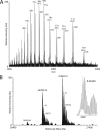
Fig. 1.
Fig. 1.
Top-down mass spectrometry of bacteriorhodopsin holoprotein. A, ESI mass spectrum of bacteriorhodopsin after purification by size exclusion chromatography in chloroform/methanol/aqueous formic acid. The spectrum shown was recorded by the FTICR operated below unit resolution to illustrate the typical charge state distribution observed for this integral membrane protein. The retinal chromophore is hydrolyzed from the polypeptide in acidic conditions such that paired signals arise from both apo- and holoforms at each charge state. Approximate average masses and charge states are labeled. Ion statistics for individual ions were poor in this full scan experiment. B, deconvolution of apo- and holoforms. The zero-charge molecular mass profile obtained after deconvolution of a selected ion monitoring experiment (m/z 100 width; 40 transients averaged) on the 11-charge ion shows both forms differing by the mass of retinal (266 Da) as well as mild oxidation (+16 Da) and formylation (+28 Da) of the polypeptide. With the instrument operated at 750,000 resolution at m/z 400, a resolution of around 60,000 was achieved for the 11-charge ions at m/z 2460. C, CAD of the holoprotein. The ion isolation on the 11-charge precursor (2460; inset top left) and its CAD tandem mass spectrum are shown. The ion isolation is widened to ensure maximal signal strength. Note that this experiment uses the extended mass range of the ion trap with useful product ions up to nearly 3200 m/z. Unit resolution was achieved on all product ions by operating the instrument at 750,000 resolution at 400 m/z. D, ion assignments for the bacteriorhodopsin holoprotein. CAD experiments were performed on 11-, 12-, 13-, 14-, 17-, 18-charge precursors, and the product ions were matched to the known primary structure of bacteriorhodopsin with its retinal cofactor at Lys-216 using ProSightPC software (version 1.0) operated at 10-ppm tolerance with the delta mass feature deactivated. Matched peak lists were pooled, and the composite list was again matched to the structure to give the ion assignments shown. 67 b- and 55 y-ions were matched, giving coverage of 79 of 247 peptide bonds (32%) and a P Score of 3.9e−150. Transmembrane domains are boxed. AU, arbitrary units; R, resolution.
Fig. 1.
Fig. 1.
Top-down mass spectrometry of bacteriorhodopsin holoprotein. A, ESI mass spectrum of bacteriorhodopsin after purification by size exclusion chromatography in chloroform/methanol/aqueous formic acid. The spectrum shown was recorded by the FTICR operated below unit resolution to illustrate the typical charge state distribution observed for this integral membrane protein. The retinal chromophore is hydrolyzed from the polypeptide in acidic conditions such that paired signals arise from both apo- and holoforms at each charge state. Approximate average masses and charge states are labeled. Ion statistics for individual ions were poor in this full scan experiment. B, deconvolution of apo- and holoforms. The zero-charge molecular mass profile obtained after deconvolution of a selected ion monitoring experiment (m/z 100 width; 40 transients averaged) on the 11-charge ion shows both forms differing by the mass of retinal (266 Da) as well as mild oxidation (+16 Da) and formylation (+28 Da) of the polypeptide. With the instrument operated at 750,000 resolution at m/z 400, a resolution of around 60,000 was achieved for the 11-charge ions at m/z 2460. C, CAD of the holoprotein. The ion isolation on the 11-charge precursor (2460; inset top left) and its CAD tandem mass spectrum are shown. The ion isolation is widened to ensure maximal signal strength. Note that this experiment uses the extended mass range of the ion trap with useful product ions up to nearly 3200 m/z. Unit resolution was achieved on all product ions by operating the instrument at 750,000 resolution at 400 m/z. D, ion assignments for the bacteriorhodopsin holoprotein. CAD experiments were performed on 11-, 12-, 13-, 14-, 17-, 18-charge precursors, and the product ions were matched to the known primary structure of bacteriorhodopsin with its retinal cofactor at Lys-216 using ProSightPC software (version 1.0) operated at 10-ppm tolerance with the delta mass feature deactivated. Matched peak lists were pooled, and the composite list was again matched to the structure to give the ion assignments shown. 67 b- and 55 y-ions were matched, giving coverage of 79 of 247 peptide bonds (32%) and a P Score of 3.9e−150. Transmembrane domains are boxed. AU, arbitrary units; R, resolution.
Fig. 2.
Fig. 2.
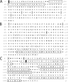
Top-down mass spectrometry for identification of post-translationally modified unknown. A, sequence tags from an unknown species of mass 11,185 Da. A top-down experiment was performed on the m/z 1245 ion (9+), corresponding to a previously unseen species within the preparation. The sequence tag compiler function of ProSightPC 1.0 was used to generate a list of potential sequence tags using default parameters. These tags were then matched to the complete Nostoc proteome database using the same software. B, result of sequence tag search. The sequence tag search identified a known subunit of the cytochrome b6f complex as the best match. Subunit 4 (PetD) was matched with two tags of 4 and 5 amino acid residues in length as highlighted. C, ion assignments without refinement of primary structure. The PetD sequence was compared with the top-down product ion peak list with 10 y-ions matched, strongly suggesting modification of the N terminus. D, ion assignments after refinement of N terminus. The first 58 amino acid residues were removed from the N terminus, resulting in close agreement of measured and calculated masses for this species (<1 ppm). An additional set of 19 b-ions was subsequently matched, confirming the N terminus with a P Score of 7.3e−35. It was concluded that the 11,185-Da molecule was an artifact resulting from DP cleavage due to brief exposure to concentrated formic acid.
Fig. 3.
Fig. 3.
Post-translational modifications of large subunits of the Nostoc cytochrome b6f complex. A, N-terminal acetylation of the Rieske Fe-S subunit (PetC). CAD was performed on a 19-charge precursor at m/z 1006. Product ions were matched to PetC, confirming N-terminal acetylation after removal of the initiating Met residue. The iron-sulfur center was apparently displaced with subsequent oxidation of Cys residues to form a second disulfide bond, giving four b- and eight y-ions matched. B, c-type cytochrome modification of the cytochrome f subunit (PetA). CAD was performed on 28- and 22-charge precursor ions at m/z 1135 and 1445, and the product ion peak lists were matched to PetA, confirming removal of residues 1–44 and covalent attachment of c-heme. To achieve the best match of measured data and primary structure, it was necessary to convert one Glu residue (Glu-136) to Gln, giving 13 b- and 23 y-ions matched. C, c-type cytochrome modification of the cytochrome b subunit (PetB). CAD was performed on the 17-charge precursor at m/z 1457, and the product ion peak list was matched to PetB, confirming removal of the initiating Met residue and covalent attachment of c-heme. As in the case of PetA (C), it was necessary to convert one Glu residue to Gln toward the N terminus to optimally match the data to the primary structure.
Fig. 4.
Fig. 4.
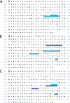
Top-down mass spectrometry of integral transmembrane porin, murine VDAC1 modified with Ro 68-3400. A, ion assignments, no modification included. VDAC1 carrying one molecule of Ro 68-3400 was analyzed by top-down mass spectrometry. CAD was performed on the 32-charge precursor at m/z 1014. The ion assignments shown were obtained by matching the product ion peak list from the singly modified VDAC1 to the unmodified sequence (with a consequent precursor delta mass of 274 Da). A set of overlapping b- and y-ions (shaded light blue; ions do not carry the modification) indicate that the data support full sequence coverage of unmodified VDAC1, alerting us to potential problems with the data set at least with respect to ions that do not carry the modification. Ions that do not carry the modification can arise from ions that do carry the modification due to neutral loss and are consequently ambiguous with respect to useful localization of a modification. Only product ions that carry the modification should be used to localize the modification site. B, ion assignments, Cys-127 modification. With the modification placed at Cys-127, a set of product y-ions appears (shaded dark blue; ions carry the modification) that localizes the modification to the C-terminal segment of the protein that contains both Cys residues. A number of unmodified product ion assignments (shaded light blue) support modification of Cys-127 but should be regarded with skepticism based upon the interpretation of A. According to the crystal structure, Cys-127 protrudes into the bilayer. C, ion assignments, Cys-232 modification. With the modification placed at Cys-232, a new set of y-ions appears that carries the modification (shaded dark blue) and excludes Cys-127. Thus, assuming only Cys residues can be modified, there is firm evidence to support the presence of Cys-232 modification. It remains possible that a substoichiometric population modified at Cys-127 is present, but this cannot be confirmed with the available ion assignments. According to the crystal structure, Cys-232 protrudes into the pore channel.

Similar articles

Cited by

References

    1. Kelleher N. L., Lin H. Y., Valaskovic G. A., Aaserud D. J., Fridriksson E. K., McLafferty F. W. (1999) Top down versus bottom up protein characterization by tandem high-resolution mass spectrometry. J. Am. Chem. Soc 121, 806–807
    1. Kelleher N. L., Zubarev R. A., Bush K., Furie B., Furie B. C., McLafferty F. W., Walsh C. T. (1999) Localization of labile posttranslational modifications by electron capture dissociation: the case of gamma-carboxyglutamic acid. Anal. Chem 71, 4250–4253 - PubMed
    1. Jebanathirajah J. A., Pittman J. L., Thomson B. A., Budnik B. A., Kaur P., Rape M., Kirschner M., Costello C. E., O'Connor P. B. (2005) Characterization of a new qQq-FTICR mass spectrometer for post-translational modification analysis and top-down tandem mass spectrometry of whole proteins. J. Am. Soc. Mass Spectrom 16, 1985–1999 - PubMed
    1. Whitelegge J. P., Gundersen C. B., Faull K. F. (1998) Electrospray-ionization mass spectrometry of intact intrinsic membrane proteins. Protein Sci 7, 1423–1430 - PMC - PubMed
    1. Wu C. C., MacCoss M. J., Howell K. E., Yates J. R., 3rd (2003) A method for the comprehensive proteomic analysis of membrane proteins. Nat. Biotechnol 21, 532–538 - PubMed

Publication types

MeSH terms