. 2010 May;156(Pt 5):1424-1438.
doi: 10.1099/mic.0.036640-0. Epub 2010 Jan 21.

Analysis of HmsH and its role in plague biofilm formation

Affiliations

Analysis of HmsH and its role in plague biofilm formation

Arwa Abu Khweek et al. Microbiology (Reading). 2010 May.

Abstract

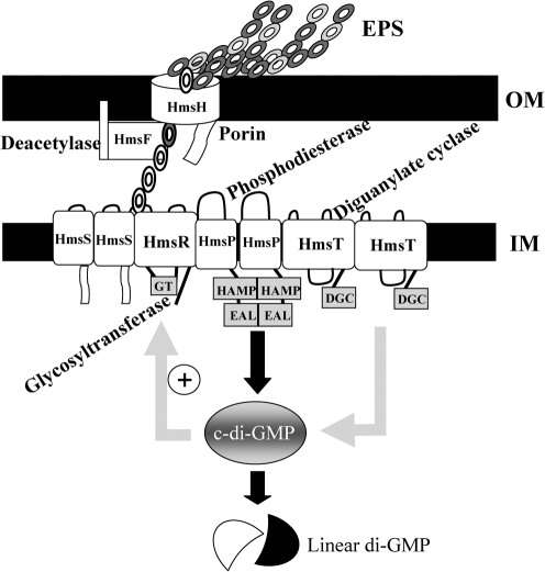
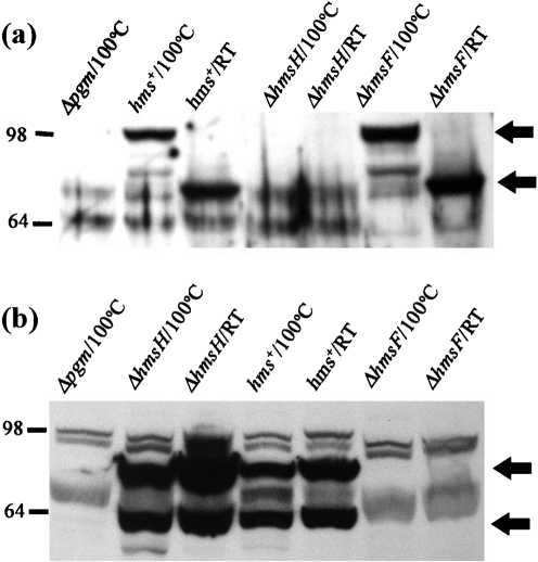
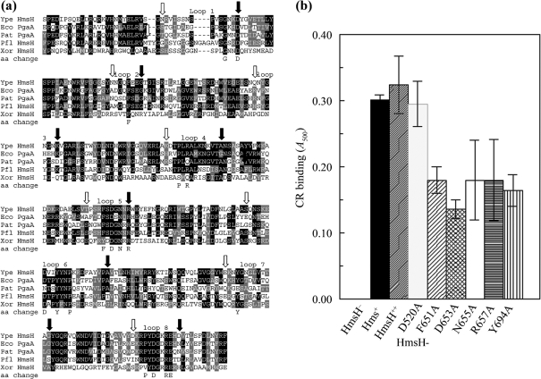
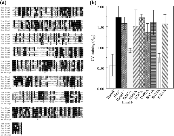
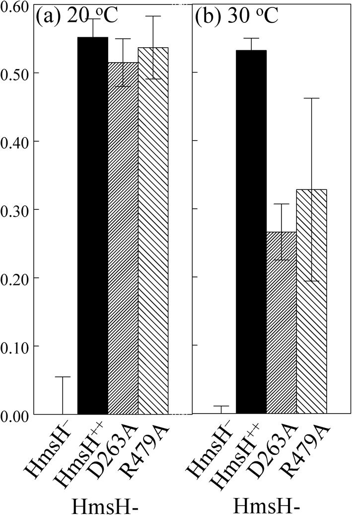
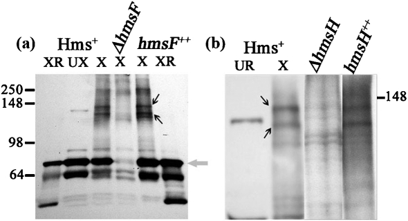
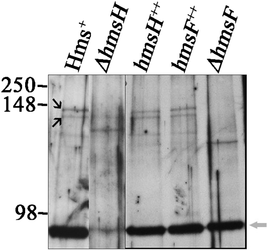
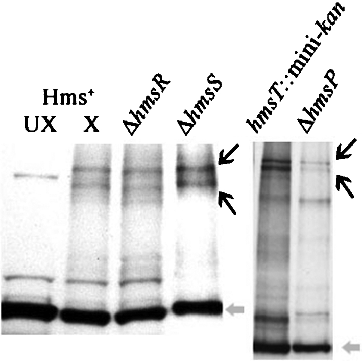
The Yersinia pestis Hms(+) phenotype is a manifestation of biofilm formation that causes adsorption of Congo red and haemin at 26 degrees C but not at 37 degrees C. This phenotype is required for blockage of the proventricular valve of the oriental rat flea and plays a role in transmission of bubonic plague from fleas to mammals. Genes responsible for this phenotype are located in three separate operons, hmsHFRS, hmsT and hmsP. HmsH and HmsF are outer membrane (OM) proteins, while the other four Hms proteins are located in the inner membrane. According to the Hidden Markov Method-based predictor, HmsH has a large N terminus in the periplasm, a beta-barrel structure with 16 beta-strands that traverse the OM, eight surface-exposed loops, and seven short turns connecting the beta-strands on the periplasmic side. Here, we demonstrate that HmsH is a heat-modifiable protein, a characteristic of other beta-barrel proteins, thereby supporting the bioinformatics analysis. Alanine scanning mutagenesis was used to identify conserved amino acids in the HmsH-like family that are critical for the function of HmsH in biofilm formation. Of 23 conserved amino acids mutated, four residues affected HmsH function and three likely caused protein instability. We used formaldehyde cross-linking to demonstrate that HmsH interacts with HmsF but not with HmsR, HmsS, HmsT or HmsP. Loss-of-function HmsH variants with single alanine substitutions retained their beta-structure and interaction with HmsF. Finally, using a polar hmsH : : mini-kan mutant, we demonstrated that biofilm development is not important for the pathogenesis of bubonic or pneumonic plague in mice.


Figures

Fig. 1.
Fig. 1.
Y. pestis Hms-dependent biofilm model. Chains of circles represent linked monomers of the EPS component of the biofilm (filled and unfilled circles, acetylated and deacetylated monomers). Labels indicate the putative or proven enzymic activities/functions of Hms proteins. The ‘+’ sign indicates likely stimulation of HmsR enzymatic activity by c-di-GMP. At 26–34 °C, all Hms proteins are highly expressed, while at 37 °C, the levels of HmsH, HmsR and HmsT are significantly reduced to varying degrees by post-translational mechanisms. HmsT degradation reduces c-di-GMP levels. This, along with low levels of HmsR and HmsH, inhibits biofilm formation.
Fig. 2.
Fig. 2.
Effect of heat treatment of HmsH and HmsF on migration in polyacrylamide gels. Samples were suspended in sample buffer containing 2 % SDS, heated at 100 °C for 10 min or unheated (RT; ∼20 °C), then resolved by 8 % SDS-PAGE. (a) Western blot probed with HmsH antisera. Arrows identify HmsH in boiled and unboiled samples. (b) Western blot probed with HmsF antisera. The upper arrow indicates the unprocessed form of HmsF, while the lower one indicates the processed form. Strains (see also Table 1): Δpgm, KIM6; hms+, KIM6+; ΔhmsH, KIM6-2115; ΔhmsF, KIM6-2116.
Fig. 3.
Fig. 3.
Analysis of changes in conserved residues in the predicted external loops between β-strands in the C-terminal region of HmsH. (a) Alignment of these regions of HmsH and HmsH-like proteins from: Y. pestis KIM10+ (Ype); P. fluorescens Pf-5 (Pfl); Pectobacterium atrosepticum SCR11043 (Pat; formerly Erwinia carotovora); E. coli K-12 substrain MG1655 (Eco); and X. oryzae KACC10331 (Xor). The black and grey boxes indicate residue identity and similarity, while arrows show the starts (open) and ends (filled) of the eight predicted loops between β-strands. The amino acid change line shows the amino acids individually changed to alanine. (b) Quantitative CR binding after 3 h incubation at 20 °C with CR-containing HIB medium by Y. pestis cells expressing amino acid substitutions in the predicted loops of HmsH. Controls are KIM6-2115 (in-frame ΔhmsH; HmsH), KIM6+ (Hms+) and KIM6-2115(pNPM22) (HmsH++). Plasmid pNPM22, which expresses hmsH from its native promoter, was also used to express HmsH with alanine substitutions. Thirteen mutants were not significantly different from positive controls (Table 2); only one of these, HmsH-D520A, along with the five mutants with significant decreases in CR binding, are shown. Data shown are the mean of two or more independent experiments with duplicate samples from each trial. Error bars, sd. For clarity, the secondary mutation (D520A) in HmsH-F651-D520A and HmsH-N655A-D520A is not shown in the labels.
Fig. 4.
Fig. 4.
Analysis of changes in conserved residues in the N terminus of HmsH. (a) Alignment of these regions of HmsH and HmsH-like proteins from Y. pestis KIM10+ (Ype); P. fluorescens Pf-5 (Pfl); Pectobacterium atrosepticum SCR11043 (Pat; formerly Erwinia carotovora); E. coli K-12 substrain MG1655 (Eco); and X. oryzae KACC10331 (Xor). The black and grey boxes indicate residue identity and similarity, respectively. Sequences start with the first residue after the SignalP predicted signal sequence. The amino acid change line shows the amino acids individually changed to alanine. (b) Quantitative CV staining by Y. pestis cells expressing amino acid substitutions in the N-terminal region of HmsH. Controls are KIM6-2115 (in-frame ΔhmsH; HmsH), KIM6+ (Hms+) and KIM6-2115(pNPM22) (HmsH++). Data shown are the mean of two or more independent experiments with duplicate samples from each trial. Error bars, sd. For clarity, the secondary mutation (D520A) and (D691A) in HmsH-E345A-D520A, HmsH-R433-D520A, HmsH-D397A-D520A and HmsH-R491-D691A is not shown in the labels.
Fig. 5.
Fig. 5.
CR binding by HmsH-D263A and HmsH-R479A variants is temperature-dependent. Quantitative CR binding is shown for Y. pestis KIM6-2115 (in-frame ΔhmsH; HmsH), KIM6-2115(pNPM22) (HmsH++) and the two variants after 3 h incubation at 20 °C (a) or 30 °C (b) with CR-containing HIB medium. Data shown are the mean of two or more independent experiments with duplicate samples from each trial. Error bars, sd.
Fig. 6.
Fig. 6.
Western blot of formaldehyde-cross-linked Y. pestis strains probed with HmsF antisera. (a) Y. pestis KIM6+ (Hms+), KIM6-2116 (ΔhmsF) and KIM6-2116(pBADhmsF) (hmsF++). Samples were cross-linked (X), not cross-linked (UX), or cross-linked and then reversed (XR). The grey arrow points to the HmsF monomer band. (b) Y. pestis KIM6+ (Hms+), KIM6-2115 (ΔhmsH) and KIM6-2115(pNPM22) (hmsH++). Except for lane 1, all samples were cross-linked. The HmsF monomer is not shown in (b). Black arrows in both panels point to the cross-linked complexes containing HmsF. Molecular masses of protein standards are shown within the range of 250–64 kDa. For (b), only the location of the 148 kDa standard is shown.
Fig. 7.
Fig. 7.
Western blot of formaldehyde-cross-linked Y. pestis strains probed with HmsH antisera. Strains: Y. pestis KIM6+ (Hms+), KIM6-2115 (ΔhmsH), KIM6-2115(pNPM22) (hmsH++), KIM6-2116(pBADhmsF) (hmsF++) and KIM6-2116 (ΔhmsF). Black arrows identify the two HmsH–HmsF cross-linked complexes, while the grey arrow shows the HmsH monomer. Molecular masses of protein standards are shown within the range of 250–98 kDa.
Fig. 8.
Fig. 8.
Western blot of formaldehyde-cross-linked Y. pestis strains KIM6+ (Hms+), KIM6-2118 (ΔhmsR), KIM6-2119 (ΔhmsS), KIM6-2051 (hmsT : : mini-kan) and KIM6-2090.2+ (ΔhmsP) probed with HmsF antisera. Except for one Hms+ control (Hms+ UX), all samples were cross-linked (X) with formaldehyde. Black arrows identify the cross-linked HmsH–HmsF complex, while the grey arrow identifies the HmsF monomer.

Similar articles

Cited by

References

    1. Ausubel, F. M. (1987). Current protocols in Molecular Biology. New York: Greene Publishing & Wiley-Interscience.
    1. Bacot, A. W. (1915). LXXXI. Further notes on the mechanism of the transmission of plague by fleas. J Hyg (Lond) 14 (Plague Suppl. 4), 744–776. - PMC - PubMed
    1. Bacot, A. W. & Martin, C. J. (1914). LXVII. Observations on the mechanism of the transmission of plague by fleas. J Hyg (Lond) 13 (Plague Suppl. 3), 423–439. - PMC - PubMed
    1. Bagos, P. G., Liakopoulos, T. D., Spyropoulos, I. C. & Hamodrakas, S. J. (2004a). A Hidden Markov Model method, capable of predicting and discriminating beta-barrel outer membrane proteins. BMC Bioinformatics 5, 29. - PMC - PubMed
    1. Bagos, P. G., Liakopoulos, T. D., Spyropoulos, I. C. & Hamodrakas, S. J. (2004b). PRED-TMBB: a web server for predicting the topology of beta-barrel outer membrane proteins. Nucleic Acids Res 32, W400–W404. - PMC - PubMed

Publication types

MeSH terms

LinkOut - more resources