

Assessment of sulcation of the fetal brain in cases of isolated agenesis of the corpus callosum using in utero MR imaging
- PMID: 20093312
- PMCID: PMC7963955
- DOI: 10.3174/ajnr.A1982
Assessment of sulcation of the fetal brain in cases of isolated agenesis of the corpus callosum using in utero MR imaging
Abstract
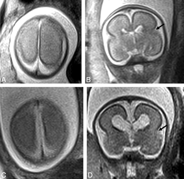
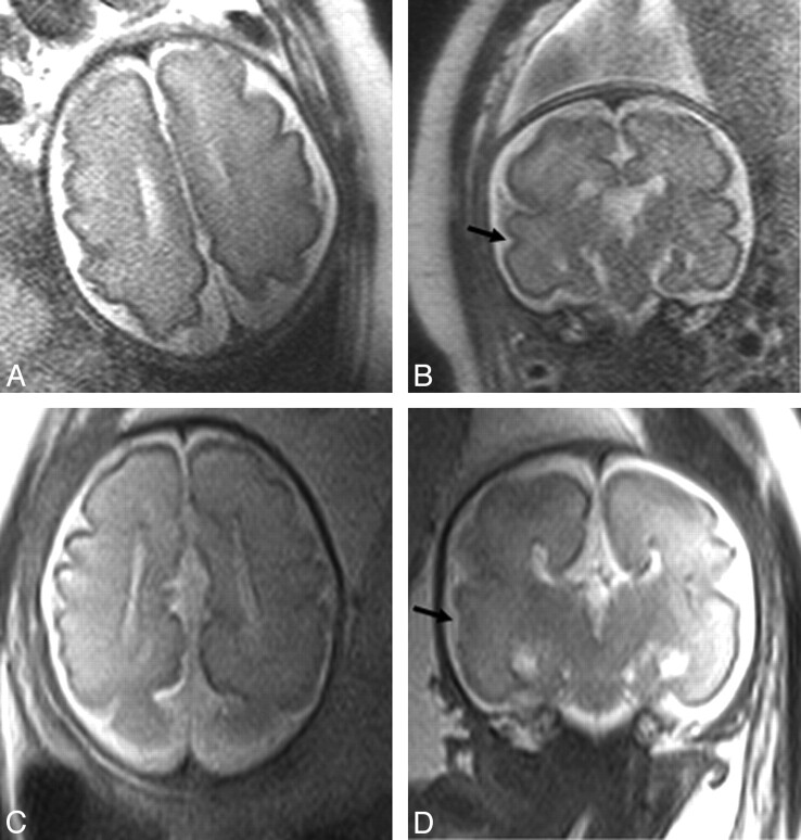
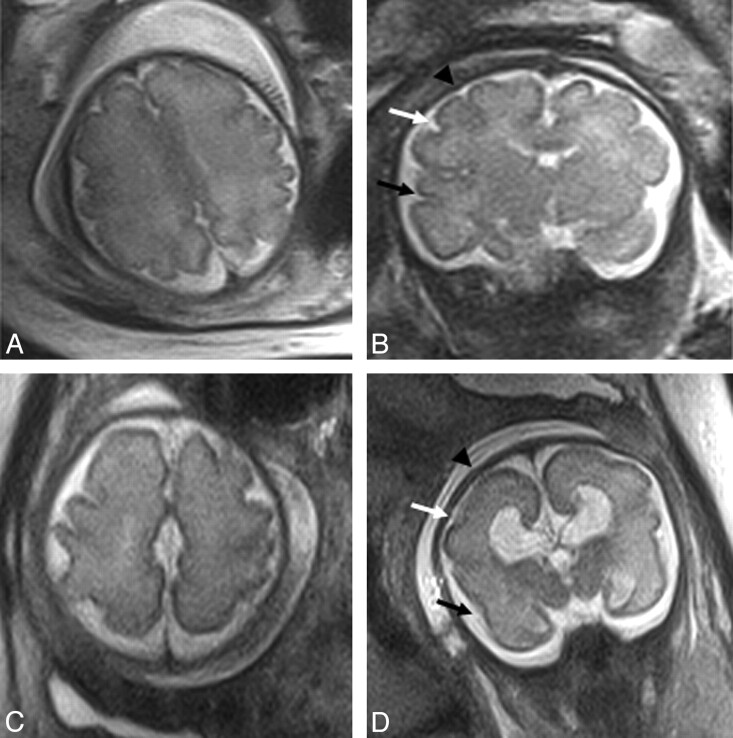
Background and purpose: There is gathering evidence to suggest that agenesis of the corpus callosum is associated with delayed fetal sulcation; it is possible that the corpus callosum facilitates normal gyral development. In this paper we sought to confirm whether delayed sulcation is found in fetuses with isolated agenesis of the corpus callosum as judged by in utero MR imaging.
Materials and methods: Retrospective analysis of 20 fetuses with isolated corpus callosum agenesis investigated by in utero MR imaging and 20 aged-matched normal fetuses was performed in the second or third trimester. All fetuses were singleton pregnancies with known gestational age, imaged on a 1.5T superconducting MR system. Estimation of sulcation maturity was made with reference to a standard atlas and subgroup analysis of earlier gestation (group 1, 21-26 weeks) and later gestation (group 2, 30-34 weeks) fetuses was performed.
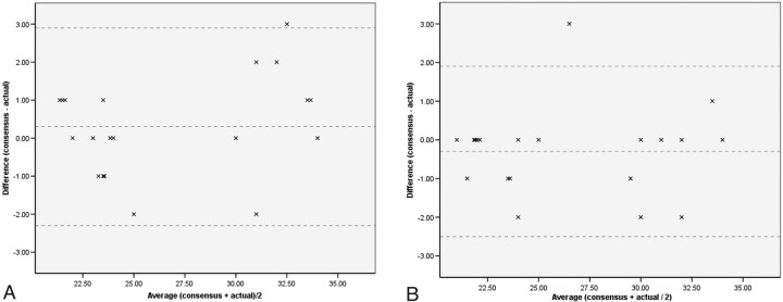
Results: Group 1 (n = 12) did not show a statistically significant difference between the 2 subgroups (P = .44) in terms of sulcation. A significant difference was demonstrated in the later gestation, group 2 (n = 8) fetal analyses; mean difference between consensus and actual gestation for normal fetuses was 0.9 weeks (SD of 1.5 weeks) versus -0.5 weeks (SD of 1.1 weeks) for the agenesis of corpus callosum cases (P = .046), suggestive of delayed sulcation in callosal agenesis.
Conclusions: Delayed sulcation encountered in third trimester fetuses with agenesis of the corpus callosum may be seen and does not in itself imply an additional brain abnormality.
Figures
Similar articles
-
Agenesis of the corpus callosum: an MR imaging analysis of associated abnormalities in the fetus.AJNR Am J Neuroradiol. 2009 Feb;30(2):257-63. doi: 10.3174/ajnr.A1331. Epub 2008 Nov 6. AJNR Am J Neuroradiol. 2009. PMID: 18988682 Free PMC article.
-
The "cortical invagination sign": a midtrimester sonographic marker of unilateral cortical focal dysgyria in fetuses with complete agenesis of the corpus callosum.Am J Obstet Gynecol MFM. 2023 Dec;5(12):101198. doi: 10.1016/j.ajogmf.2023.101198. Epub 2023 Oct 20. Am J Obstet Gynecol MFM. 2023. PMID: 37866717
-
Agenesis of the corpus callosum in fetuses with mild ventriculomegaly: role of MR imaging.Radiol Med. 2010 Mar;115(2):301-12. doi: 10.1007/s11547-009-0474-7. Epub 2009 Dec 16. Radiol Med. 2010. PMID: 20017009 English, Italian.
-
Role of prenatal magnetic resonance imaging in fetuses with isolated agenesis of corpus callosum in the era of fetal neurosonography: A systematic review and meta-analysis.Acta Obstet Gynecol Scand. 2021 Jan;100(1):7-16. doi: 10.1111/aogs.13958. Epub 2020 Aug 24. Acta Obstet Gynecol Scand. 2021. PMID: 32652537
-
Role of magnetic resonance imaging in fetuses with mild or moderate ventriculomegaly in the era of fetal neurosonography: systematic review and meta-analysis.Ultrasound Obstet Gynecol. 2019 Aug;54(2):164-171. doi: 10.1002/uog.20197. Epub 2019 Jul 11. Ultrasound Obstet Gynecol. 2019. PMID: 30549340
Cited by
-
Speech Disorders in Children with Congenital Heart Disease Attending a Tertiary Institution in South East Nigeria.Niger Med J. 2025 Jun 16;66(2):735-745. doi: 10.71480/nmj.v66i2.757. eCollection 2025 Mar-Apr. Niger Med J. 2025. PMID: 40703897 Free PMC article.
-
Quantification of sulcal emergence timing and its variability in early fetal life: Hemispheric asymmetry and sex difference.Neuroimage. 2022 Nov;263:119629. doi: 10.1016/j.neuroimage.2022.119629. Epub 2022 Sep 14. Neuroimage. 2022. PMID: 36115591 Free PMC article.
-
Improved neurodevelopmental prognostication in isolated corpus callosal agenesis: fetal magnetic resonance imaging-based scoring system.Ultrasound Obstet Gynecol. 2021 Jul;58(1):34-41. doi: 10.1002/uog.22102. Ultrasound Obstet Gynecol. 2021. PMID: 32484578 Free PMC article.
-
Disorganized Patterns of Sulcal Position in Fetal Brains with Agenesis of Corpus Callosum.Cereb Cortex. 2018 Sep 1;28(9):3192-3203. doi: 10.1093/cercor/bhx191. Cereb Cortex. 2018. PMID: 30124828 Free PMC article.
-
Corpus callosum agenesis and rehabilitative treatment.Ital J Pediatr. 2010 Sep 17;36:64. doi: 10.1186/1824-7288-36-64. Ital J Pediatr. 2010. PMID: 20849621 Free PMC article. Review.
References
-
- Garel C. MRI of the Fetal Brain: Normal Development and Cerebral Pathologies. Berlin: Springer; 2004
-
- Lan LM, Yamashita Y, Tang Y, et al. . Normal fetal brain development: MR imaging with a half-Fourier rapid acquisition with relaxation enhancement sequence. Radiology. 2000;215:205–10 - PubMed
-
- Rich P, Jones R, Britton J, et al. . MRI of the foetal brain. Clin Radiol 2007;62:303–13 - PubMed
-
- Levine D, Barnes PD. Cortical maturation in normal and abnormal fetuses as assessed with prenatal MR imaging. Radiology 1999;210:751–58 - PubMed
MeSH terms
LinkOut - more resources
Full Text Sources
Medical