. 2010 Mar 19;285(12):8572-84.
doi: 10.1074/jbc.M109.083394. Epub 2010 Jan 21.

A new type of signal peptidase cleavage site identified in an RNA virus polyprotein

Affiliations

A new type of signal peptidase cleavage site identified in an RNA virus polyprotein

Ioana Bintintan et al. J Biol Chem. .

Abstract

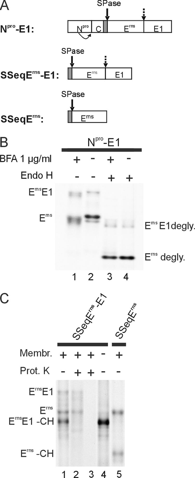
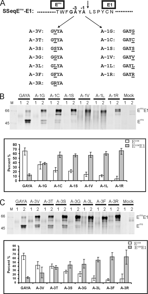
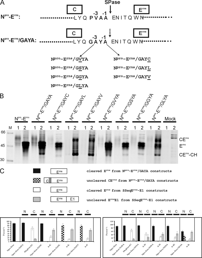
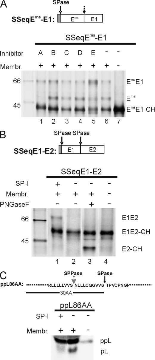
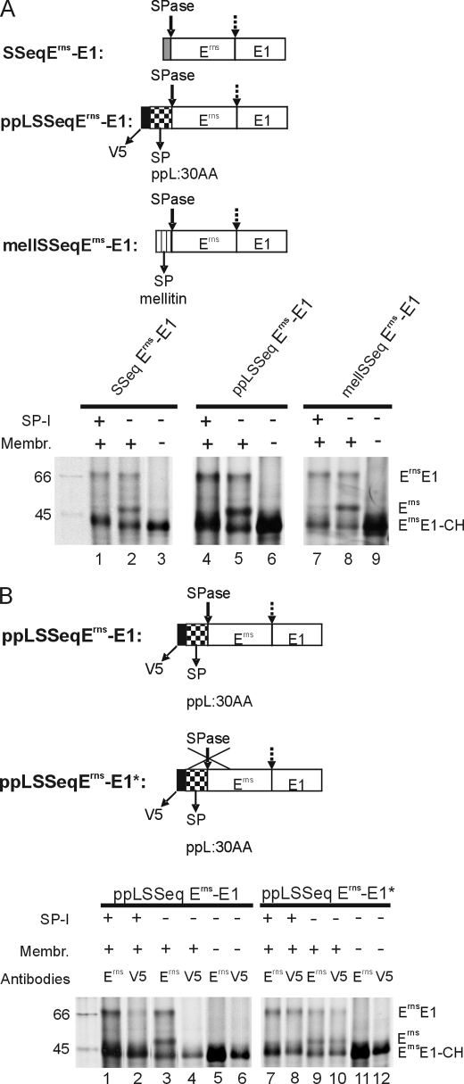
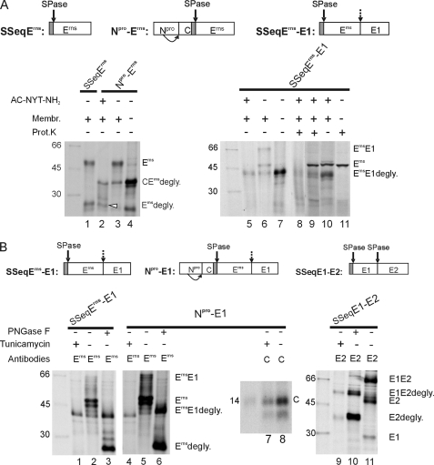
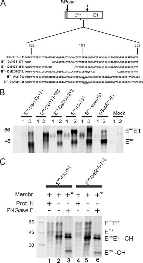
Pestiviruses, a group of enveloped positive strand RNA viruses belonging to the family Flaviviridae, express their genes via a polyprotein that is subsequently processed by proteases. The structural protein region contains typical signal peptidase cleavage sites. Only the site at the C terminus of the glycoprotein E(rns) is different because it does not contain a hydrophobic transmembrane region but an amphipathic helix functioning as the E(rns) membrane anchor. Despite the absence of a hydrophobic region, the site between the C terminus of E(rns) and E1, the protein located downstream in the polyprotein, is cleaved by signal peptidase, as demonstrated by mutagenesis and inhibitor studies. Thus, E(rns)E1 is processed at a novel type of signal peptidase cleavage site showing a different membrane topology. Prevention of glycosylation or introduction of mutations into the C-terminal region of E(rns) severely impairs processing, presumably by preventing proper membrane interaction or disturbing a conformation critical for the protein to be accepted as a substrate by signal peptidase.


Figures

FIGURE 1.
FIGURE 1.
ErnsE1 is cleaved in the ER. A, a schematic drawing of the cDNA constructs used in the expression studies presented in B and C. The names of the constructs are given on the left. The bars show the expressed regions of the viral polyprotein with the designations of the mature viral proteins. Signal peptides are shown as gray bars. A bent arrow indicates autocatalytic cleavage of Npro at its C terminus. A solid arrow points to known SPase cleavage sites, whereas the site investigated here is marked with a broken arrow. B, SDS-PAGE with products precipitated with a rabbit serum against Erns from cells transiently expressing the Npro-E1 construct in the absence or presence of BFA as indicated at the top of the gel. The precipitated proteins were loaded directly onto the gel or pretreated with endoglycosidase H (Endo H) as indicated above the gel. C, SDS-PAGE with products precipitated with the Erns-specific mAb 24/16 after in vitro translation of the constructs specified at the top. Translation was done in the presence or absence of canine microsomal membranes as indicated. To prove translocation of proteins into membrane vesicles, aliquots of the translation products obtained after translation in the presence of membranes were treated with Proteinase K (lane 2) or Proteinase K and Triton X-100 (lane 3) as described (32). The names of the different products are indicated on the left and right of gels in B and C. degly., the removal of carbohydrate side chains by deglycosylation; -CH, glycosylation did not occur because of absence of membranes or inefficient translocation. Prot. K, Proteinase K.
FIGURE 2.
FIGURE 2.
ErnsE1 processing efficiency of constructs with mutations at positions −1 and −3 of the cleavage site. A, mutations introduced into construct SSeqErns-E1. The sequence around the Erns/E1 cleavage site (broken arrow) is given, and positions −1 and −3 are indicated. Below, the names of the different constructs with the indicated mutations at −3 or −1 are underlined. B and C, results obtained after expression of the mutations at position −1 (B) or −3 (C). Top, SDS-PAGE with the proteins precipitated with the rabbit Erns serum from cell culture fluid (lanes 1) or the membrane fractions of cells (lanes 2) expressing the constructs given above or no plasmid (Mock). Lane M contains a protein size marker, and the size of the visible bands in kDa is given on the left. On the right, the names of the precipitated proteins are given. Below, a diagram shows the quantification of the results with the intracellular amounts of cleaved Erns (white bars) and uncleaved ErnsE1 precursor (gray bars) given here as a percentage of the total recovered Erns protein. Mean values of at least three independent experiments are given, and the S.D. is indicated. Note that the construct designated “GAYA” represents the wild type.
FIGURE 3.
FIGURE 3.
Processing efficiency of constructs with mutations at positions −1 and −3 in a standard signal peptide cleavage site. A, the original sequence context in the internal signal peptide located between capsid protein C and Erns in the CSFV polyprotein (construct Npro-Erns, upper part) and the sequence of the mutant Npro-Erns/GAYA with the C-terminal part of the signal peptide replaced by the GAYA motif found at the Erns C terminus. Below, the names of the different constructs with the indicated mutations at −3 or −1 of Npro-Erns/GAYA are given. B, the results obtained after transient expression, immunprecipitation, and SDS-PAGE of the mutated proteins. C, diagrams showing the processing efficiency determined for the different mutants presented in B in comparison with the results obtained for the equivalent mutations in the context of the Erns C terminus demonstrated in Fig. 2. On top, an explanation of the different types of bars is given. Below, the results for mutations affecting positions −1 (left diagram) or −3 (right diagram) are shown. Each section of the diagrams presents one type of mutation with its effect on processing of the standard SPase cleavage site at the N terminus of Erns (N) or on processing at the Erns C terminus (C). See also the legend to Fig. 2.
FIGURE 4.
FIGURE 4.
Influence of different protease inhibitors on ErnsE1 processing. A, SDS-PAGE with products of in vitro translation experiments with construct SSeqErns-E1 (shown on top) in the presence of the protease inhibitors specified by letters above the gel (A, SP-I, MeOSuc-Ala-Ala-Pro-Val chloromethyl ketone used at 1.5–2.5 mm; B, signal peptide peptidase inhibitor, 1,3-di-(N-carboxybenzoyl-l-leucyl-l-leucyl)amino acetone, used at 10 μm; C, γ-secretase inhibitor, N-[N-(3,5-difluorophenacetyl)-l-alanyl]-S-phenylglycine t-butyl ester, used at 100 μm; D, serine/cysteine protease inhibitor, leupeptin, used at 25 μm; E, site-2 protease-metalloprotease inhibitor, 1,10-phenanthroline, used at 5 mm). Note that the concentration at which 1,10-phenanthroline was used in this study was shown to inhibit also the site 1 intramembrane protease (71). Translation of the RNA in the absence of xinhibitor or in the absence of inhibitor and microsomal membranes, as specified by the code at the top of the gel, served as controls. Lane 5, showing the products obtained in the presence of inhibitor E, was exposed 5 times longer than the other lanes. See also the legend to Fig. 1. Effects of SP-I on processing of construct SSeqE1-E2 (B) or a preprolactin construct (C) were also determined. For the preprolactin construct, which includes the first 86 amino acids of the precursor protein, the signal peptide of 30 amino acids with the SPase and the signal peptide peptidase (SPPase) cleavage sites are specified. Protein size marker bands are shown with the molecular masses given in kDa.
FIGURE 5.
FIGURE 5.
Importance of the type of signal peptide and of signal peptide cleavage on ErnsE1 processing. A, the upper part shows schemes of the analyzed constructs in which ErnsE1 was combined with different signal peptides. Below, SDS-PAGE with proteins precipitated with mAb 24/16 after in vitro translation of the constructs indicated above the gels. Translation was done in the presence or absence of microsomal membranes or SP-I, as indicated. B, top, constructs pplSSeqErns-E1 and variant pplSSeqErns-E1* with the cleavage site SP/Erns blocked by mutation, are shown. Below, SDS-PAGE with products of in vitro translation precipitated with mAb 24/16 against Erns or an anti-V5 mAb, as specified above the gel. See also the legend to Fig. 1.
FIGURE 6.
FIGURE 6.
ErnsE1 processing under prevention of glycosylation. A, results obtained after in vitro translation. Top, the constructs used in the studies are presented schematically. Below, SDS-PAGE gels are shown with products precipitated with mAb 24/16 after in vitro translation of the indicated products in the presence or absence of the N-glycosylation acceptor (competitive inhibitor) AC-NYT-NH2 (lanes 1–8). Lanes 8–11 show the results of Proteinase K protection assays, with lanes 9–11 containing products of in vitro translation without immunoprecipitation. The presence or absence of Proteinase K is indicated at the top of the gel. Lane 11 shows a control established by translation in the absence of membranes and inhibitor followed by Proteinase K treatment. Please note that in lanes 9–11 a strong unspecific band migrating a bit faster than Erns. The white arrow marks unglycosylated Erns cleaved by SPase from the Npro/C/Erns precursor. B, results of transient expression assays conducted with the constructs shown on top. The gels show the proteins precipitated with the given antibodies (Erns, mAb 24/16 against Erns; C, rabbit serum against C protein; E2, mAb A18 against E2) from the extracts of cells transfected with the indicated constructs. The transfected cells were treated with tunicamycin as indicated above the gels. Some of the precipitated proteins were deglycosylated with PNGase F (lanes 3, 6, and 10). See also the legends to Figs. 1 and 2. Please note that E1 is visible in lane 11 due to coprecipitation with E2 because of formation of a disulfide-linked heterodimer in infected cells (13). Prot. K, Proteinase K.
FIGURE 7.
FIGURE 7.
Effect of short deletions or alanine insertions in the Erns C-terminal region on ErnsE1 processing. A, construct SSeqErns-E1 is shown with the C-terminal region of Erns presented as an enlargement with the sequence given in one-letter code (residue number in the mature Erns given at the top). The deletions and the positions of the inserted alanine residues (underlined) are demonstrated with the names of the corresponding expression constructs specified on the left. In analogy to the bovine viral diarrhea virus Erns, the amphipathic helix should start around position 180, as defined by an alanine insertion scanning approach (32). B, SDS-PAGE with the proteins precipitated with mAb 24/16 from tissue culture fluid (lanes 1) and cell extracts (lanes 2) of cells transfected with the indicated constructs. C, controls that were either done with Proteinase K protection assays using products of in vitro translation (lanes 1, 2, 4, and 5) or proteins precipitated from transfected cells (similar to B) that were deglycosylated with PNGase F. *, the membranes were not added as in the in vitro translation assays but were present because of expression in cells. See also the legends to Figs. 1 and 2.
FIGURE 8.
FIGURE 8.
Schematic presentation of the membrane topology and processing scheme of the pestivirus polyprotein region encompassing Erns, E1, and the N-terminal part of E2 (not drawn to scale). Transmembrane regions and the Erns C-terminal amphipathic helix are shown as cylinders, whereas the rest of the proteins are indicated by black lines. The two gray horizontal lines represent the ER- or cytosol-facing surfaces of the ER membrane. The translocon is indicated as a light gray structure. SPase cleavage sites are marked by arrows and SPase. The signal sequence upstream of Erns responsible for Erns translocation is not shown. Note that the transmembrane region upstream of the E2 protein is a regular signal sequence with a typical transmembrane topology that stands in marked contrast to the in plane configuration of the Erns amphipathic helix preceding the Erns/E1 SPase cleavage site.

Similar articles

Cited by

References

    1. Heinz F. X., Collett M. S., Purcell R. H., Gould E. A., Howard C. R., Houghton M., Moormann J. M., Rice C. M., Thiel H. J. (2000) in Virus Taxonomy: Seventh Report of the International Committee on Taxonomy of Viruses (van Regenmortel M. H. V., Fauquet C. M., Bishop D. H. L., Carstens E. B., Estes M. K., Lemon S. M., Maniloff J., Mayo M. A., McGeoch D. J., Pringle C. R., Wickner R. B. eds) pp. 859–878, Academic Press, Inc., San Diego, CA
    1. Lindenbach B. D., Thiel H.-J., Rice C. M. (2007) in Fields Virology (Knipe D. M., Howley P. M. eds) pp. 1101–1152, Lippincott-Raven Publishers, Philadelphia
    1. Collett M. S., Larson R., Belzer S. K., Retzel E. (1988) Virology 165, 200–208 - PubMed
    1. Collett M. S., Moennig V., Horzinek M. C. (1989) J. Gen. Virol. 70, 253–266 - PubMed
    1. Elbers K., Tautz N., Becher P., Stoll D., Rümenapf T., Thiel H. J. (1996) J. Virol. 70, 4131–4135 - PMC - PubMed

Publication types

LinkOut - more resources