. 2010 Mar 26;285(13):9749-9761.
doi: 10.1074/jbc.M109.043117. Epub 2010 Jan 21.

Pituitary adenylate cyclase-activating polypeptide (PACAP)/PAC1HOP1 receptor activation coordinates multiple neurotrophic signaling pathways: Akt activation through phosphatidylinositol 3-kinase gamma and vesicle endocytosis for neuronal survival

Affiliations

Pituitary adenylate cyclase-activating polypeptide (PACAP)/PAC1HOP1 receptor activation coordinates multiple neurotrophic signaling pathways: Akt activation through phosphatidylinositol 3-kinase gamma and vesicle endocytosis for neuronal survival

Victor May et al. J Biol Chem. .

Abstract

MAPK and Akt pathways are predominant mediators of trophic signaling for many neuronal systems. Among the vasoactive intestinal peptide/secretin/glucagon family of related peptides, pituitary adenylate cyclase-activating polypeptide (PACAP) binding to specific PAC(1) receptor isoforms can engage multiple signaling pathways and promote neuroprotection through mechanisms that are not well understood. Using a primary sympathetic neuronal system, the current studies demonstrate that PACAP activation of PAC(1)HOP1 receptors engages both MAPK and Akt neurotrophic pathways in an integrated program to facilitate neuronal survival after growth factor withdrawal. PACAP not only stimulated prosurvival ERK1/2 and ERK5 activation but also abrogated SAPK/JNK and p38 MAPK signaling in parallel. In contrast to the potent and rapid effects of PACAP in ERK1/2 phosphorylation, PACAP stimulated Akt phosphorylation in a late phase of PAC(1)HOP1 receptor signaling. From inhibitor and immunoprecipitation analyses, the PACAP/PAC(1)HOP1 receptor-mediated Akt responses did not represent transactivation mechanisms but appeared to depend on G alpha(q)/phosphatidylinositol 3-kinase gamma activity and vesicular internalization pathways. Phosphatidylinositol 3-kinase gamma-selective inhibitors blocked PACAP-stimulated Akt phosphorylation in primary neuronal cultures and in PAC(1)HOP1-overexpressing cell lines; RNA interference-mediated knockdown of the receptor effectors attenuated PACAP-mediated Akt activation. Similarly, perturbation of endocytic pathways also blocked Akt phosphorylation. Between ERK and Akt pathways, PACAP-stimulated Akt signaling was the primary cascade that attenuated cultured neuron apoptosis after growth factor withdrawal. The partitioning of PACAP-mediated Akt signaling in endosomes may be a key mechanism contributing to the high spatial and temporal specificity in signal transduction necessary for survival pathways.


Figures

FIGURE 1.
FIGURE 1.
PACAP peptides promote sympathetic neuron survival after growth factor withdrawal. A–C, immature 5-day primary sympathetic neuronal cultures were withdrawn from NGF-containing serum-free medium for 24 h before fixation and Hoechst 33258 nuclear staining as described under “Experimental Procedures.” Neurons undergoing apoptosis after growth factor withdrawal (B) demonstrated nuclear condensation profiles (arrowheads), whereas cultures receiving 100 nm PACAP replacement presented far fewer nuclear apoptotic features (C). D, neuronal apoptosis after NGF deprivation was accentuated after 48 h as measured by MTS survival assays. Even under these circumstances, PACAP27 (P27) and PACAP38 (P38) demonstrated similar efficacy in protecting ∼50% of the population. VIP was less effective consistent with the prominence in PAC1 receptor expression in sympathetic neurons. Data represent a mean of 5–6 culture replicates for each treatment ±S.E. *, different from control; +, different from NGF-deficient cultures; ‡, different from VIP-treated cultures.
FIGURE 2.
FIGURE 2.
PACAP regulates MAPK and Akt neurotrophic pathways with distinct temporal profiles. Mature primary sympathetic neuronal cultures (9 days in vitro) were acutely withdrawn from NGF for 4 h before 100 nm PACAP27 replacement for the times shown. The cultures were extracted in the presence of phosphatase/protease inhibitors for SDS-PAGE fractionation and Western blotting analyses as described under “Experimental Procedures.” In separate experiments the order of the different phospho-specific antibody applications to the blots was varied, and the results were identical. Acute NGF withdrawal diminished endogenous ERK1/2, ERK5, and Akt phosphorylation levels, whereas JNK and p38 MAPK activation was enhanced. The PACAP-induced changes in effector phosphorylation demonstrated distinct temporal profiles and in patterns consistent with neuronal survival and abrogation of proapoptotic signals. Blots were hybridized for actin immunoreactivity as a final step to demonstrate near equal sample loading. Data are representative of three independent experiments.
FIGURE 3.
FIGURE 3.
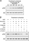
PACAP potently increases sympathetic Akt phosphorylation. A, primary sympathetic neuronal cultures were withdrawn from NGF (as in Fig. 2) and treated with different concentrations of PACAP for 2 h before extraction and Western blot analyses. As with ERK, PACAP potently increased Akt phosphorylation. B, for studies with signaling pathway inhibitors, NGF-deprived cultures were pretreated with the indicated inhibitors for 15 min before peptide addition for 2 h. PI3K inhibitor LY294002 (25 μm) blocked PACAP-mediated Akt phosphorylation. MEK and PKC inhibitors, PD98059 (25 μm), and bisindolylmaleimide I (BimI, 15 μm) and the inactive LY294002 analog LY303511 (25 μm) had little or no effects. Application of the PKA inhibitor H89 (25 μm) consistently potentiated PACAP-stimulated Akt phosphorylation. Data are representative of four independent experiments.
FIGURE 4.
FIGURE 4.
Inhibition of sympathetic receptor tyrosine kinase activity or protein synthesis does not block PACAP-stimulated Akt phosphorylation. A, mature primary sympathetic cultures were acutely withdrawn from NGF and pretreated with 200 nm K252a for 15 min before 100 nm PACAP addition for 3 h. The cultures were harvested in lysis buffer containing phosphatase/protease inhibitors for SDS-PAGE and Western analyses using antibodies for phosphorylated TrkA or Akt. The major form of TrkA in sympathetic neurons is the 140-kDa mature protein. Acute NGF deprivation diminished endogenous TrkA phosphorylation levels. Although the addition of PACAP27 activated Akt, TrkA phosphorylation in the same samples was not enhanced from the low basal levels. Although treatments with the Trk tyrosine kinase inhibitor further diminished culture TrkA phosphorylation, co-treatment with K252a had no effects on PACAP-mediated Akt phosphorylation. B, these results were corroborated in immunoprecipitation experiments. Sympathetic neuronal cultures were acutely withdrawn from NGF to attenuate endogenous TrkA phosphorylation levels (4 h) before PACAP addition for 2 h. The cultures lysed in buffer containing phosphatase/protease inhibitors and the extracts were immunoprecipitated with TrkA antibodies for Western analyses using a phosphotyrosine antibody. PACAP did not stimulate TrkA phosphorylation from the low basal levels. C, similar to experimental designs in panel A, the presence of 5 μg/ml cycloheximide (CHX) for the entire 3-h PACAP treatment period did not abrogate PACAP-stimulated Akt activation. Data are representative of three independent experiments.
FIGURE 5.
FIGURE 5.
Inhibition of Gαq and PI3Kγ signaling attenuates PACAP-stimulated Akt activation. A, sympathetic neuronal cultures were acutely withdrawn from NGF for 4 h to attenuate cellular Akt phosphorylation levels, then pretreated with 10 μm AS252424 (15 min) to selectively inhibit PI3Kγ activity before 100 nm PACAP27 addition for 4 h. AS252424 blocked PACAP/PAC1 receptor-stimulated Akt activation but did not attenuate NGF/TrkA-stimulated Akt phosphorylation (not shown). Identical results were obtained with PI3Kγ-selective inhibitor AS605240. B, treatment of stable AtT-20/PAC1HOP1 cultures with 100 nm PACAP27 for 4 h stimulated Akt phosphorylation; as in panel A with primary sympathetic neurons, the response was blocked upon inhibition with PI3Kγ-selective AS252424 (10 μm) treatment. AtT-20/PAC1HOP1 cultures were transfected at 70% confluency with mouse siRNA oligonucleotides to affect Gαq (C) or PI3Kγ (D) knockdown. After 48 h, the cultures were treated with 100 nm PACAP27 for 4 h. PACAP-stimulated Akt phosphorylation was attenuated when Gαq or PI3Kγ expression was diminished. Culture total Akt levels demonstrate loading equivalency across all samples. Representative data are from 2–4 experiments.
FIGURE 6.
FIGURE 6.
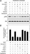
PACAP-stimulated Akt phosphorylation is blocked by clathrin pathway inhibitors. A, acute NGF-deprived cultures as described in Fig. 2 were pretreated with inhibitors for clathrin (chlorpromazine (CPZ), monodansylcadaverine (MDC)) or caveolae (filipin, nystatin) for 30 min before the addition of 100 nm PACAP27 for 3 h. As before, the cultures were harvested for SDS-PAGE and Western analyses for activated Akt. B, quantitative Western analyses of data in panel A were normalized to total Akt and expressed as -fold change from −NGF control. Reagents associated with clathrin-mediated vesicle internalization blocked PACAP-stimulated Akt phosphorylation. n = 3, data represent the mean ± S.E. *, different from PACAP-treated cultures at p < 0.05.
FIGURE 7.
FIGURE 7.
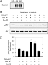
Sympathetic neuron expression of dominant negative dynamin inhibits PACAP-stimulated Akt phosphorylation. A, control sympathetic neurons (CTL) and cultures infected with wild type (WT) or dominant negative dynamin K44A (DN) were extracted for Western analyses of dynamin protein expression. The two dynamin preparations yielded comparable levels of protein expression. B, sympathetic neuronal cultures infected with either dynamin (Dyn) WT or dynamin DN (48 h) were deprived of NGF for 4 h before the addition of 100 nm PACAP27 (4 h) for Western analyses of phosphorylated Akt levels. Cultures expressing dominant negative dynamin blocked PACAP-stimulated Akt activation. C, quantitative Western analyses of data in panel B were normalized to total culture Akt levels and expressed as -fold change from untreated control. n = 3, data represent the mean ± S.E. *, different from untreated controls at p < 0.05.
FIGURE 8.
FIGURE 8.
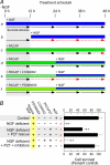
PACAP-mediated Akt signaling is neuroprotective in sympathetic neuronal cultures. A, shown is an experimental schematic for PACAP-mediated sympathetic neuronal survival. After NGF withdrawal (0 h) from immature sympathetic neuronal cultures, there is a 12–15-h survival window delaying neuronal commitment to apoptosis. If NGF is returned to the culture within the survival window (a), the cultures continue to live (black solid arrows) and do not undergo apoptosis programs. But if NGF is returned to the cultures outside of window parameters (b), the neurons are committed to apoptosis, and subsequent NGF addition is unable to affect rescue (red broken arrows). The addition of PACAP in place of NGF during the survival window extends the survival period (c) such that a much delayed addition of NGF (blue bar) can fully protect the neurons (d). Accordingly, the addition of signaling inhibitors (hatched bars, e and f) with PACAP during the window period will block the extended survival period and can be diagnostic of PACAP neurotrophic pathways. B, in this paradigm using the MTS survival assay (lanes a–f correspond to those in panel A), NGF replacement outside of the survival window did not offer neuroprotection when survival was assessed 48 h later (b). PACAP alone offered 50% protection (c) in NGF-deprived cultures, whereas the sequential addition of PACAP + NGF as described above was fully protective (d). Between ERK and Akt pathways, only LY294002 blocked the abilities for PACAP to promote neuronal survival in this experimental paradigm (e). As the MEK inhibitor PD98059 had no apparent effects (f), these results suggested that PI3K/Akt signaling may be more immediate in mediating PACAP survival signaling. Data represent the mean of 5–6 culture replicates ± S.E.; *, significantly different from control; +, different from PACAP-rescued cultures at p < 0.05.
FIGURE 9.
FIGURE 9.
Schematic of coordinate PACAP receptor signaling for sympathetic neuronal survival. The ability for the PAC1 receptor to engage Gαs/Gαq signaling pathways represents a multifaceted but coordinate means for neurotrophic signaling. PAC1 receptor Gαs signaling in sympathetic neurons can stimulate cAMP mechanisms to promote sustained ERK activation. In addition to rapid PAC1 receptor-mediated Gαq activation of phospholipase C /PKC pathways, the internalization of PAC1 receptors through clathrin-coated vesicles may facilitate scaffold assembly to engage PI3kγ/Akt signaling for survival mechanisms. Whether ERK, Akt, or other PAC1 receptor-stimulated mechanisms attenuate JNK/p38 MAPK activation after NGF-deprivation remains to be elucidated. PDK, phosphoinositide-dependent protein kinase; AC, adenylyl cyclase.

Similar articles

Cited by

References

    1. Sherwood N. M., Krueckl S. L., McRory J. E. (2000) Endocr. Rev. 21, 619–670 - PubMed
    1. Spengler D., Waeber C., Pantaloni C., Holsboer F., Bockaert J., Seeburg P. H., Journot L. (1993) Nature 365, 170–175 - PubMed
    1. Lutz E. M., Sheward W. J., West K. M., Morrow J. A., Fink G., Harmar A. J. (1993) FEBS Lett. 334, 3–8 - PubMed
    1. Ishihara T., Shigemoto R., Mori K., Takahashi K., Nagata S. (1992) Neuron. 8, 811–819 - PubMed
    1. Inagaki N., Yoshida H., Mizuta M., Mizuno N., Fujii Y., Gonoi T., Miyazaki J.., Seino S. (1994) Proc. Natl. Acad. Sci. U.S.A. 91, 2679–2683 - PMC - PubMed

Publication types

MeSH terms

Substances