. 2010 Mar 26;285(13):9780-9791.
doi: 10.1074/jbc.M109.065466. Epub 2010 Jan 21.

Energy restriction as an antitumor target of thiazolidinediones

Affiliations

Energy restriction as an antitumor target of thiazolidinediones

Shuo Wei et al. J Biol Chem. .

Abstract

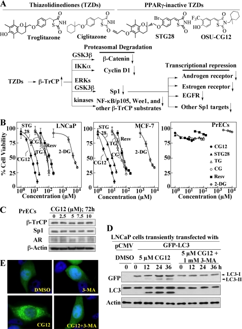
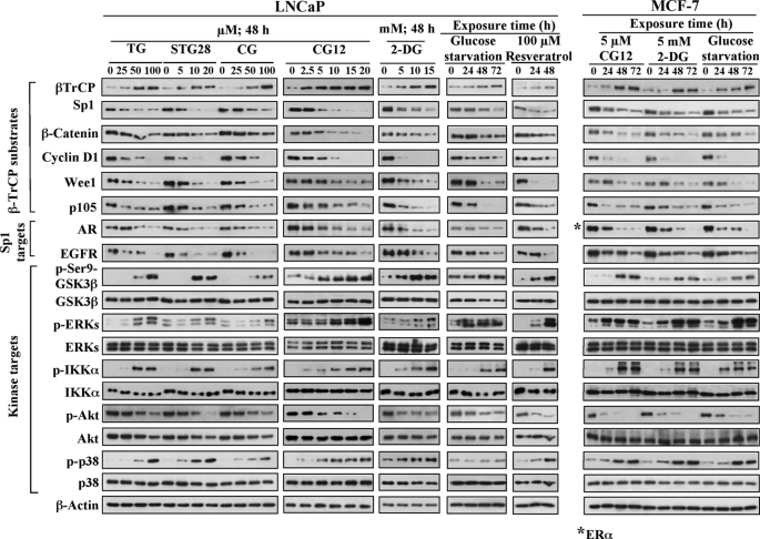
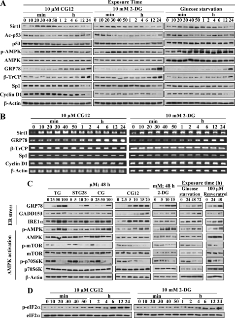
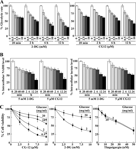
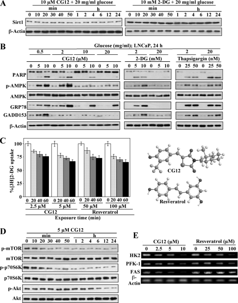
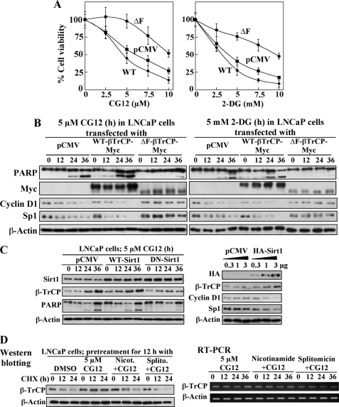
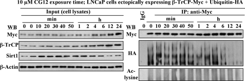
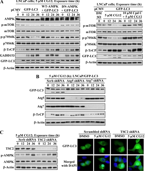
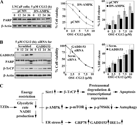
Cancer cells gain growth advantages in the microenvironment by shifting cellular metabolism to aerobic glycolysis, the so-called Warburg effect. There is a growing interest in targeting aerobic glycolysis for cancer therapy by exploiting the differential susceptibility of malignant versus normal cells to glycolytic inhibition, of which the proof-of-concept is provided by the in vivo efficacy of dietary caloric restriction and natural product-based energy restriction-mimetic agents (ERMAs) such as resveratrol and 2-deoxyglucose in suppressing carcinogenesis in animal models. Here, we identified thiazolidinediones as a novel class of ERMAs in that they elicited hallmark cellular responses characteristic of energy restriction, including transient induction of Sirt1 (silent information regulator 1) expression, activation of the intracellular fuel sensor AMP-activated protein kinase, and endoplasmic reticulum stress, the interplay among which culminated in autophagic and apoptotic death. The translational implications of this finding are multifold. First, the novel function of troglitazone and ciglitazone in targeting energy restriction provides a mechanistic basis to account for their peroxisome proliferator-activated receptor gamma-independent effects on a broad spectrum of signaling targets. Second, we demonstrated that Sirt1-mediated up-regulation of beta-transducin repeat-containing protein-facilitated proteolysis of cell cycle- and apoptosis-regulatory proteins is an energy restriction-elicited signaling event and is critical for the antitumor effects of ERMAs. Third, it provides a molecular rationale for using thiazolidinediones as scaffolds to develop potent ERMAs, of which the proof-of-principle is demonstrated by OSU-CG12. OSU-CG12, a peroxisome proliferator-activated receptor gamma-inactive ciglitazone derivative, exhibits 1- and 3-order of magnitude higher potency in eliciting starvation-like cellular responses relative to resveratrol and 2-deoxyglucose, respectively.


Figures

FIGURE 1.
FIGURE 1.
PPARγ-independent antiproliferative effects of TZDs. A, upper panel, structures of troglitazone, ciglitazone, and their respective PPARγ-inactive derivatives STG28 and OSU-CG12. Lower panel, a schematic diagram depicting the mode of action of these TZDs in activating β-TrCP-mediated proteolysis and the consequent transcriptional repression of Sp1 target genes. B, dose-dependent effects of troglitazone (TG), ciglitazone (CG), STG28, and OSU-CG12 (CG12) vis-à-vis resveratrol (Resv) and 2-DG on the viability of LNCaP and MCF-7 cells versus PrECs in 10% fetal bovine serum-supplemental medium for 72 h. The MTT data are expressed as the means ± S.D. (n = 6). C, lack of effect of OSU-CG12 on the expression levels of β-TrCP, Sp1, and AR in PrECs. D, evidence that TZDs induce autophagy. Time-dependent effect of 5 μm OSU-CG12 on the conversion of both GFP-tagged and endogenous LC3-I to LC3-II, which could be blocked by 3-methyladenine (3-MA, 1 mm). E, microscopic analysis of the effect of 5 μm OSU-CG12, alone or in the presence of 1 mm 3-methyladenine, on the pattern of GFP-LC3 fluorescence.
FIGURE 2.
FIGURE 2.
Energy restriction and TZDs share the ability to facilitate β-TrCP-mediated proteolysis. Endpoints included the expression of β-TrCP, β-TrCP substrates, and Sp1 target gene products, and the phosphorylation status of kinases involved in facilitating β-TrCP-substrate recognition. TG, troglitazone; CG, ciglitazone; CG12, OSU-CG12; EGFR, epidermal growth factor receptor; IKKα, IκB kinase α.
FIGURE 3.
FIGURE 3.
TZDs share the ability of 2-DG and glucose starvation to elicit energy restriction-associated cellular responses in LNCaP cells. A, Western blot analysis of the time-dependent effects of 10 μm OSU-CG12 vis-à-vis 10 mm 2-DG and glucose starvation on various markers associated with energy restriction (induction of Sirt1 expression, p53 deacetylation, AMPK phosphorylation, and the expression of ER stress indicator GRP78) and with β-TrCP-dependent proteolysis (expression of β-TrCP, Sp1, and cyclin D1). B, parallel analysis of the mRNA expression levels of Sirt1, GRP78, β-TrCP, Sp1, and cyclin D1 by RT-PCR in cells treated with OSU-CG12 and 2-DG as described above. C, Western blot analysis of the effect of TZDs at different doses, relative to 2-DG, resveratrol, and glucose starvation on ER stress and AMPK/mTOR/p70S6K signaling. D, Western blot analysis of the time-dependent effect of 10 μm OSU-CG12 versus 10 mm 2-DG on the phosphorylation of eIF-2α, a marker associated with energy restriction. TG, troglitazone; CG, ciglitazone; CG12, OSU-CG12; IRE1α, inositol-requiring enzyme 1α.
FIGURE 4.
FIGURE 4.
Evidence that OSU-CG12 targets glucose metabolism in LNCaP cells. A, time- and dose-dependent effect of OSU-CG12 (right panel) vis-à-vis 2-DG (left panel) on glycolytic rate. The data are expressed as the means ± S.D. (n = 3). B, time-dependent effect of 5 μm OSU-CG12 vis-à-vis 5 mm 2-DG on the intracellular levels of NADH (left panel) and lactate (right panel). The data are expressed as the means ± S.D. (n = 3). C, supplemental glucose provides dose-dependent protection against the antiproliferative activity of OSU-CG12 (left panel) and 2-DG (middle panel), but not the ER stress inducer thapsigargin (right panel). The data are expressed as the means ± S.D. (n = 6). CG12, OSU-CG12.
FIGURE 5.
FIGURE 5.
Mechanisms underlying OSU-CG12-mediated inhibition of glucose metabolism in LNCaP cells. A, supplemental glucose (20 mg/ml) reversed the transient induction of Sirt1 by OSU-CG12 (CG12) and 2-DG. B, dose-dependent protective effects of supplemental glucose against the induction of PARP cleavage, AMPK activation, and the expression of GRP78 and GADD153 in cells treated with different doses of OSU-CG12 or 2-DG. The ER stress-inducing agent thapsigargin was used as a negative control. C, left panel, dose- and time-dependent effects of OSU-CG12 and resveratrol on [3H]2DG uptake. The data are expressed as the means ± S.D. (n = 3). Right panel, ball-and-stick structures of OSU-CG12 and resveratrol. D, Western blot analysis of the time-dependent suppressive effects of 5 μm OSU-CG12 on the phosphorylation of mTOR, p70S6K, and Akt. E, RT-PCR analysis of the dose-dependent effects of OSU-CG12 vis-à-vis resveratrol on the mRNA levels of hexokinase 2 (HK2) and phosphofructokinase-1 (PFK-1), the first two enzymes in the glycolytic pathway, and fatty acid synthase (FAS) after 24 h of exposure.
FIGURE 6.
FIGURE 6.
β-TrCP expression is important for the antiproliferative effects of ERMAs and is up-regulated by Sirt1-mediated stabilization of β-TrCP protein in LNCaP cells. A, effects of ectopic expression of WT-β-TrCP-Myc (WT) and ΔF-β-TrCP-Myc (ΔF) versus pCMV control on the dose-dependent inhibition of cell viability by OSU-CG12 (left panel) and 2-DG (right panel). The data are expressed as the means ± S.D. (n = 6). B, effects of ectopic expression of WT-β-TrCP-Myc and ΔF-β-TrCP-Myc on the ability of OSU-CG12 (5 μm) and 2-DG (5 mm) to facilitate PARP cleavage and the degradation of the β-TrCP substrates cyclin D1 and Sp1. C, Sirt1 up-regulation elevates β-TrCP expression levels in OSU-CG12-treated LNCaP cells. Left panel, dominant-negative (DN) inhibition of Sirt1 blocked OSU-CG12-mediated β-TrCP induction and PARP cleavage. Right panel, ectopic expression of HA-Sirt1 increased β-TrCP expression in a dose-dependent manner with corresponding decreases in the expression of cyclin D1 and Sp1. D, Sirt1 increased β-TrCP expression via protein stabilization. Left panel, inhibition of Sirt1 deacetylase activity by nicotinamide or splitomicin reversed the ability of OSU-CG12 to enhance β-TrCP protein stability. Right panel, RT-PCR analysis showed that β-TrCP mRNA levels in LNCaP cells treated as described above remained unchanged. DMSO, dimethyl sulfoxide; CHX, cycloheximide; CG12, OSU-CG12.
FIGURE 7.
FIGURE 7.
OSU-CG12-mediated β-TrCP accumulation results from reduced ubiquitination via an acetylation-independent mechanism. LNCaP cells transiently transfected with plasmids encoding β-TrCP-Myc and ubiquitin-HA were exposed to 10 μm OSU-CG12 for different time intervals. Equal amounts of cell lysates were immunoblotted with anti-Myc, anti-β-TrCP, and anti-Sirt1 antibodies (Input, left panel) or immunoprecipitated (IP) with anti-Myc antibodies, followed by Western blot analysis (WB) with anti-Myc, anti-HA, and anti-acetyl (Ac)-lysine antibodies.
FIGURE 8.
FIGURE 8.
OSU-CG12 induces autophagy by targeting the AMPK/TSC2/mTOR/p70S6K pathway. A, dominant-negative or pharmacological inhibition of AMPK blocked OSU-CG12-mediated autophagy but had no effect on ER stress. Left panel, effects of the ectopic expression of the WT versus the K45R kinase-dead, dominant-negative (DN) form of AMPK on the ability of OSU-CG12 to modulate the expression levels of p-mTOR, p-p70S6K, β-TrCP, and GADD153, and the conversion of GFP-LC3 in GFP-LC3-expressing LNCaP cells. Right panel, parallel analysis of the effects of the AMPK inhibitor compound C. B, shRNA-mediated silencing of Atg5 and Atg7 was used as a positive control to confirm the protective effect of AMPK inhibition on OSU-CG12-induced GFP-LC3 II conversion. C, shRNA-mediated knockdown of TSC2 protected cells from OSU-CG12-induced autophagy. Left panel, validation of the effectiveness of shRNA-mediated knockdown of TSC2 by Western blot analysis of TSC2 and p-AMPK expression in OSU-CG12-treated cells. Right panel, GFP-LC3-expressing LNCaP cells transfected with scrambled or TSC2 shRNA were exposed to Me2SO (DMSO) or OSU-CG12 for 36 h and then examined by fluorescence microscopy to assess patterns of GFP-LC3 fluorescence. CG12, OSU-CG12.
FIGURE 9.
FIGURE 9.
Relative roles of AMPK activation and GADD153 in mediating the effect of OSU-CG12 on apoptosis and viability in LNCaP cells. A, dominant-negative (DN) inhibition of AMPK had no effect on OSU-CG12-induced PARP cleavage (left), but partially protected LNCaP cells from OSU-CG12-mediated suppression of cell viability (central) and release of LDH (right). Data are expressed as means ± S.D. (n = 6). B, siRNA-mediated knockdown of GADD153 had no effect on OSU-CG12-induced PARP cleavage or β-TrCP induction (left), OSU-CG12-mediated antiproliferative activity (central), or OSU-CG12-mediated LDH release (right). Viability and LDH data are expressed as means ± S.D. (n = 6). C, schematic diagram depicting the mechanism by which TZDs act as ERMAs by perturbing glucose homeostasis, resulting in the hallmark cellular responses, including transient Sirt1 induction, AMPK activation, and ER stress. Our data indicate a mechanistic link between Sirt1 induction and β-TrCP protein accumulation, culminating in apoptosis through the proteasomal degradation and transcriptional repression of a series of apoptosis-regulatory proteins. The AMPK activation results in autophagy via the conventional AMPK-TSCs-mTOR-p70S6K pathway. The ER stress signal triggers the up-regulation of sensor proteins, such as GRP78, GADD153 and inositol-requiring enzyme 1α (IRE1α), which, might also play a role in down-regulating cell growth.

Similar articles

Cited by

References

    1. Yki-Järvinen H. (2004) N. Engl. J. Med. 351, 1106–1118 - PubMed
    1. Panchapakesan U., Chen X. M., Pollock C. A. (2005) Nat. Clin. Pract. Nephrol. 1, 33–43 - PubMed
    1. Olefsky J. M. (2000) J. Clin. Invest. 106, 467–472 - PMC - PubMed
    1. Sharma A. M., Staels B. (2007) J. Clin. Endocrinol. Metab. 92, 386–395 - PubMed
    1. Grommes C., Landreth G. E., Heneka M. T. (2004) Lancet Oncol. 5, 419–429 - PubMed

Publication types

LinkOut - more resources