. 2010 Feb 12;327(5967):873-876.
doi: 10.1126/science.1183173. Epub 2010 Jan 21.

Repulsion of superinfecting virions: a mechanism for rapid virus spread

Affiliations

Repulsion of superinfecting virions: a mechanism for rapid virus spread

Virginie Doceul et al. Science. .

Abstract

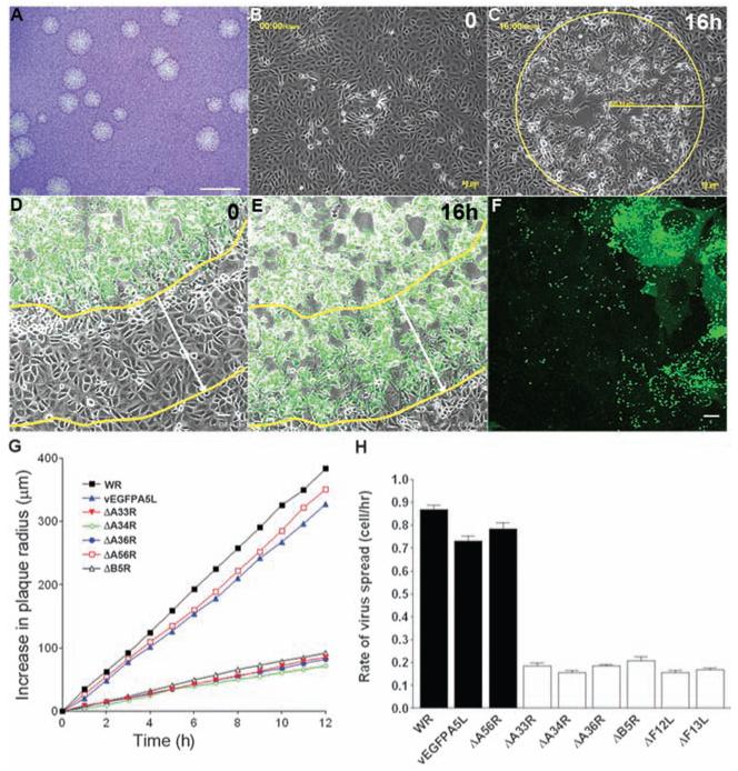
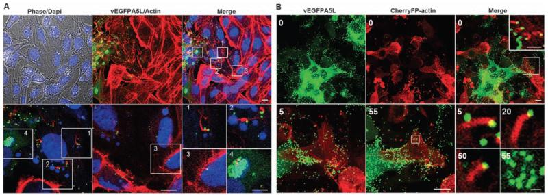
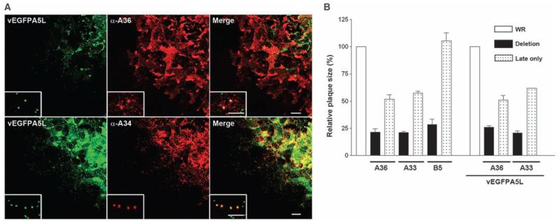
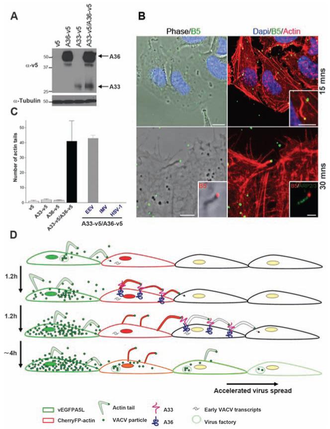
Viruses are thought to spread across susceptible cells through an iterative process of infection, replication, and release, so that the rate of spread is limited by replication kinetics. Here, we show that vaccinia virus spreads across one cell every 75 minutes, fourfold faster than its replication cycle would permit. To explain this phenomenon, we found that newly infected cells express two surface proteins that mark cells as infected and, via exploitation of cellular machinery, induce the repulsion of superinfecting virions away toward uninfected cells. Mechanistically, early expression of proteins A33 and A36 was critical for virion repulsion and rapid spread, and cells expressing these proteins repelled exogenous virions rapidly. Additional spreading mechanisms may exist for other viruses that also spread faster than predicted by replication kinetics.


Figures

Fig. 1
Fig. 1
VACV spreads more rapidly than predicted. (A) VACV plaques 3 days after infection in BSC-1 cells. Scale bar, 5 mm. (B and C) Live cell imaging recording plaque formation at 0 (B) and 16 (C) hours later. (D and E) Live cell imaging of vEGFPA5L-infected cells confirmed the correlation between cytopathic effect (cpe) and virus infection. Yellow lines indicate the boundary between infected and uninfected cells and white arrows indicate the distance this has moved over 16 hours. (F) Confocal image showing the spread of EGFP-tagged virus particles (single green dots) far from the center of infection. (G) Increase in plaque radius formed by VACV WR and mutants with time; n = 6 to 11 plaques. (H) Diagram showing the rate of spread (BSC-1 cell per hour) with indicated viruses. White bars indicate viruses with a defect in actin tail formation. Error bars are SEM, with n = 6 to 11 plaques. Scale bar, (A) 5 mm, (B) to (E) 50 μm, and (F), 10 μm.
Fig. 2
Fig. 2
Cells form actin tails before production of new virions. (A) Confocal images showing the edge of vEGFPA5L plaque (green) on BSC-1 cells stained for actin (red) or DNA (blue). Bottom panel shows zoomed areas (white squares 1 to 4). Actin tails are on cells with nascent factories (cytoplasmic blue) but that are not producing any virus particles (green) (squares 1 and 2), and on a cell with no virus factory (square 3), whereas square 4 shows a productive virus factory (green). Scale bars, top row, 10 μm; bottom row, 10 μm; and insets 1 to 4, 5 μm. (B) Actin tails (red) present at the surface of a cell expressing cherryFP-actin but with no green virus factory [time (t) = 0, white square and zoomed inset]. Bottom panels show zoomed images of this cell with actin tails detected 5, 20, and 50 min later, before the appearance of virus factories at 55 min as indicated by the white square. Scale bars, top row, 10 μm; top right inset, 5 μm; bottom row, 10 μm; and bottom right inset, 1 μm.
Fig. 3
Fig. 3
Early expression of A33 and A36 is important for VACV spread. (A) Images of edge of plaque showing A36, but not A34, is expressed early during infection. A36 was detected in cells where no late protein A5 (green) was present, whereas A34 was expressed late during infection in cells that also express A5. (Insets) Zoomed images of virions (single green dots) relative to A36 and A34 distribution. (B) Graph showing the size of plaques formed by recombinant viruses in which A33R, A36R, or B5R are under a late promoter only (4b) or deleted (Δ) as compared with parental viruses WR or vEGFPA5L. Error bars are SEM mean values from three experiments with n = 11 to 12 plaques. Scale bars, 20 μm; insets, 5 μm.
Fig. 4
Fig. 4
Expression of A33 and A36 is sufficient for actin tail formation. (A) Immunoblot showing A33 and A36 expression. (B) Actin tails present 15 and 30 min after spinoculation of EEV particles onto HeLa cells expressing A33 and A36 proteins. Staining for Arp2/3 shows actin polymerisation machinery. Scale bars, top row, 10 μm; inset, 5 μm; bottom row, 5 μm; inset, 1 μm. (C) Graph showing the mean number of actin tails detected per coverslip in the different cell lines. Actin tails were not formed by IMV or HSV-1. Error bars are SEM; n = 3 experiments. (D) Model showing how VACV spreads rapidly. The first infected cell expresses EGFP-A5 late during infection and releases green virions, which infect an adjacent cell expressing cherry actin (red). Early after infection, A33 and A36 are expressed at the cell surface and mark the cell as infected. Upon contact with new CEV/EEV particles, the A33/A36 complex induces the formation of red actin tails, which repel these virions toward uninfected cells. Superinfecting virions may be repelled from multiple infected cells before an uninfected cell is found that can be infected.

Comment in

  • Cell biology. Propelling progeny.
    Pickup DJ. Pickup DJ. Science. 2010 Feb 12;327(5967):787-8. doi: 10.1126/science.1187160. Science. 2010. PMID: 20150470 No abstract available.

References

    1. Moss B. In: Fields Virology. Knipe DM, editor. Vol. 2. Lippincott Williams & Wilkins; Philadelphia: 2007. pp. 2905–2946.
    1. Condit RC, Moussatche N, Traktman P. Adv. Virus Res. 2006;66:31. - PubMed
    1. Smith GL, Vanderplasschen A, Law M. J. Gen. Virol. 2002;83:2915. - PubMed
    1. Roberts KL, Smith GL. Trends Microbiol. 2008;16:472. - PubMed
    1. Sanderson CM, Way M, Smith GL. J. Virol. 1998;72:1235. - PMC - PubMed

Publication types

MeSH terms