. 2010 Mar;176(3):1104-12.
doi: 10.2353/ajpath.2010.090956. Epub 2010 Jan 21.

Human peripheral lymphoid tissues contain autoimmune regulator-expressing dendritic cells

Affiliations

Human peripheral lymphoid tissues contain autoimmune regulator-expressing dendritic cells

Pietro Luigi Poliani et al. Am J Pathol. 2010 Mar.

Erratum in

  • Am J Pathol. 2010 May;176(5):2581

Abstract

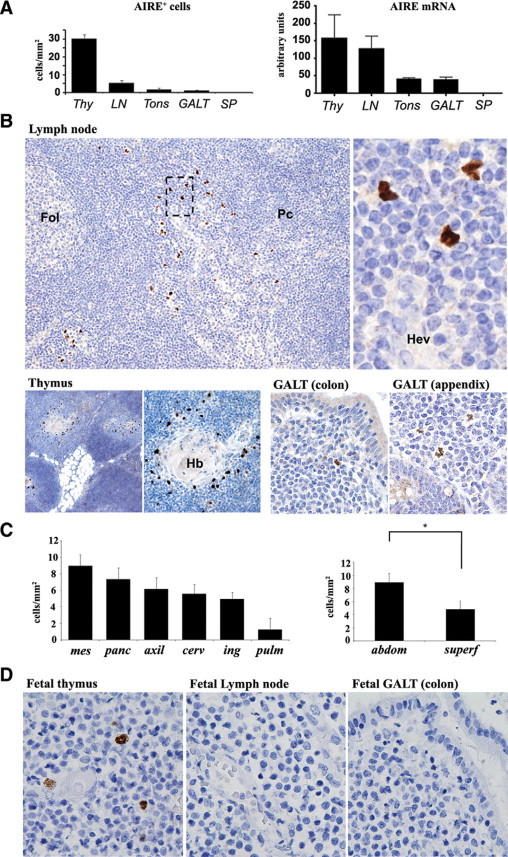
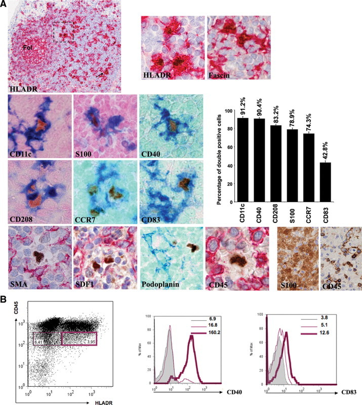
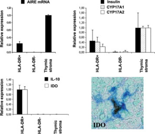
Autoimmune regulator (AIRE) modulates the expression of tissue-restricted antigens (TSAs) and promotes central tolerance in the thymus. However, few autoreactive T cells escape negative selection and reach the periphery, where peripheral tolerance is required to avoid autoimmunity. Murine lymph nodes (LNs) have been shown to contain "stromal" cells expressing AIRE and TSAs. Here we report the occurrence of AIRE-expressing cells in human peripheral lymphoid tissues, including LNs, tonsils, and gut-associated lymphoid tissue, with the exception of the spleen. Notably, AIRE+ cells are absent in fetal LNs and, in postnatal life, they are more numerous in abdominal than in superficial LNs, thus suggesting that their development in periphery may depend on instructive signals from microenvironment and antigen challenge. Extrathymic AIRE+ cells show a dendritic morphology, consistently express human leukocyte antigen-DR (HLADR) and fascin, and are largely positive for CD11c and S100 and for the dendritic cell-activation markers CD40, CD83, DC-LAMP/CD208, and CCR7. Lymphoid, myelomonocytic, mesenchymal, and epithelial cell lineage markers are negative. The HLADRhigh/AIRE+ cell fraction isolated from mesenteric LNs expressed TSAs (insulin, CYP17A1, and CYP21A2), as well as molecules associated with tolerogenic functions, such as interleukin-10 and indoleamine 2,3-dioxygenase. Data indicate that AIRE+ cells in human peripheral lymphoid tissues correspond to a subset of activated interdigitating dendritic cells expressing TSAs and the tolerogenic molecules indoleamine 2,3-dioxygenase and interleukin-10, suggestive of a potential tolerogenic function.


Figures

Figure 1
Figure 1
Expression of AIRE in human thymus and peripheral lymphoid tissues. A: The number of AIRE+ cells/mm2 evaluated by immunohistochemistry (left plot) and AIRE mRNA levels analyzed by quantitative RT-PCR (right plot) show that extrathymic AIRE expression occurs particularly in LNs, albeit at lower level than in the thymus (Thy), tonsils (Tons), and GALT but not in the spleen (SP). B: In LNs (top panel) AIRE+ cells are distributed as scattered cells in the interfollicular area/outer paracortex (Fol, follicle; Pc, paracortex), in the vicinity of high endothelial venules (Hev), as indicated in the higher magnification from the dotted box area (original magnification: ×10 and ×80). In the thymus (bottom panel, left), AIRE+ cells occur in the medulla and correspond to medullary epithelial cells around Hassall's bodies (Hb) (original magnification: ×4 and ×10). Rare cells are present in the colon, appendix, and GALT (bottom panel, right) (original magnification: ×20). C: The number of nodal AIRE+ cells differs in anatomical regions (left plot). The difference between AIRE+ cells in abdominal (mesenteric plus peripancreatic) compared with superficial (axillary, inguinal, and cervical) LNs is statistically significant (right plot; *P < 0.001). mes, mesenteric; panc, peripancreatic; axil, axillary; cerv, cervical; ing, inguinal; pulm, pulmonary; abdom, abdominal; superf, superficial. D: In the fetus, only the thymus contains AIRE+ cells in the medulla, whereas no positive cells occur in LNs and GALT (all panels, original magnification: ×40). All immunostains for AIRE were performed using diaminobenzidine/hydrogen peroxide as the chromogen and hematoxylin as the nuclear counterstain. Bar graphs show averages and SEs of at least three independent measurements.
Figure 2
Figure 2
A: Characterization of nodal AIRE-expressing cells by double immunostains. AIRE+ cells consistently express HLADR and are distributed in the outer paracortex and in the interfollicular areas (arrow and dotted square), admixed with HLADR+AIRE cells corresponding to IDCs. They coexpress other molecules normally found on IDCs, such as fascin, CD11c, and S100, as well as the DC activation and maturation antigens CD40, CD208/DC-LAMP, CCR7, and CD83. The plot shows the percentages of double-positive/total AIRE+ cells. In all figures the dendritic morphology of the AIRE+ cells is obvious. In the bottom row examples of molecules normally expressed by fibroblastic reticulum cells (smooth muscle actin [SMA], stromal cell-derived factor-1 [SDF1], and podoplanin) and lacking on the AIRE+ cells are shown. AIRE+ cells are also negative for CD45; this finding is not in contrast with their dendritic cell nature, because IDCs in dermatopathic lymphadenitis are composed of S100+ IDCs that largely lack CD45, as shown in the bottom right serial sections. In double stains AIRE is developed in brown with diaminobenzidine/hydrogen peroxide, whereas the other antigen is developed either in blue or red, and nuclear counterstain with methyl green or hematoxylin, respectively. All figures, original magnification: ×40, except for the upper left figure (×4) and the lower right two figures (×20). B: Dot plot of cell suspension (left) obtained from mesenteric LNs after low-density gradient enrichment shows distribution of the CD45lowHLADRhigh (purple heavy line) in comparison with CD45lowHLADR (purple normal line). The CD45lowHLADRhigh cell fraction displays higher level of CD40 (center) and a light increase of CD83 (right). Gray histogram represents the isotype control. Numbers indicate the mean fluorescence intensity for each population. Representative profiles of three independent experiments are shown.
Figure 3
Figure 3
Nodal AIRE+ cells express tissue-restricted antigens and show a tolerogenic phenotype. Quantitative RT-PCR analysis on cell suspensions from mesenteric LNs previously enriched for AIRE expressing DCs (top left): the enriched HLADR+ but not the HLADR cell fraction expresses AIRE mRNA and the AIRE-dependent tissue-restricted antigens insulin, CYP17A1, and CYP21A2 (top right). Quantitative RT-PCR shows that the HLADR+AIRE+-enriched cell fraction has an increased expression of IDO and IL-10 transcripts (bottom left), and double immunostain confirms the coexpression of IDO by the AIRE+ cells, suggesting a tolerogenic phenotype (bottom right). mRNA is expressed as relative gene expression levels. Averages and SEs (bars) of three independent experiments are shown. For double stain, AIRE is developed in brown with diaminobenzidine/hydrogen peroxide; IDO is developed in blue; nuclear counterstain is with methyl green. Original magnification, ×40.

Similar articles

Cited by

References

    1. Kyewski B, Klein L. A central role for central tolerance. Annu Rev Immunol. 2006;24:571–606. - PubMed
    1. Hamazaki Y, Fujita H, Kobayashi T, Choi Y, Scott HS, Matsumoto M, Minato N. Medullary thymic epithelial cells expressing Aire represent a unique lineage derived from cells expressing claudin. Nat Immunol. 2007;8:304–311. - PubMed
    1. Peterson P, Org T, Rebane A. Transcriptional regulation by AIRE: molecular mechanisms of central tolerance. Nat Rev Immunol. 2008;8:948–957. - PMC - PubMed
    1. Anderson MS, Venanzi ES, Chen Z, Berzins SP, Benoist C, Mathis D. The cellular mechanism of Aire control of T cell tolerance. Immunity. 2005;23:227–239. - PubMed
    1. Mathis D, Benoist C. Aire. Annu Rev Immunol. 2009;27:287–312. - PubMed

Publication types