

ApoE-deficient mice on cholate-containing high-fat diet reveal a pathology similar to lung sarcoidosis
- PMID: 20093498
- PMCID: PMC2832138
- DOI: 10.2353/ajpath.2010.090857
ApoE-deficient mice on cholate-containing high-fat diet reveal a pathology similar to lung sarcoidosis
Abstract
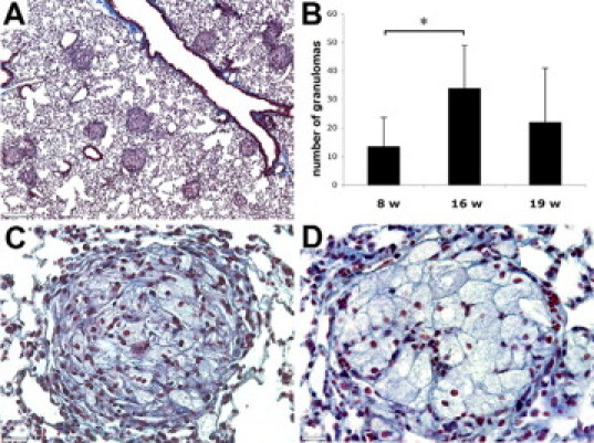
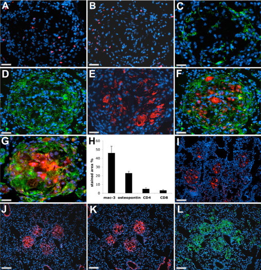
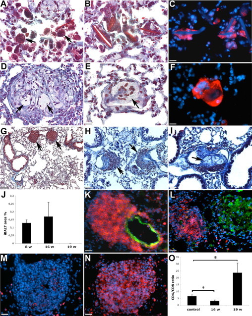
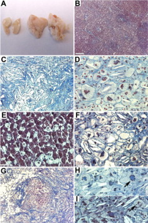
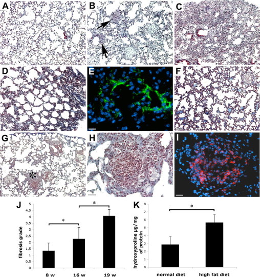
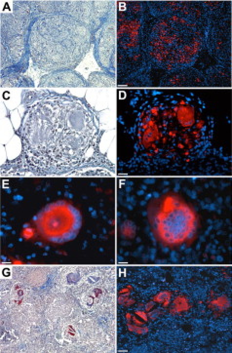
Sarcoidosis is a chronic disease of unknown etiology characterized by the formation of non-necrotizing epithelioid granulomas in various organs, especially in the lungs. The lack of an adequate animal model reflecting the pathogenesis of the human disease is one of the major impediments in studying sarcoidosis. In this report, we describe ApoE-/- mice on a cholate-containing high-fat diet that exhibit granulomatous lung inflammation similar to human sarcoidosis. Histological analysis revealed well-defined and non-necrotizing granulomas in about 40% of mice with the highest number of granulomas after 16 weeks on a cholate-containing high-fat diet. Granulomas contained CD4+ and CD8+ T cells, and the majority of the cells in granulomas showed immunoreactivity for the macrophage marker Mac-3. Cells with morphological features of epithelioid cells expressed angiotensin-converting enzyme, osteopontin, and cathepsin K, all characteristics of epithelioid and giant cells in granulomas of human sarcoidosis. Giant cells and nonspecific inclusions such as Schaumann's bodies and crystalline deposits were also detected in some lungs. Granulomatous inflammation resulted in progressive pulmonary fibrosis. Removal of cholate from the diet prevented the formation of lung granulomas. The observed similarities between the analyzed mouse lung granulomas and granulomas of human sarcoidosis, as well as the chronic disease character leading to fibrosis, suggest that this mouse model might be a useful tool to study sarcoidosis.
Figures
Similar articles
-
Lack of cathepsin activities alter or prevent the development of lung granulomas in a mouse model of sarcoidosis.Respir Res. 2011 Jan 20;12(1):13. doi: 10.1186/1465-9921-12-13. Respir Res. 2011. PMID: 21251246 Free PMC article.
-
The pathology of pulmonary sarcoidosis: update.Semin Diagn Pathol. 2007 Aug;24(3):150-61. doi: 10.1053/j.semdp.2007.06.002. Semin Diagn Pathol. 2007. PMID: 17882899 Review.
-
[Granulomatous diseases and pathogenic microorganism].Kekkaku. 2008 Feb;83(2):115-30. Kekkaku. 2008. PMID: 18326339 Japanese.
-
Reprint of: The pathology of pulmonary sarcoidosis: update.Semin Diagn Pathol. 2018 Sep;35(5):324-333. doi: 10.1053/j.semdp.2018.09.001. Epub 2018 Sep 24. Semin Diagn Pathol. 2018. PMID: 30262157
-
Molecular Mechanism in the Development of Pulmonary Fibrosis in Patients with Sarcoidosis.Int J Mol Sci. 2023 Jun 28;24(13):10767. doi: 10.3390/ijms241310767. Int J Mol Sci. 2023. PMID: 37445947 Free PMC article. Review.
Cited by
-
Autophagy and Obesity-Related Lung Disease.Am J Respir Cell Mol Biol. 2016 May;54(5):636-46. doi: 10.1165/rcmb.2016-0045PS. Am J Respir Cell Mol Biol. 2016. PMID: 26900794 Free PMC article. Review.
-
Transcriptional survey of alveolar macrophages in a murine model of chronic granulomatous inflammation reveals common themes with human sarcoidosis.Am J Physiol Lung Cell Mol Physiol. 2018 Apr 1;314(4):L617-L625. doi: 10.1152/ajplung.00289.2017. Epub 2017 Dec 6. Am J Physiol Lung Cell Mol Physiol. 2018. PMID: 29212802 Free PMC article.
-
Paigen diet-fed apolipoprotein E knockout mice develop severe pulmonary hypertension in an interleukin-1-dependent manner.Am J Pathol. 2011 Oct;179(4):1693-705. doi: 10.1016/j.ajpath.2011.06.037. Epub 2011 Aug 9. Am J Pathol. 2011. PMID: 21835155 Free PMC article.
-
Göttingen minipig model of diet-induced atherosclerosis: influence of mild streptozotocin-induced diabetes on lesion severity and markers of inflammation evaluated in obese, obese and diabetic, and lean control animals.J Transl Med. 2015 Sep 22;13:312. doi: 10.1186/s12967-015-0670-2. J Transl Med. 2015. PMID: 26394837 Free PMC article.
-
Apolipoprotein E deficiency and high-fat diet cooperate to trigger lipidosis and inflammation in the lung via the toll-like receptor 4 pathway.Mol Med Rep. 2015 Aug;12(2):2589-97. doi: 10.3892/mmr.2015.3774. Epub 2015 May 12. Mol Med Rep. 2015. PMID: 25975841 Free PMC article.
References
-
- Verleden GM, du Bois RM, Bouros D, Drent M, Millar A, Muller-Quernheim J, Semenzato G, Johnson S, Sourvino G, Olivier D, Pietinalho A, Xaubet A. Genetic predisposition and pathogenetic mechanisms of interstitial lung diseases of unknown origin. Eur Respir J Suppl. 2001;32:17S–29S. - PubMed
-
- Lynch JP, 3rd, Ma YL, Koss MN, White ES. Pulmonary sarcoidosis. Semin Respir Crit Care Med. 2007;28:53–74. - PubMed
-
- Nagai S, Handa T, Ito Y, Ohta K, Tamaya M, Izumi T. Outcome of sarcoidosis. Clin Chest Med. 2008;29:565–574. - PubMed
-
- Sharma OP. Sarcoidosis: a historical perspective. Clin Dermatol. 2007;25:232–241. - PubMed
Publication types
MeSH terms
Substances
Grants and funding
LinkOut - more resources
Full Text Sources
Medical
Molecular Biology Databases
Research Materials
Miscellaneous
