. 2010 May 1;181(9):935-46.
doi: 10.1164/rccm.200907-1128OC. Epub 2010 Jan 21.

Neutrophil alpha-defensins cause lung injury by disrupting the capillary-epithelial barrier

Affiliations

Neutrophil alpha-defensins cause lung injury by disrupting the capillary-epithelial barrier

Khalil Bdeir et al. Am J Respir Crit Care Med. .

Abstract

Rationale: The involvement of neutrophil activation in the sentinel, potentially reversible, events in the pathogenesis of acute lung injury (ALI) is only partially understood. alpha-Defensins are the most abundant proteins secreted by activated human neutrophils, but their contribution to ALI in mouse models is hindered by their absence from murine neutrophils and the inability to study their effects in isolation in other species.

Objectives: To study the role of alpha-defensins in the pathogenesis of ALI in a clinically relevant setting using mice transgenic for polymorphonuclear leukocyte expression of alpha-defensins.

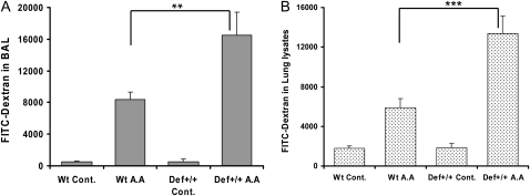
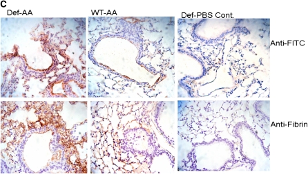
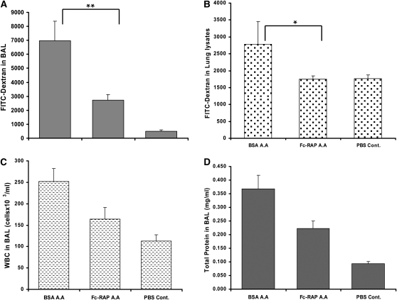
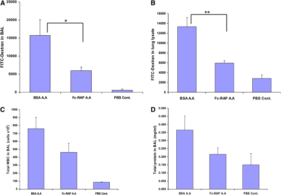
Methods: Transgenic mice expressing polymorphonuclear leukocyte alpha-defensins were generated. ALI was induced by acid aspiration. Pulmonary vascular permeability was studied in vivo using labeled dextran and fibrin deposition. The role of the low-density lipoprotein-related receptor (LRP) in permeability was examined.

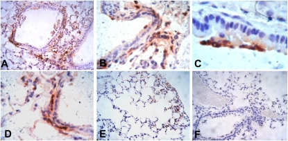
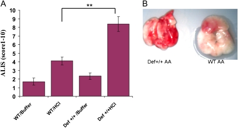
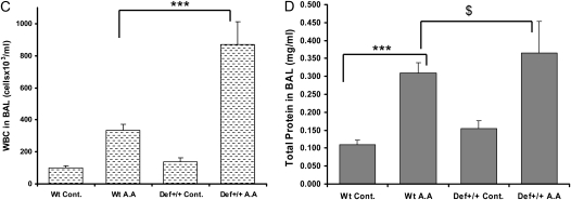
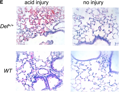
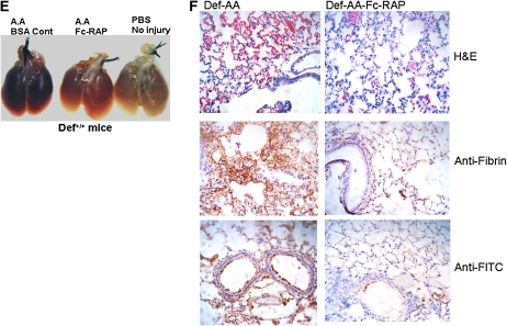
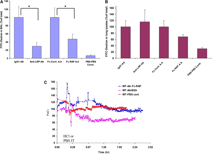
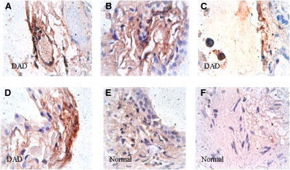
Measurements and main results: Acid aspiration induced neutrophil migration and release of alpha-defensins into lung parenchyma and airways. ALI was more severe in alpha-defensin-expressing mice than in wild-type mice, as determined by inspection, influx of neutrophils into the interstitial space and airways, histological evidence of epithelial injury, interstitial edema, extravascular fibrin deposition, impaired oxygenation, and reduced survival. Within 4 hours of insult, alpha-defensin-expressing mice showed greater disruption of capillary-epithelial barrier function and ALI that was attenuated by systemic or intratracheal administration of specific inhibitors of the LRP.

Conclusions: alpha-Defensins mediate ALI through LRP-mediated loss of capillary-epithelial barrier function, suggesting a potential new approach to intervention.


Figures

Figure 1.
Figure 1.
α-Defensin is released into the lungs of Def+/+ mice during acute lung injury (ALI). ALI was induced by intratracheal administration of HCl (0.1 N) or saline. After 4 hours, the lungs were inflated with 10% buffered formalin phosphate, isolated, washed, and fixed. Paraffin-embedded sections from Def+/+ mice exposed to acid aspiration (AD) or saline (E) were stained using rabbit anti-human defensin sera or preimmune rabbit sera as the negative control. Wild-type mice exposed to acid aspiration show no staining for α-defensin (F). Sections shown at 100× (A, E, and F), 200× (B and D), and 400× (C) magnifications. Results are representative of three mice from each exposure group.
Figure 2.
Figure 2.
Acute lung injury (ALI) is more severe in Def+/+ mice after acid aspiration. Acid aspiration was induced in Def+/+ and wild-type (WT) mice, as described previously here. After 4 hours, the lungs were inspected visually and scored for severity of injury (as in Reference 49). (A) ALI score (scale, 1–10). Mean (±SEM) of 5–10 mice in each experimental group from two different experiments are shown. **P = 0.0012; Student's t test. (B) Visual appearance of injured lungs from Def+/+ and WT mice 4 hours after acid aspiration (AA). Blood was flushed from the pulmonary vasculature with 20 ml of cold saline by perfusing the right ventricle, and the lungs were photographed under a dissecting microscope. Results shown are representative of three mice from each group. Total white blood cells (WBCs) and protein are increased in bronchoalveolar lavage (BAL) fluid from Def+/+ mice after acid aspiration. Acid aspiration was induced in Def+/+ and WT mice. After 4 hours, BAL fluid was analyzed for total WBCs (C) and total protein content (D). Means (±SEM) from 8–12 mice in each experimental group from three different experiments are shown. ***P < 0.0001, $P = 0.42. (E) Def+/+ mice exhibit more intense histological evidence of tissue injury after acid aspiration. Acid aspiration was induced in Def+/+ and WT mice. After 4 hours, the lungs were inflated with formalin, and fixed in 10% buffered formalin phosphate. Paraffin-embedded sections were stained with hematoxylin and eosin (H&E). Low-power (40×) and medium-power (200×) images of H&E–stained Def+/+ (upper panels) and WT (lower panels) mouse lungs after saline or acid aspiration. Tissues from acid-injured Def+/+ lungs show increased alveolar septal thickening and congestion compared with the acid-injured WT control animals. Lungs from saline-injected Def+/+ (left upper panel) and WT mice (left lower panel) show normal parenchymal architecture. The images are representative of three to four mice from each treatment group.
Figure 2.
Figure 2.
Acute lung injury (ALI) is more severe in Def+/+ mice after acid aspiration. Acid aspiration was induced in Def+/+ and wild-type (WT) mice, as described previously here. After 4 hours, the lungs were inspected visually and scored for severity of injury (as in Reference 49). (A) ALI score (scale, 1–10). Mean (±SEM) of 5–10 mice in each experimental group from two different experiments are shown. **P = 0.0012; Student's t test. (B) Visual appearance of injured lungs from Def+/+ and WT mice 4 hours after acid aspiration (AA). Blood was flushed from the pulmonary vasculature with 20 ml of cold saline by perfusing the right ventricle, and the lungs were photographed under a dissecting microscope. Results shown are representative of three mice from each group. Total white blood cells (WBCs) and protein are increased in bronchoalveolar lavage (BAL) fluid from Def+/+ mice after acid aspiration. Acid aspiration was induced in Def+/+ and WT mice. After 4 hours, BAL fluid was analyzed for total WBCs (C) and total protein content (D). Means (±SEM) from 8–12 mice in each experimental group from three different experiments are shown. ***P < 0.0001, $P = 0.42. (E) Def+/+ mice exhibit more intense histological evidence of tissue injury after acid aspiration. Acid aspiration was induced in Def+/+ and WT mice. After 4 hours, the lungs were inflated with formalin, and fixed in 10% buffered formalin phosphate. Paraffin-embedded sections were stained with hematoxylin and eosin (H&E). Low-power (40×) and medium-power (200×) images of H&E–stained Def+/+ (upper panels) and WT (lower panels) mouse lungs after saline or acid aspiration. Tissues from acid-injured Def+/+ lungs show increased alveolar septal thickening and congestion compared with the acid-injured WT control animals. Lungs from saline-injected Def+/+ (left upper panel) and WT mice (left lower panel) show normal parenchymal architecture. The images are representative of three to four mice from each treatment group.
Figure 3.
Figure 3.
Def+/+ mice show greater vascular permeability after acid aspiration: kinetics. Acute lung injury (ALI) was induced by acid aspiration. At 5 minutes after acid or saline aspiration, a 200-μl solution containing 1% fluorescein isothiocyanate (FITC)-dextran solution in phosphate-buffered saline was injected intravenously. At 4 hours after aspiration, the lungs were lavaged and the blood was flushed from the lung vasculature by perfusing the right ventricle with 20 ml cold saline. The lungs were homogenized and FITC-dextran content was measured in the bronchoalveolar lavage (BAL) (A) and lung lysates (B). BAL and lung lysates from Def+/+ mice contained increased FITC-dextran (**P = 0.0023, ***P = 0.0008, respectively). Mean (±SEM) from 9–12 mice in each experimental group from three independent experiments is shown. (C) Def+/+ mice show greater vascular permeability after acid aspiration/parenchymal deposition of fibrin and FITC-dextran. Paraffin-embedded sections, prepared from formalin-fixed lungs of Def+/+ and wild-type (WT) control mice 4 hours after acid aspiration and intravenous injection of FITC-dextran, were subjected to antigen retrieval and immunostained using rabbit anti-FITC antibodies (5 μg/ml) to detect the tissue distribution of FITC-dextran (upper panels) as a measure of barrier function, or with goat anti-fibrin(ogen) antibodies (2 μg/ml) (lower panels). This was followed by incubation with biotinylated secondary antibodies and horseradish peroxidase–conjugated streptavidin. Color was detected using the avidin-biotin complex ABC Kit and counterstained with hematoxylin. Medium power (200×) images showing acid-injured Def+/+ (left panels) and WT mouse (middle panels) lungs and uninjured Def+/+ mouse lung controls (right panels). Sections from acid-injured Def+/+ lungs shows strong immunopositivity, with anti-FITC distributed diffusely in the bronchiolar mucosa and alveolar septa, and strong immunopositivity with anti-fibrin involving peribronchiolar and perivascular tissues. In contrast, acid-injured WT mouse lungs show weak mucosal positivity with anti-FITC and weak alveolar septal positivity with anti-fibrin. Uninjured Def+/+ mouse lung controls are negative for FITC and fibrin(ogen). Results are representative of two to three mice from each treatment group.
Figure 3.
Figure 3.
Def+/+ mice show greater vascular permeability after acid aspiration: kinetics. Acute lung injury (ALI) was induced by acid aspiration. At 5 minutes after acid or saline aspiration, a 200-μl solution containing 1% fluorescein isothiocyanate (FITC)-dextran solution in phosphate-buffered saline was injected intravenously. At 4 hours after aspiration, the lungs were lavaged and the blood was flushed from the lung vasculature by perfusing the right ventricle with 20 ml cold saline. The lungs were homogenized and FITC-dextran content was measured in the bronchoalveolar lavage (BAL) (A) and lung lysates (B). BAL and lung lysates from Def+/+ mice contained increased FITC-dextran (**P = 0.0023, ***P = 0.0008, respectively). Mean (±SEM) from 9–12 mice in each experimental group from three independent experiments is shown. (C) Def+/+ mice show greater vascular permeability after acid aspiration/parenchymal deposition of fibrin and FITC-dextran. Paraffin-embedded sections, prepared from formalin-fixed lungs of Def+/+ and wild-type (WT) control mice 4 hours after acid aspiration and intravenous injection of FITC-dextran, were subjected to antigen retrieval and immunostained using rabbit anti-FITC antibodies (5 μg/ml) to detect the tissue distribution of FITC-dextran (upper panels) as a measure of barrier function, or with goat anti-fibrin(ogen) antibodies (2 μg/ml) (lower panels). This was followed by incubation with biotinylated secondary antibodies and horseradish peroxidase–conjugated streptavidin. Color was detected using the avidin-biotin complex ABC Kit and counterstained with hematoxylin. Medium power (200×) images showing acid-injured Def+/+ (left panels) and WT mouse (middle panels) lungs and uninjured Def+/+ mouse lung controls (right panels). Sections from acid-injured Def+/+ lungs shows strong immunopositivity, with anti-FITC distributed diffusely in the bronchiolar mucosa and alveolar septa, and strong immunopositivity with anti-fibrin involving peribronchiolar and perivascular tissues. In contrast, acid-injured WT mouse lungs show weak mucosal positivity with anti-FITC and weak alveolar septal positivity with anti-fibrin. Uninjured Def+/+ mouse lung controls are negative for FITC and fibrin(ogen). Results are representative of two to three mice from each treatment group.
Figure 4.
Figure 4.
The low-density lipoprotein–related receptor antagonist, Fc–receptor-associated protein (RAP), reduces vascular permeability and acute lung injury (ALI) in wild-type (WT) mice. A 200-μl solution containing 3 μM Fc-RAP or bovine serum albumin (BSA; 1.9 μg/g body weight) in phosphate-buffered saline was injected intraperitoneally 5 minutes before induction of acid injury in WT mice. An additional 10% of this dose (100 μl of a 0.3-μM solution) was injected intravenously as a bolus immediately before acid aspiration. Acid aspiration and intravenous injection of fluorescein isothiocyanate (FITC)–dextran were performed as described in the legend to Figure 3. At 4 after acid aspiration, mice were killed, and the content of FITC-dextran in bronchoalveolar lavage (BAL) (A) and lung lysates (B) was measured, as described in the legend to Figure 3. Fc-RAP significantly reduced FITC-dextran content in the BAL fluid (**P = 0.0043) and in lung lysates (*P = 0.05). Total white blood cells (WBCs) (C) and total protein (D) in the BAL fluid were measured as described in the legend to Figure 3. The mean (±SEM) from six to nine mice in each experimental group from two independent experiments is shown.
Figure 5.
Figure 5.
Fc–receptor-associated protein (RAP) reduces vascular permeability and acute lung injury (ALI) in Def+/+ mice. Def+/+ mice received intraperitoneal and intravenous injections of Fc-RAP or bovine serum albumin (BSA) control, followed by acid injury and intravenous injection of fluorescein isothiocyanate (FITC)–dextran, as described in the legend to Figure 3. At 4 hours after acid aspiration, mice were killed, and the content of FITC-dextran in bronchoalveolar lavage (BAL) (A) and lung lysates (B) was measured. Fc-RAP significantly reduced FITC-dextran in BAL fluid (*P = 0.043) and lung lysates (**P = 0.004). Total white blood cells (WBCs) (C) and total protein (D) in the BAL fluid were measured as described in the legend to Figure 2. The mean (±SEM) from six to eight mice in each experimental group from two independent experiments is shown. Another cohort of Def+/+ and wild-type mice were treated with Fc-RAP or BSA control, as described previously here, and acid aspiration was induced, followed by intravenous injection of FITC-dextran, as described in the legend to Figure 3. At 4 hours after aspiration, the lungs were inflated with formalin, washed, and fixed with 10% formalin. (E) Gross appearance. The lungs were harvested and photographed. Lungs of Def+/+ mice given BSA l (left panel) or Fc-RAP (middle panel) 4 hours after injury are shown; no injury control (right panel). (F) Histology and immunostaining. Paraffin-embedded sections prepared from the same lungs were stained by hematoxylin and eosin (H&E) (upper panels), or immunostained with goat anti-fibrin(ogen) antibodies (middle panels) or rabbit anti-FITC antibodies (lower panels), and binding was detected as described in the legend to Figure 3. H&E staining of lung from a BSA-treated, acid-injured Def+/+ mouse at 10× magnification showing more extensive edema and alveolar septal thickening compared with lung from a Fc-RAP–treated, acid-injured Def+/+ mouse. Strong anti-FITC and anti-fibrin immunopositivity is seen in the lung from the BSA-treated mouse compared with focal and weak immunopositivity in the Fc-RAP–treated mouse lung. Results are representative of three mice from each treatment group.
Figure 5.
Figure 5.
Fc–receptor-associated protein (RAP) reduces vascular permeability and acute lung injury (ALI) in Def+/+ mice. Def+/+ mice received intraperitoneal and intravenous injections of Fc-RAP or bovine serum albumin (BSA) control, followed by acid injury and intravenous injection of fluorescein isothiocyanate (FITC)–dextran, as described in the legend to Figure 3. At 4 hours after acid aspiration, mice were killed, and the content of FITC-dextran in bronchoalveolar lavage (BAL) (A) and lung lysates (B) was measured. Fc-RAP significantly reduced FITC-dextran in BAL fluid (*P = 0.043) and lung lysates (**P = 0.004). Total white blood cells (WBCs) (C) and total protein (D) in the BAL fluid were measured as described in the legend to Figure 2. The mean (±SEM) from six to eight mice in each experimental group from two independent experiments is shown. Another cohort of Def+/+ and wild-type mice were treated with Fc-RAP or BSA control, as described previously here, and acid aspiration was induced, followed by intravenous injection of FITC-dextran, as described in the legend to Figure 3. At 4 hours after aspiration, the lungs were inflated with formalin, washed, and fixed with 10% formalin. (E) Gross appearance. The lungs were harvested and photographed. Lungs of Def+/+ mice given BSA l (left panel) or Fc-RAP (middle panel) 4 hours after injury are shown; no injury control (right panel). (F) Histology and immunostaining. Paraffin-embedded sections prepared from the same lungs were stained by hematoxylin and eosin (H&E) (upper panels), or immunostained with goat anti-fibrin(ogen) antibodies (middle panels) or rabbit anti-FITC antibodies (lower panels), and binding was detected as described in the legend to Figure 3. H&E staining of lung from a BSA-treated, acid-injured Def+/+ mouse at 10× magnification showing more extensive edema and alveolar septal thickening compared with lung from a Fc-RAP–treated, acid-injured Def+/+ mouse. Strong anti-FITC and anti-fibrin immunopositivity is seen in the lung from the BSA-treated mouse compared with focal and weak immunopositivity in the Fc-RAP–treated mouse lung. Results are representative of three mice from each treatment group.
Figure 6.
Figure 6.
Fc–receptor-associated protein (RAP) and anti–low-density lipoprotein–related receptor (LRP) antibody attenuate aspiration-induced vascular permeability. A 50-μl solution of phosphate-buffered saline (PBS) containing 1 μM Fc-RAP (0.16 μg/g body weight) or human-Fc control, anti-LRP antibody (0.5 μg/g body weight) or an IgG1 isotype was injected intratracheally in wild-type (WT) mice. Acid injury was induced 20–30 minutes later, followed by intravenous injection of fluorescein isothiocyanate (FITC)–dextran, as described in the legend to Figure 5. At 4 hours after aspiration, mice were killed, and the content of FITC-dextran in bronchoalveolar lavage (BAL) fluid (A) and lung lysates (B) was measured, as described in the legend to Figure 5. Fc-RAP caused a significant reduction in FITC-dextran content of the BAL fluid and lung lysates (*P = 0.026 and *P = 0.027, respectively). The mean (±SEM) from five to eight mice in each experimental group from two independent experiments is shown. Fc-RAP attenuates hypoxemia after acute lung injury (ALI). Oxygen tension in the systemic blood (PaO2) before and after induction of acid injury was measured using phosphorescence quenching. A-100 μl solution containing 200 μM Oxyphor G3 was injected intravenously into WT mice. Phosphorescent lifetimes, which correlate directly with PaO2, were measured at 1-minute intervals using a phosphorometer in a transillumination setup placed on the mouse paw. After initial baseline measurements for 5–10 minutes, mice were given either intratracheally Fc-RAP or bovine serum albumin (BSA), as described in the legend to Figure 4. After 20 minutes, HCl (0.5 N HCl, 2 μl/g body weight) or PBS was administered and PaO2 was measured for an additional 3 hours. Results representative of three mice from each group are shown.
Figure 7.
Figure 7.
α-Defensin is released into human lung parenchyma after lung injury. Paraffin-embedded sections from human lung showing diffuse alveolar damage (DAD) (AD) and normal-appearing section of control lung (E and F) were stained using rabbit anti-human α-defensin antisera or preimmune rabbit sera as the negative control, as described in the legend to Figure 1. High-power (400×) images of DAD and normal-appearing lung showing bronchiolar epithelial, endothelial, and subendothelial immunopositivity with α-defensin. In contrast, control sections showed no staining of bronchiolar epithelium, endothelium, or subendothelial tissue. Results shown are representative of three DAD cases and two normal control sections.

Similar articles

Cited by

References

    1. Matthay MAG, Zimmerman GA, Esmon C, Bhattacharya J, Coller B, Doerschuk CM, Floros J, Gimbrone MA, Hoffman E, Hubmayr M, et al. Future research directions in acute lung injury: summary of a National Heart, Lung, and Blood Institute working group. Am J Respir Crit Care Med 2003;167:1027–1035. - PubMed
    1. Rubenfeld GD, Caldwell E, Peabody E, Weaver J, Martin DP, Neff M, Stern EJ, Hudson LD. Incidence and outcomes of acute lung injury. N Engl J Med 2005;353:1685–1693. - PubMed
    1. Marik PE, Kaplan D. Aspiration pneumonia and dysphagia in the elderly. Chest 2003;124:328–336. - PubMed
    1. Ware LB, Matthay MA. The acute respiratory distress syndrome. N Engl J Med 2000;342:1334–1349. - PubMed
    1. Davidson TA, Caldwell ES, Curtis JR, Hudson LD, Steinberg KP. Reduced quality of life in survivors of acute respiratory distress syndrome compared with critically ill control patients. JAMA 1999;281:354–360. - PubMed

Publication types

Substances