. 2010 Feb;120(2):593-606.
doi: 10.1172/JCI38030. Epub 2010 Jan 19.

c-Myb binds MLL through menin in human leukemia cells and is an important driver of MLL-associated leukemogenesis

Affiliations

c-Myb binds MLL through menin in human leukemia cells and is an important driver of MLL-associated leukemogenesis

Shenghao Jin et al. J Clin Invest. 2010 Feb.

Abstract

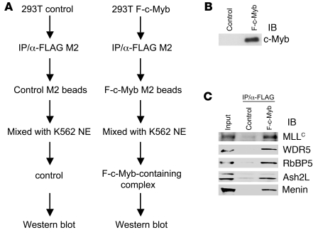
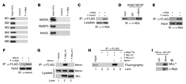
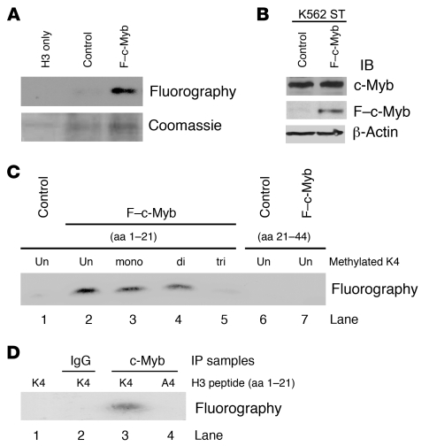
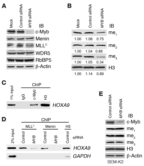
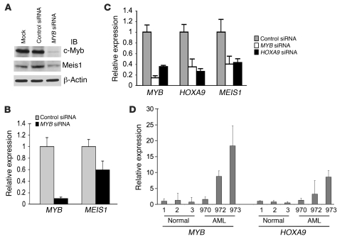
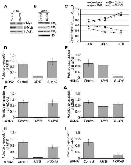
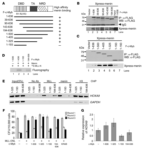
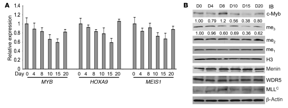
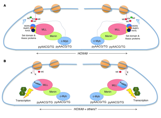
Mixed-lineage leukemia (MLL) is a proto-oncogene frequently involved in chromosomal translocations associated with acute leukemia. These chromosomal translocations commonly result in MLL fusion proteins that dysregulate transcription. Recent data suggest that the MYB proto-oncogene, which is an important regulator of hematopoietic cell development, has a role in leukemogenesis driven by the MLL-ENL fusion protein, but exactly how is unclear. Here we have demonstrated that c-Myb is recruited to the MLL histone methyl transferase complex by menin, a protein important for MLL-associated leukemic transformation, and that it contributes substantially to MLL-mediated methylation of histone H3 at lysine 4 (H3K4). Silencing MYB in human leukemic cell lines and primary patient material evoked a global decrease in H3K4 methylation, an unexpected decrease in HOXA9 and MEIS1 gene expression, and decreased MLL and menin occupancy in the HOXA9 gene locus. This decreased occupancy was associated with a diminished ability of an MLL-ENL fusion protein to transform normal mouse hematopoietic cells. Previous studies have shown that MYB expression is regulated by Hoxa9 and Meis1, indicating the existence of an autoregulatory feedback loop. The finding that c-Myb has the ability to direct epigenetic marks, along with its participation in an autoregulatory feedback loop with genes known to transform hematopoietic cells, lends mechanistic and translationally relevant insight into its role in MLL-associated leukemogenesis.


Figures

Figure 1
Figure 1. c-Myb coprecipitates with the menin and MLL complex.
(A) Schematic depiction of experimental scheme. HEK 293T cells were transiently transfected with expression vector encoding FLAG-tagged c-Myb. FLAG-tagged empty vector was used as a control. Cell lysates prepared from transfectants were mixed with α-FLAG antibody affinity M2 agarose to purify FLAG–c-Myb protein. After washing with lysis buffer (1% NP-40), FLAG–c-Myb–containing M2 beads were incubated with nuclear extracts (NE) from K562 cells to isolate the c-Myb–containing complex. Beads were washed twice with nuclear extract buffer (0.05% NP-40). (B and C) Isolated samples were probed by immunoblotting with α–c-Myb (B), α-menin, α-MLLC, α-WDR5, α-RbBP5, and α-Ash2L antibodies (C).
Figure 2
Figure 2. c-Myb associates with MLL through menin.
(A) In vitro translated, 35S-labeled MLL fragments (indicated on the left) and F–c-Myb (indicated at the top) were mixed, followed by IP with anti-FLAG M2 agarose. Samples were resolved on SDS-PAGE, then amplified, dried, and fluorographed. (B) In vitro protein-binding assay of WDR5, RbBP5, and Ash2L with F–c-Myb as described in A. (C) Cell lysates prepared from 293T cells cotransfected with c-Myb and FLAG-menin were subjected to IP with anti-FLAG M2 agarose, followed by immunoblotting with α–c-Myb antibody. (D) Nuclear extracts prepared from K562 cells were subjected to IP with α-menin antibody or control IgG. Immunoprecipitates were analyzed by immunoblot with α–c-Myb antibody. (E and F) c-Myb and menin proteins were in vitro translated with or without a FLAG tag separately. The various proteins were mixed as indicated in the blots of E and F and then subjected to IP with anti-FLAG M2 agarose. Immunoprecipitates were analyzed by immunoblotting with α–c-Myb (E) or α-menin (F) antibody. (G) FLAG-tagged MLL deletion mutants (F-MLL-N: aa 1–400; F-MLLd11: aa 12–400) and menin were transiently expressed in 293T cells. Cell lysates were subjected to IP with α–FLAG M2 agarose. Immunoprecipitates were analyzed by immunoblotting with α-menin antibody. (H) In vitro translated proteins were mixed as indicated on the right, followed by IP with α-FLAG M2 agarose. Samples were resolved on SDS-PAGE, then amplified, dried, and fluorographed. (I) Nuclear extracts from human leukemia patient (UPN no. 391) were immunoprecipitated by α-menin antibody or general IgG as a control. Samples were resolved on 5% SDS-PAGE, followed by immunoblotting with α-MLLN or α–c-Myb antibody.
Figure 3
Figure 3. The c-Myb–containing complex methylates histone H3 on lysine 4.
(A) F–c-Myb or control immunoprecipitate (described in Figure 1A) was incubated with recombinant human histone H3 and the methyl donor [3H]SAM. Samples were resolved on 15% SDS-PAGE, stained with Coomassie blue (bottom panel), amplified, dried, and flourographed (top panel). (B) Cell lysates (top panel), α-FLAG immunoprecipitates (middle panel) prepared from F–c-Myb stably transfected K562 cells, or empty vector control K562 stable cells were immunoblotted with α–c-Myb and α-FLAG antibodies. (C) Methylation of histone H3 peptides with different levels of K4 premethylation (aa 1–21) or without methylation (aa 21–44) by F–c-Myb–containing complexes prepared from K562 stable cells. (D) Mutation of H3 peptide from K4 to A4 abrogated the methylation activity of the c-Myb complex.
Figure 4
Figure 4. Loss of c-Myb affects global H3K4 methylation level in K562 cells but not in human SEM-K2 leukemic cells.
(A and B) K562 cells were nucleofected with control siRNA or MYB siRNA. Whole-cell extracts were prepared 3 days after the initial nucleofection according to the cell number counts and probed by immunoblotting with α–c-Myb, α-menin, α-WDR5, α-MLLC, α-RbBP5, α–β-actin (A), α-H3K4(Me)1-3, and α-H3 (B) antibodies. Mock indicates K562 cells nucleofected with the same amount of nuclease-free water without siRNA. (C) ChIP assay was performed to assess the occupancy of c-Myb on the HOXA9 gene locus of HL-60 cells. Chromatin was immunoprecipitated with α–c-Myb antibody. The presence of the HOXA9 locus DNA in the chromatin precipitates was assessed by standard PCR. Negative and positive controls consisted of IgG and α–histone H3 antibody, respectively. (D) ChIP analysis of HL-60 cells nucleofected with control siRNA or MYB siRNA was performed using antibodies specific for MLLC and menin. The presence of the HOXA9 locus DNA in the chromatin precipitates was assessed by standard PCR. ChIP using α–histone H3 antibody served as a positive control. (E) SEM-K2 cells were nucleofected with control siRNA or MYB siRNA. Whole-cell extracts were prepared 3 days after the initial nucleofection according to the cell number counts and probed by immunoblotting with α–c-Myb, α-H3K4(Me)1-3, α-H3, and α–β-actin antibodies.
Figure 5
Figure 5. Loss of c-Myb results in a decrease in HOXA9 and MEIS1 gene expression.
(A) K562 cells were nucleofected with control siRNA or MYB siRNA. Whole-cell extracts were prepared 3 days after the initial nucleofection. Proteins were subjected to immunoblot analysis with α–c-Myb, α-Meis1, and α–β-actin antibodies. Mock indicates K562 cells nucleofected with the same amount of nuclease-free water without siRNA. (B) K562 cells were nucleofected with control siRNA or MYB siRNA. RNA samples, prepared 2 days after the initial nucleofection, were reverse transcribed and used for qRT-PCR analysis for MYB and MEIS1 expression, determined in triplicate. Relative expression of MYB and MEIS1 to GAPDH is shown, with error bars indicating standard deviations. (C) Human ML-2 leukemia cells were nucleofected with control, MYB, or HOXA9 siRNA. RNA samples were prepared 2 days after initial nucleofection and subjected to qRT-PCR using GAPDH as an internal standard. Error bars represent standard deviations based on assays performed in triplicate. (D) RNA samples prepared from normal bone marrow donors (nos. 1, 2, and 3) and AML patients (UPN nos. 970, 972, and 973) were subjected to qRT-PCR for MYB and HOXA9 mRNA expression levels as described in B. Error bars represent the standard deviation of assays performed in triplicate.
Figure 6
Figure 6. Decrease in trimethylation of H3K4 and HOXA9 gene expression is specific for MYB silencing but not secondary to the antiproliferation effect of MYB silencing.
(A and B) KCL-22 cells were nucleofected with control siRNA or B-MYB siRNA. Whole-cell extracts were prepared 3 days after the initial nucleofection according to the cell number counts and probed by immunoblotting with α–c-Myb, α–B-Myb, and α–β-actin (A), α-H3K4(Me)1-3, and α-H3 (B) antibodies. (C) Cell proliferation assays were carried out with KCL-22 cells nucleofected with mock, control, MYB, and B-MYB siRNAs at indicated time points. Silencing MYB or B-MYB inhibited cell proliferation. (DG) KCL-22 cells were nucleofected with control, MYB, or B-MYB siRNA. RNA samples, prepared 2 days after the initial nucleofection, were reverse transcribed and used for qRT-PCR analysis for MYB (D), B-MYB (E), HOXA9 (F), and MEIS1 (G) gene expression determined in triplicate as described in Figure 5B. (H and I) KCL-22 cells were nucleofected with control, MYB, or HOXA9 siRNA. RNA samples, prepared 2 days after the initial nucleofection, were reverse transcribed and used for qRT-PCR analysis for MYB (H) and HOXA9 (I) gene expression determined in triplicate as described in Figure 5B. Error bars represent the standard deviation of assays performed in triplicate (CI).
Figure 7
Figure 7. The interaction between c-Myb and the menin-MLL complex is required for localization of MLL and menin on the HOXA9 gene, transformation of myeloid progenitors, and HOXA9 gene expression.
(A) Schematic representation of the constructions used. DBD, DNA binding domain; TA, transactivation domain; NRD, negative regulation domain. Various c-Myb mutants containing FLAG tag at their N termini and Xpress-tagged menin were transiently transfected in 293T cells. Cell lysates prepared from transfectants were subjected to IP with anti-FLAG M2 agarose. High-affinity menin-binding capacities are indicated on the right. (B and C) Immunoprecipitated proteins were separated by 10% SDS-PAGE and immunoblotted with anti-FLAG antibody (upper panel) or anti-Xpress antibody (bottom panel) antibody. (D) F–c-Myb mutants, menin, and 35S-labeled F–MLL-N were generated by in vitro transcription/translation. c-Myb was incubated with protein reaction mixtures as indicated on the right in the presence of α-FLAG M2 agarose. Samples were resolved on 10% SDS-PAGE, amplified, dried, and fluorographed. (E) ChIP assay of HL-60 cells transfected with c-Myb mutants was performed using the antibodies indicated at the top. Amplicons upstream of the HOXA9 and GAPDH genes were analyzed. (F) CFU per 104 plated cells are shown for each round of plating. Error bars represent standard deviations of 3 independent experiments. (G) Relative expression levels of HOXA9 gene are shown in SEM-K2 cells transfected with c-Myb or its mutants. Error bars represent the standard deviation of assays performed in triplicate.
Figure 8
Figure 8. Treatment of AML patient no. 866 with MYB antisense oligonucleotides resulted in a reduction in HOXA9 and MEIS1 gene expression, as well as a decrease in trimethylation of H3K4.
(A) RNA samples were prepared from cells isolated from patient no. 866 at the indicated days and subjected to qRT-PCR for MYB, HOXA9, and MEIS1 mRNA expression levels as described in Figure 5D. Error bars represent the standard deviation of assays performed in triplicate. (B) Whole-cell lysates prepared from the same patient at indicated days were analyzed by immunoblotting with a variety of antibodies indicated on the right. Numbers between the top rows are densitometry measurements of scanned bands relative to the day 0 value, which was arbitrarily set at 1.00.
Figure 9
Figure 9. c-Myb binds to menin-MLL and directs the complex to canonical c-Myb–binding sites in the HOXA9 promoter locus.
(A) Proposed interaction of c-Myb with wild-type menin-MLL in the HOXA9 promoter locus. c-Myb binds to its canonical recognition sites and holds the complex in place, leading to maintenance of HOXA9 expression. Changes in surrounding chromatin, specifically H3K4 methylation in neighboring nucleosomes (orange circles), is brought about by the MLLC SET domain–associating proteins, most likely WDR5, whose function may be enhanced by an as-yet-uncharacterized interaction with c-Myb, as indicated by the thin blue arrow. (B) c-Myb brings the menin-MLL fusion protein complex to the HOXA9 locus, where, as in A, it serves to anchor the complex and sustain HOXA9 expression. Sustained fusion protein expression leads to other, as-yet-uncharacterized transcriptional changes that likely play a role in leukemic transformation. As indicated, other unidentified cofactors are postulated to assist in transformation.

Similar articles

Cited by

References

    1. Klempuauer KH, Gonda TJ, Bishop JM. Nucleotide sequence of the retroviral leukemia gene v-myb and its cellular progenitor c-myb: the architecture of a transduced oncogene. . Cell. 1982;31(2 pt 1):453–463. doi: 10.1016/0092-8674(82)90138-6. - DOI - PubMed
    1. Duprey SP, Boettiger D. Developmental regulation of c-myb in normal myeloid progenitor cells. . Proc Natl Acad Sci U S A. 1985;82(20):6937–6941. doi: 10.1073/pnas.82.20.6937. - DOI - PMC - PubMed
    1. Kastan MB, Slamon DJ, Civin CI. Expression of protooncogene c-myb in normal human hematopoietic cells. . Blood. 1989;73(6):1444–1451. - PubMed
    1. Graf T. Myb: a transcriptional activator linking proliferation and differentiation in hematopoietic cells. Curr Opin Genet Dev. 1992;2(2):249–255. doi: 10.1016/S0959-437X(05)80281-3. - DOI - PubMed
    1. Gewirtz AM, Calabretta B. A c-myb antisense oligodeoxynucleotide inhibits normal human hematopoiesis in vitro. . Science. 1988;242(4883):1303–1306. doi: 10.1126/science.2461588. - DOI - PubMed

Publication types

MeSH terms