. 2010 Feb;120(2):521-32.
doi: 10.1172/JCI40706. Epub 2010 Jan 19.

The tobacco-specific carcinogen NNK induces DNA methyltransferase 1 accumulation and tumor suppressor gene hypermethylation in mice and lung cancer patients

Affiliations

The tobacco-specific carcinogen NNK induces DNA methyltransferase 1 accumulation and tumor suppressor gene hypermethylation in mice and lung cancer patients

Ruo-Kai Lin et al. J Clin Invest. 2010 Feb.

Abstract

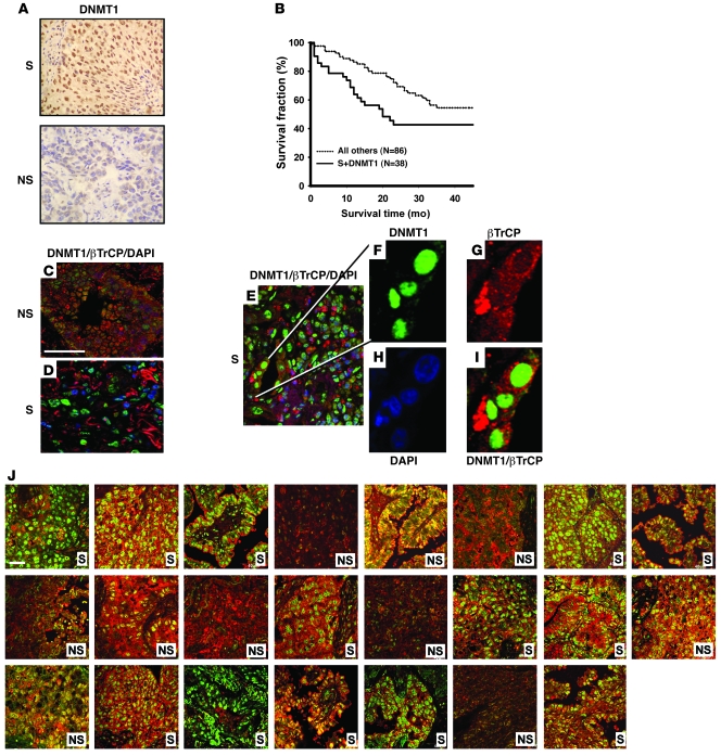
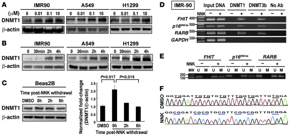
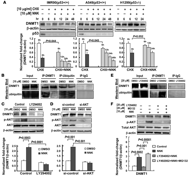
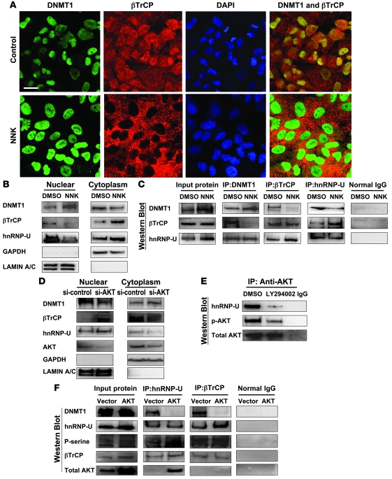
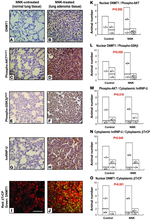
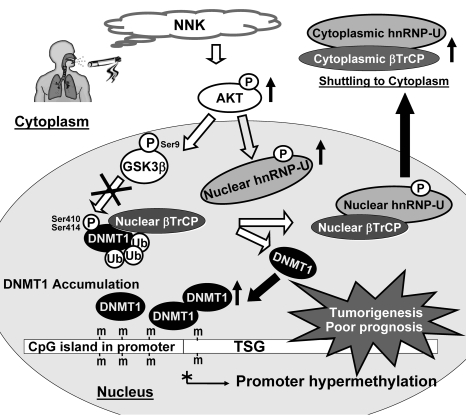
DNA methyltransferase 1 (DNMT1) catalyzes DNA methylation and is overexpressed in many human diseases, including cancer. The tobacco-specific carcinogen NNK also induces DNA methylation. However, the role of DNMT1-mediated methylation in tobacco carcinogenesis remains unclear. Here we used human and mouse lung cancer samples and cell lines to determine a mechanism whereby NNK induced DNMT1 expression and activity. We determined that in a human lung cell line, glycogen synthase kinase 3beta (GSK3beta) phosphorylated DNMT1 to recruit beta-transducin repeat-containing protein (betaTrCP), resulting in DNMT1 degradation, and that NNK activated AKT, inhibiting GSK3beta function and thereby attenuating DNMT1 degradation. NNK also induced betaTrCP translocation to the cytoplasm via the heterogeneous nuclear ribonucleoprotein U (hnRNP-U) shuttling protein, resulting in DNMT1 nuclear accumulation and hypermethylation of the promoters of tumor suppressor genes. Fluorescence immunohistochemistry (IHC) of lung adenomas from NNK-treated mice and tumors from lung cancer patients that were smokers were characterized by disruption of the DNMT1/betaTrCP interaction and DNMT1 nuclear accumulation. Importantly, DNMT1 overexpression in lung cancer patients who smoked continuously correlated with poor prognosis. We believe that the NNK-induced DNMT1 accumulation and subsequent hypermethylation of the promoter of tumor suppressor genes may lead to tumorigenesis and poor prognosis and provide an important link between tobacco smoking and lung cancer. Furthermore, this mechanism may also be involved in other smoking-related human diseases.


Figures

Figure 1
Figure 1. DNMT1 nuclear overexpression correlates with poor prognosis and shows mutually exclusive localization with βTrCP expression in lung cancer patients who continuously smoked.
(A) Representative IHC analysis of DNMT1 in paraffin sections from lung cancer patients who were smokers (S) and those who did not smoke (NS). Increased DNMT1 nuclear positive immunoreactivity was found in the patient who smoked. Original magnification, ×200. (B) Kaplan-Meier survival curves for overall postoperative survival showed that lung cancer patients who continuously smoked and had overexpression of DNMT1 protein had a worse prognosis compared with all other patient groups (P = 0.007, log-rank test). (CI) Representative fluorescence IHC and confocal microscopy of DNMT1 (green), βTrCP (red), and DAPI (blue) in paraffin sections of tumors from lung cancer patients who did not (C) and did continuously smoke (DI). βTrCP protein was mainly localized in the cytoplasm, which was distinct from the nuclear localization of DNMT1 protein in patients who smoked. Original magnification, ×630 (CE); ×1,500 (FI). Scale bar: 40 μm. (J) Representative figures of double fluorescence IHC for DNMT1 and βTrCP in a tissue array including 23 lung cancer patients. Original magnification, ×630. Scale bar: 40 μm.
Figure 2
Figure 2. NNK-induced DNMT1 protein binds to promoters and results in hypermethylation of TSGs.
(A and B) Expression levels of DNMT1 in human lung cell lines were analyzed by Western blotting. NNK increased protein levels of DNMT1 in (A) dose-dependent and (B) time-dependent manners in the 3 cell lines tested. β-Actin antibody was used as an internal control. (C) DNM1 protein returned to basal levels 2–6 hours after discontinuation of NNK treatment in Beas-2B bronchial epithelial cells. (D) ChIP-PCR using DNMT1 and DNMT3b antibodies for amplification of FHIT, p16INK4a, and RARB promoters before and after NNK treatment for 24 hours in IMR90 cells. Binding of DNMT1 and DNMT3b proteins to their target TSG promoters was induced by NNK. (E and F) MSP for promoter hypermethylation status in the FHIT, p16INK4a, and RARB promoters (E) and bisulfite sequencing of the p16INK4a promoter (F) before and after NNK treatment for 48 hours in IMR90 cells. Hypermethylated genes were defined as those that produced amplified methylation products in MSP or CG-dinucleotide sequences in bisulfite sequencing assays. M, methylated; U, unmethylated.
Figure 3
Figure 3. NNK enhances DNMT1 protein stability through the AKT signaling pathway, which is associated with the ubiquitin-proteosome system.
(A) NNK prolonged DNMT1 protein half-life. DNMT1 protein levels were determined by Western blotting and densitometry. β-Actin was used as a loading control, and p53 served as a positive control for CHX treatment because its half-life is known to be short. White bars, cells treated with DMSO; dark gray bars, cells treated with CHX for 6 hours; light gray bars, cells treated with CHX and NNK for the indicated times. (B) Ubiquitination of DNMT1 decreased upon NNK treatment. A549 cells were treated with NNK or DMSO control for 2 hours, after which cell lysates were immunoprecipitated with anti-DNMT1 or anti-ubiquitin antibody and then Western blotted. Normal IgG was used as a negative control. (C and D) A549 cells were pretreated with or without the PI3K/AKT pathway inhibitor LY294002 (C) or AKT siRNA (D), then treated with NNK. Western blotting and densitometry showed that each reduced levels of NNK-induced DNMT1. (E) LY294002 treatment increased ubiquitination of DNMT1, leading to low levels of DNMT1 protein in A549 cells. (F) Western blotting and densitometry of A549 cells treated with NNK with or without pretreatment with LY294002 and the proteasome inhibitor MG132 showed altered DNMT1 protein levels. Data are mean ± SEM (n = 3).
Figure 4
Figure 4. GSK3β and βTrCP interact with DNMT1 protein and enhance DNMT1 degradation.
(A) A549 cells were transfected with pCMV-SPORT6-GSK3β, then treated with or without NNK. The GSK3β pathway promoted DNMT1 protein degradation, which was attenuated by NNK treatment. Data are mean ± SEM (n = 3). (B) Cell lysates were immunoprecipitated with anti-DNMT1 or anti–phospho-Ser antibody and then Western blotted. DNMT1 protein formed complexes with GSK3β and βTrCP proteins. Increase DNMT1 phosphorylation was mediated by exogenous GSK3β, which increased the interaction between DNMT1 and βTrCP. Normal IgG was used as a negative control. (C) βTrCP increased the ubiquitination level of DNMT1. A549 cells were transfected with pCMV-SPORT6-βTrCP for 24 hours, after which cell lysates were immunoprecipitated with anti-DNMT1 and then western blotted. (D) Site-directed mutagenesis of both Ser410 (S410A) and Ser414 (S414A) on DNMT1 protein decreased the phosphorylation level of DNMT1 protein by GSK3β and disrupted the interaction between βTrCP and DNMT1. Cells were transfected with WT or mutant His-tag DNMT1 expression vector and exogenous GSK3β or vector control. Cell lysates were immunoprecipitated with anti–His-tag antibody and then Western blotted.
Figure 5
Figure 5. NNK enhances hnRNP-U/βTrCP translocation to the cytoplasm and induces DNMT1 accumulation in the nucleus, but this effect is attenuated by AKT inhibition.
(A) Immunofluorescence confocal microscopy to localize DNMT1 (green), βTrCP (red), and nucleus (blue; DAPI). The merged figures showed colocalization of DNMT1 with βTrCP protein in A549 cells treated with DMSO control. Mutually exclusive localization of DNMT1 and βTrCP was observed in cells treated with NNK. Original magnification, ×630. Scale bar: 20 μm. (B) Cell fractionation assay to analyze distribution of nuclear and cytoplasmic proteins, using anti-DNMT1, anti-βTrCP, anti–hnRNP-U, and anti-GSK3βSer9 antibodies. Anti-GAPDH and anti–lamin A/C antibodies were used as cytoplasmic and nuclear protein markers, respectively. The distribution of βTrCP and hnRNP-U proteins was contrary to that of DNMT1 protein. (C) DNMT1 and βTrCP interaction was interrupted by NNK treatment. In contrast, βTrCP and hnRNP interaction was increased by NNK treatment. (D) Cell fractionation assay was performed in A549 cells with or without AKT siRNA. Inhibition of AKT induced both hnRNP-U and βTrCP proteins predominantly located in the nucleus, where DNMT1 protein level decreased. (E) IP assay using anti-AKT antibody showed that hnRNP formed a complex with AKT, and the interaction was attenuated by LY294002. (F) An increase of hnRNP-U phosphorylation was mediated by exogenous AKT, which increased the interaction between hnRNP-U and βTrCP. In addition, AKT decreased the interaction of DNMT1 with hnRNP-U and βTrCP.
Figure 6
Figure 6. Representative IHC and fluorescence IHC of mouse tissue slides.
IHC and fluorescence IHC were performed on paraffin sections of normal lung tissue from untreated mice and lung adenoma of NNK-treated mice. Protein expression levels of nuclear DNMT1 (A and B), p-AKTSer473 (C and D), the inactive form of p-GSK3βSer9 (E and F), cytoplasmic hnRNP (G and H), and cytoplasmic βTrCP (red, I and J; merged image with DNMT1, green) increased in lung adenoma tissue of NNK-treated mice compared with the normal lung tissue of untreated mice. Original magnification, ×200 (AH); ×630 (I and J). Scale bar: 40 μm. (KO) Concordance analysis by Pearson c2 test showed a strong correlation between nuclear DNMT1 and p-AKTSer473 (K), p-GSK3βSer9 (L), and cytoplasmic βTrCP (O); between p-AKTSer473 and cytoplasmic hnRNP (M); and between cytoplasmic hnRNP and cytoplasmic βTrCP (N) in control and NNK-treated mice. Symbols within bars denote expression of the respective proteins; for example, “+/–” in K denotes mice with positive nuclear DNMT1 expression and negative p-AKTSer473 expression.
Figure 7
Figure 7. Proposed model to illustrate the accumulation of nuclear DNMT1 by NNK-induced AKT/GSK3β/βTrCP/hnRNP-U signaling leading to promoter hypermethylation and tumorigenesis.
NNK induces DNMT1 protein accumulation in the nucleus through AKT/GSK3β/βTrCP signaling and AKT/hnRNP-U/βTrCP nucleocytoplasmic shuttling. βTrCP is an E3 ubiquitin ligase that specifically interacts with and degrades DNMT1. NNK induces activation of AKT, then promotes GSK3β phosphorylation at Ser9 to form inactive GSK3β, which subsequently attenuates the ability of βTrCP to degrade DNMT1 protein. In addition, NNK activates AKT to induce interaction between phosphorylated hnRNP-U and βTrCP, which disrupts βTrCP/DNMT1 interaction. The hnRNP-U/βTrCP complex translocates to the cytoplasm, leading to DNMT1 accumulation in the nucleus. Furthermore, these NNK-induced DNMT1 proteins bind to promoters of various TSGs and result in promoter hypermethylation, which ultimately leads to tumorigenesis and poor prognosis.

References

    1. Jamaluddin MS, Yang X, Wang H. Hyperhomocysteinemia, DNA methylation and vascular disease. Clin Chem Lab Med. 2007;45(12):1660–1666. doi: 10.1515/CCLM.2007.350. - DOI - PubMed
    1. Costa E, Dong E, Grayson DR, Guidotti A, Ruzicka W, Veldic M. Reviewing the role of DNA (cytosine-5) methyltransferase overexpression in the cortical GABAergic dysfunction associated with psychosis vulnerability. Epigenetics. 2007;2(1):29–36. - PubMed
    1. Kanai Y. Alterations of DNA methylation and clinicopathological diversity of human cancers. Pathol Int. 2008;58(9):544–558. doi: 10.1111/j.1440-1827.2008.02270.x. - DOI - PubMed
    1. Egger G, Liang G, Aparicio A, Jones PA. Epigenetics in human disease and prospects for epigenetic therapy. Nature. 2004;429(6990):457–463. doi: 10.1038/nature02625. - DOI - PubMed
    1. Abdolmaleky HM, et al. Methylomics in psychiatry: Modulation of gene-environment interactions may be through DNA methylation. Am J Med Genet B Neuropsychiatr Genet. 2004;127B(1):51–59. doi: 10.1002/ajmg.b.20142. - DOI - PubMed

Publication types

Substances