. 2010 Feb;120(2):559-69.
doi: 10.1172/JCI40008. Epub 2010 Jan 19.

Conventional DCs reduce liver ischemia/reperfusion injury in mice via IL-10 secretion

Affiliations

Conventional DCs reduce liver ischemia/reperfusion injury in mice via IL-10 secretion

Zubin M Bamboat et al. J Clin Invest. 2010 Feb.

Abstract

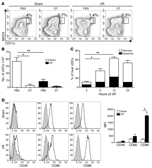
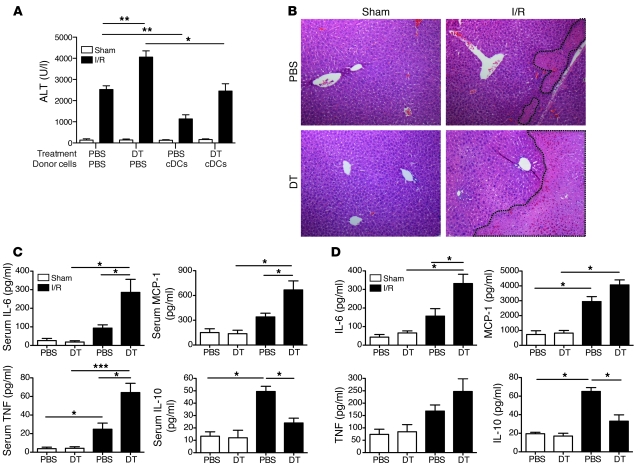
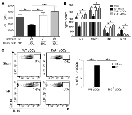
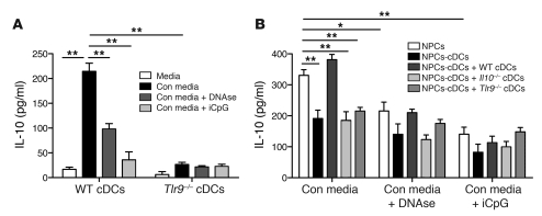
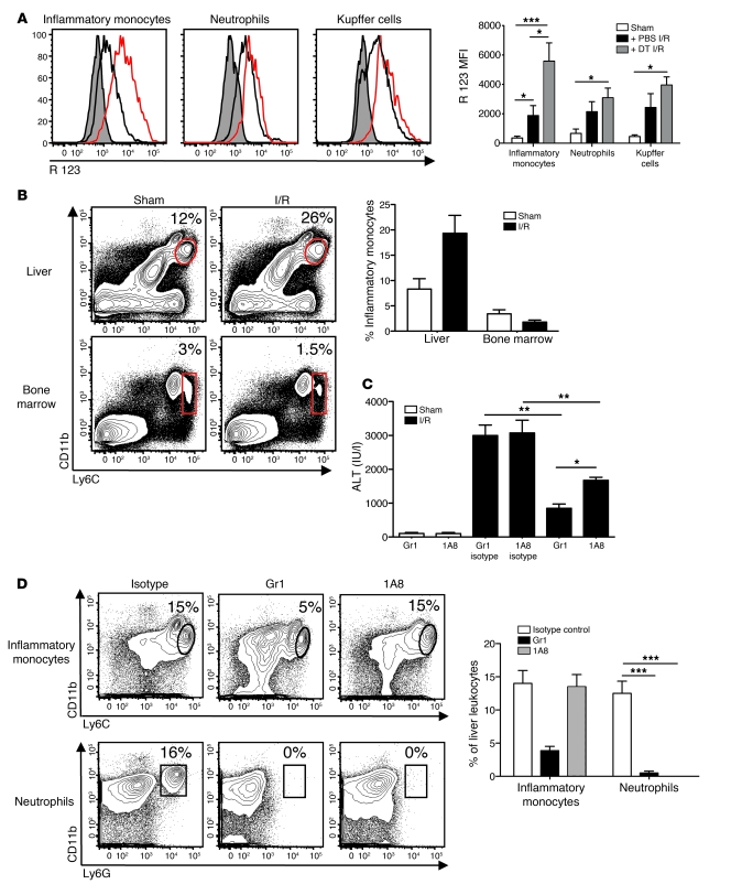
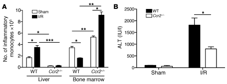
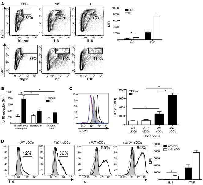
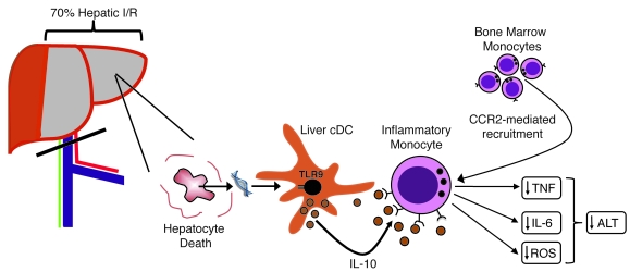
TLRs are recognized as promoters of tissue damage, even in the absence of pathogens. TLR binding to damage-associated molecular patterns (DAMPs) released by injured host cells unleashes an inflammatory cascade that amplifies tissue destruction. However, whether TLRs possess the reciprocal ability to curtail the extent of sterile inflammation is uncertain. Here, we investigated this possibility in mice by studying the role of conventional DCs (cDCs) in liver ischemia/reperfusion (I/R) injury, a model of sterile inflammation. Targeted depletion of mouse cDCs increased liver injury after I/R, as assessed by serum alanine aminotransferase and histologic analysis. In vitro, we identified hepatocyte DNA as an endogenous ligand to TLR9 that promoted cDCs to secrete IL-10. In vivo, cDC production of IL-10 required TLR9 and reduced liver injury. In addition, we found that inflammatory monocytes recruited to the liver via chemokine receptor 2 were downstream targets of cDC IL-10. IL-10 from cDCs reduced production of TNF, IL-6, and ROS by inflammatory monocytes. Our results implicate inflammatory monocytes as mediators of liver I/R injury and reveal that cDCs respond to DAMPS during sterile inflammation, providing the host with protection from progressive tissue damage.


Figures

Figure 1
Figure 1. Liver I/R induces cDC death.
CD11c-DTR mice, which were pretreated with DT or PBS 18 hours earlier, underwent a sham laparotomy (Sham) or 1 hour of ischemia, followed by 12 hours of reperfusion (I/R). Ischemic liver cDCs were assessed by flow cytometry and are shown as (A) the percentage of hepatic leukocytes within each gated region or (B) the absolute number of cDCs within the ischemic liver. Ischemic liver cDCs from PBS-treated CD11c-DTR mice were analyzed for (C) the percentage of apoptosis (AnnexinV+PI) and necrosis (Annexin V+PI+) by flow cytometry or (D) CD40, CD80, and CD86 expression 12 hours after the sham procedure or I/R. Isotype controls are shown as shaded histograms. The bar graph represents cDC maturation data pooled from 3 independent experiments. Data represent mean ± SEM (BD). Data in all panels are representative of at least 3 independent experiments; n = 5 mice per group. *P < 0.05; **P < 0.01.
Figure 2
Figure 2. cDCs confer protection in I/R.
CD11c-DTR mice were administered PBS or DT via i.p. injection. Twelve hours later, mice received an i.v. injection of 1 × 10⁷ WT cDCs or PBS and then underwent sham laparotomy or I/R. (A) Serum ALT levels and (B) representative liver H&E staining (original magnification, ×100) are shown at 12 hours after sham procedure or liver I/R. Nonviable patches of ischemic lobes are demarcated by dashed lines. CD11c-DTR mice were pretreated with DT or PBS 12 hours earlier and then subjected to sham laparotomy or 12 hours of I/R. (C) Serum cytokines levels were measured or (D) ischemic liver CD45+ NPCs were harvested and cultured overnight in media prior to measurement of supernatant cytokines. Data represent mean ± SEM (A, C, and D). Data in all panels are representative of at least 3 independent experiments; n = 4–6 mice per group. *P < 0.05; **P < 0.01; ***P < 0.001.
Figure 3
Figure 3. cDC-mediated protection in liver I/R is TLR9 and IL-10 dependent.
CD11c-DTR mice were administered DT via i.p. injection. Twelve hours later, mice received an i.v. injection of either PBS or 1 × 10⁷ WT, Tlr9–/–, or Il10–/– cDCs just prior to I/R. (A) Serum ALT levels and (B) cytokines levels were measured 12 hours later. (C) WT or Tlr9–/– (both CD45.2) cDCs were injected into WT (CD45.1) recipients just prior to sham laparotomy or I/R. Twelve hours later, ischemic liver CD45+ NPCs were isolated and cultured in media in the presence of Brefeldin A. Intracellular IL-10 production by CD45.2+CD11chi hepatic cDCs was assayed 6 hours later and is expressed as the percentage within each gated region based on IL-10 isotype control antibody staining. The bar graph represents pooled data from 3 independent experiments. Data represent mean ± SEM and are representative of at least 2 independent experiments; n = 4–6 mice per group. *P < 0.05; **P < 0.01; ***P < 0.001.
Figure 4
Figure 4. Endogenous DNA promotes cDC IL-10 production through TLR9.
(A) Immunomagnetic bead-purified cDCs from WT or Tlr9–/– mice were cultured in media alone or with conditioned media (Con media) from necrotic hepatocytes. Some wells containing conditioned media were pretreated with DNAse I prior to coculture with cDCs, while iCpG was added to other wells. Supernatant IL-10 was measured 18 hours later using a cytometric bead array. (B) Liver CD45+ NPCs were isolated from WT mice and cultured in conditioned media, with or without DNAse I pretreatment or iCpG, as in A. In selected wells, NPCs were depleted of cDCs prior to the addition of conditioned media. WT, Tlr9–/–, or Il10–/– cDCs were added back to certain wells containing NPCs-cDCs. Supernatant IL-10 was measured 18 hours later. Data depict mean ± SEM from triplicate wells and are representative of 3 independent experiments. *P < 0.05; **P < 0.01.
Figure 5
Figure 5. Inflammatory monocytes exacerbate liver I/R injury.
CD45+ NPCs were isolated from the ischemic livers of CD11c-DTR mice after PBS or DT pretreatment and 12 hours of I/R or sham procedure. (A) Histograms depict inflammatory monocyte (CD11bintLy6Chi), neutrophil (CD11bhiLy6G+), and Kupffer cell (CD11bloF4/80+) ROS production, based on the conversion of dihydrorhodamine 123 to its oxidized form, rhodamine (R) 123 (shaded areas indicate sham procedure; black lines indicate PBS and I/R treatment; red lines indicate DT and I/R treatment). The bar graph represents ROS data pooled from 3 experiments. (B) Inflammatory monocyte frequencies within the ischemic liver and bone marrow of WT mice after 12 hours of I/R or sham surgery are represented as a percentage of CD45+ hepatic leukocytes or of live bone marrow cells. The bar graph represents pooled data from 3 experiments. (C and D) WT mice received anti-Gr1, 1A8, or isotype controls, 24 and 2 hours before I/R or sham procedure. Percentages of cells within selected gates are indicated. (C) Twelve hours later, serum ALT levels and (D) inflammatory monocyte and neutrophil composition within the ischemic liver were determined. Percentages of cells within selected gates are indicated. The bar graph represents pooled data from 3 independent experiments. Data represent mean ± SEM. *P < 0.05; **P < 0.01; ***P < 0.001.
Figure 6
Figure 6. Inflammatory monocytes depend on CCR2 to promote liver I/R injury.
WT and Ccr2–/– mice underwent 12 hours of I/R or sham laparotomy, at which time (A) the number of inflammatory monocytes within the ischemic liver and bone marrow (i.e., per femur) and (B) serum ALT levels were determined. Data represent mean ± SEM and are representative of at least 2 independent experiments; n = 4–6 mice per group. *P < 0.05; **P < 0.01; ***P < 0.001.
Figure 7
Figure 7. cDC IL-10 suppresses inflammatory monocyte function during I/R.
Ischemic liver CD45+ NPCs from CD11c-DTR mice treated with DT or PBS prior to 12 hours of I/R were cultured with Brefeldin A. (A) Intracellular IL-6 and TNF was determined 6 hours later. Numbers indicate the percentages within each gated region of cytokine staining in excess of isotype controls. More than 95% of the Ly6Chi cells depicted are CD11b+ (data not shown). The bar graph depicts inflammatory monocyte cytokine production as MFI above isotype pooled from 3 experiments. (B) IL-10 receptor expression was measured on inflammatory monocytes, neutrophils, and Kupffer cells from ischemic livers of PBS-treated CD11c-DTR mice 12 hours after I/R or sham procedure. MFI values above isotype controls are shown and pooled from 3 experiments. Recipient CD11c-DTR mice pretreated with DT were injected with WT or Il10–/– cDCs just prior to hepatic ischemia or sham procedure. (C) Twelve hours later, oxidative burst was measured (red line indicates sham treatment with WT cDCs; blue line indicates sham treatment with Il10–/– cDCs; shaded area indicates I/R treatment with WT cDCs; bold line indicates I/R treatment with Il10–/– cDCs). (D) Intracellular IL-6 and TNF production by inflammatory monocytes from liver CD45+ NPCs was measured and expressed as a percentage above isotype controls (black lines indicate isotype control; shaded area indicates sham treatment; bold lines indicate I/R treatment). The bar graphs in C and D depict data pooled from 3 experiments. (D) MFI values above isotype controls are shown only for I/R mice. Data represent mean ± SEM and are representative of at least 2 experiments; n = 4–6 mice per group. *P < 0.05; **P < 0.01.
Figure 8
Figure 8. Role of cDCs in hepatic I/R.
Vascular occlusion involving 70% of the liver for 1 hour followed by 12 hours of reperfusion induces hepatocyte death. The release of host DNA activates hepatic cDCs through TLR9. The ensuing production of IL-10 by cDCs causes suppression of inflammatory monocytes that are recruited to the ischemic liver via CCR2. Reduced production of IL-6, TNF, and ROS by inflammatory monocytes lowers ALT levels and reduces liver injury.

References

    1. Lotze MT, et al. The grateful dead: damage-associated molecular pattern molecules and reduction/oxidation regulate immunity. Immunol Rev. 2007;220:60–81. doi: 10.1111/j.1600-065X.2007.00579.x. - DOI - PubMed
    1. Clavien PA, Harvey PR, Strasberg SM. Preservation and reperfusion injuries in liver allografts. An overview and synthesis of current studies. Transplantation. 1992;53(5):957–978. - PubMed
    1. Scaffidi P, Misteli T, Bianchi ME. Release of chromatin protein HMGB1 by necrotic cells triggers inflammation. Nature. 2002;418(6894):191–195. doi: 10.1038/nature00858. - DOI - PubMed
    1. Cavassani KA, et al. TLR3 is an endogenous sensor of tissue necrosis during acute inflammatory events. J Exp Med. 2008;205(11):2609–2621. doi: 10.1084/jem.20081370. - DOI - PMC - PubMed
    1. Millar DG, et al. Hsp70 promotes antigen-presenting cell function and converts T-cell tolerance to autoimmunity in vivo. Nat Med. 2003;9(12):1469–1476. doi: 10.1038/nm962. - DOI - PubMed

Publication types