Role of myo-inositol by magnetic resonance spectroscopy in early diagnosis of Alzheimer's disease in APP/PS1 transgenic mice
- PMID: 20093832
- PMCID: PMC2837893
- DOI: 10.1159/000261646
Role of myo-inositol by magnetic resonance spectroscopy in early diagnosis of Alzheimer's disease in APP/PS1 transgenic mice
Abstract
Background/aims: To explore the potential value of myo-inositol (mIns), which is regarded as a biomarker for early diagnosis of Alzheimer's disease, in APP/PS1 transgenic (tg) mice detected by (1)H-MRS.
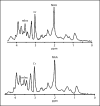
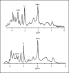
Methods: (1)H-MRS was performed in 30 APP/PS1 tg mice and 20 wild-type (wt) littermates at 3, 5 and 8 months of age. Areas under the peak of N-acetylaspartate (NAA), mIns and creatine (Cr) in the frontal cortex and hippocampus were measured, and the NAA/Cr and mIns/Cr ratios were analyzed quantitatively.
Results: Compared with the wt mice, the mIns/Cr ratio of the 3-month-old tg mice was significantly higher (p < 0.05), and pathology showed activation and proliferation of astrocytes in the frontal cortex and hippocampus. The concentration of NAA was significantly lower at 8 and 8 months of age (p < 0.05). According to the threshold of mIns/Cr that was adopted to separate the tg from the wt mice, the rate of correct predictions was 82, 94 and 95%, respectively, for 3, 5 and 8 months.
Conclusion: Of the early AD metabolites as detected by (1)H-MRS, mIns is the most valuable marker for assessment of AD. Quantitative analysis of mIns may provide important clues for early diagnosis of AD.
Figures


Similar articles
-
Age-related changes in brain metabolites and cognitive function in APP/PS1 transgenic mice.Behav Brain Res. 2012 Nov 1;235(1):1-6. doi: 10.1016/j.bbr.2012.07.016. Epub 2012 Jul 22. Behav Brain Res. 2012. PMID: 22828014
-
MICEST: a potential tool for non-invasive detection of molecular changes in Alzheimer's disease.J Neurosci Methods. 2013 Jan 15;212(1):87-93. doi: 10.1016/j.jneumeth.2012.09.025. Epub 2012 Oct 3. J Neurosci Methods. 2013. PMID: 23041110 Free PMC article.
-
Treatment effects in a transgenic mouse model of Alzheimer's disease: a magnetic resonance spectroscopy study after passive immunization.Neuroscience. 2014 Feb 14;259:94-100. doi: 10.1016/j.neuroscience.2013.11.052. Epub 2013 Dec 4. Neuroscience. 2014. PMID: 24316473 Free PMC article.
-
Multifaceted assessment of the APP/PS1 mouse model for Alzheimer's disease: Applying MRS, DTI, and ASL.Brain Res. 2018 Nov 1;1698:114-120. doi: 10.1016/j.brainres.2018.08.001. Epub 2018 Aug 3. Brain Res. 2018. PMID: 30077647
-
Regional metabolic alteration of Alzheimer's disease in mouse brain expressing mutant human APP-PS1 by 1H HR-MAS.Behav Brain Res. 2010 Jul 29;211(1):125-31. doi: 10.1016/j.bbr.2010.03.026. Epub 2010 Mar 20. Behav Brain Res. 2010. PMID: 20307581
Cited by
-
Saturation Transfer MRI for Detection of Metabolic and Microstructural Impairments Underlying Neurodegeneration in Alzheimer's Disease.Brain Sci. 2021 Dec 30;12(1):53. doi: 10.3390/brainsci12010053. Brain Sci. 2021. PMID: 35053797 Free PMC article. Review.
-
Progressive Pathological Changes in Neurochemical Profile of the Hippocampus and Early Changes in the Olfactory Bulbs of Tau Transgenic Mice (rTg4510).Neurochem Res. 2017 Jun;42(6):1649-1660. doi: 10.1007/s11064-017-2298-5. Epub 2017 May 18. Neurochem Res. 2017. PMID: 28523532 Free PMC article.
-
Metabolic Alterations Associated to Brain Dysfunction in Diabetes.Aging Dis. 2015 Oct 1;6(5):304-21. doi: 10.14336/AD.2014.1104. eCollection 2015 Sep. Aging Dis. 2015. PMID: 26425386 Free PMC article. Review.
-
Inositols Depletion and Resistance: Principal Mechanisms and Therapeutic Strategies.Int J Mol Sci. 2021 Jun 24;22(13):6796. doi: 10.3390/ijms22136796. Int J Mol Sci. 2021. PMID: 34202683 Free PMC article. Review.
-
Higher visceral fat is associated with lower cerebral N-acetyl-aspartate ratios in middle-aged adults.Metab Brain Dis. 2017 Jun;32(3):727-733. doi: 10.1007/s11011-017-9961-z. Epub 2017 Jan 31. Metab Brain Dis. 2017. PMID: 28144886 Free PMC article.
References
-
- Frey HJ, Mattila KM, Korolainen MA, Pirttilä T. Problems associated with biological markers of Alzheimer's disease. Neurochem Res. 2005;30:1501–1510. - PubMed
-
- Villemagne VL, Rowe CC, Macfarlane S, Novakovic KE, Masters CL. Imaginem oblivionis: the prospects of neuroimaging for early detection of Alzheimer's disease. J Clin Neurosci. 2005;12:221–230. - PubMed
-
- Pennanen C, Kivipelto M, Tuomainen S, Hartikainen P, Hänninen T, Laakso MP, Hallikainen M, Vanhanen M, Nissinen A, Helkala EL, Vainio P, Vanninen R, Partanen K, Soininen H. Hippocampus and entorhinal cortex in mild cognitive impairment and early AD. Neurobiol Aging. 2004;25:303–310. - PubMed
-
- Basso M, Yang J, Warren L, MacAvoy MG, Varma P, Bronen RA, van Dyck CH. Volumetry of amygdala and hippocampus and memory performance in Alzheimer's disease. Psychiatry Res. 2006;146:251–261. - PubMed
-
- den Heijer T, Geerlings MI, Hoebeek FE, Hofman A, Koudstaal PJ, Breteler MM. Use of hippocampal and amygdalar volumes on magnetic resonance imaging to predict dementia in cognitively intact elderly people. Arch Gen Psychiatry. 2006;63:57–62. - PubMed
Publication types
MeSH terms
Substances
LinkOut - more resources
Full Text Sources
Medical
Molecular Biology Databases
Miscellaneous

