. 2010 Feb 17;29(4):749-60.
doi: 10.1038/emboj.2009.397. Epub 2010 Jan 21.

The intronic splicing code: multiple factors involved in ATM pseudoexon definition

Affiliations

The intronic splicing code: multiple factors involved in ATM pseudoexon definition

Ashish Dhir et al. EMBO J. .

Abstract

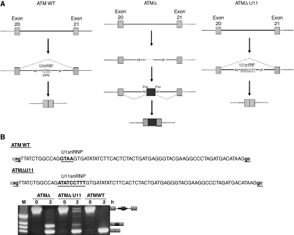
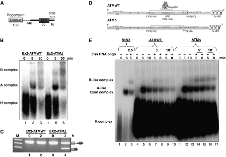
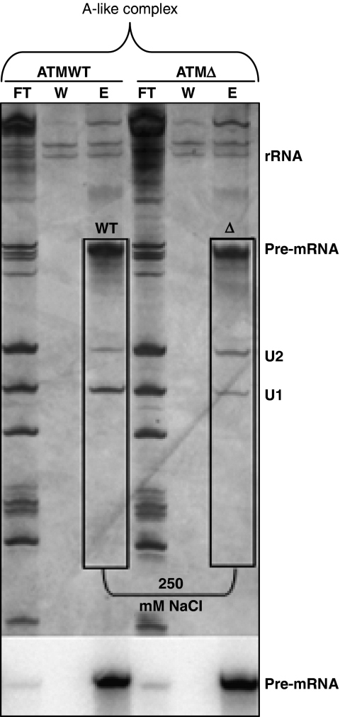
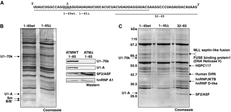
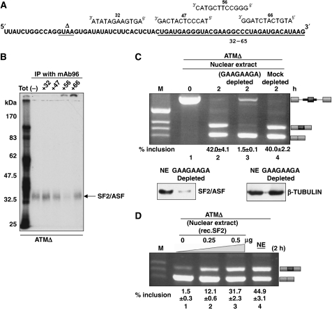
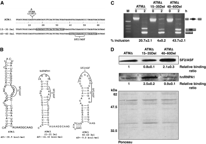
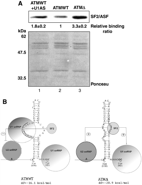
Abundance of pseudo splice sites in introns can potentially give rise to innumerable pseudoexons, outnumbering the real ones. Nonetheless, these are efficiently ignored by the splicing machinery, a process yet to be understood completely. Although numerous 5' splice site-like sequences functioning as splicing silencers have been found to be enriched in predicted human pseudoexons, the lack of active pseudoexons pose a fundamental challenge to how these U1snRNP-binding sites function in splicing inhibition. Here, we address this issue by focusing on a previously described pathological ATM pseudoexon whose inhibition is mediated by U1snRNP binding at intronic splicing processing element (ISPE), composed of a consensus donor splice site. Spliceosomal complex assembly demonstrates inefficient A complex formation when ISPE is intact, implying U1snRNP-mediated unproductive U2snRNP recruitment. Furthermore, interaction of SF2/ASF with its motif seems to be dependent on RNA structure and U1snRNP interaction. Our results suggest a complex combinatorial interplay of RNA structure and trans-acting factors in determining the splicing outcome and contribute to understanding the intronic splicing code for the ATM pseudoexon.


Conflict of interest statement

The authors declare that they have no conflict of interest.

Figures

Figure 1
Figure 1
ATM pre-mRNA splicing of exon 20 and exon 21 and effect on U11snRNP binding as opposed to U1snRNP in the ATM WT sequence. (A) Exons 20 and 21 are shown with light grey boxes, whereas the ATMΔ pseudoexon with black. Introns are depicted with a straight line. ‘ag' and ‘gc' represent the intronic splice sites flanking the pseudoexon. In normal splicing, the ISPE sequence is present and can bind an U1snRNP molecule at this position. In the A-T patient, deletion of a GTAA sequence in the ISPE abrogates U1snRNP binding and activates inclusion of the ATMΔ pseudoexon. Introduction of an U11snRNP-binding site restores inhibition. (B) Schematic representations of the binding positions of U1snRNP and of U11snRNP on the ATM WT and ATMΔ U11 pseudoexons, respectively (the sequences are shown with bold underlined letters). The sequence of the pseudoexon is shown with capital letters whereas splice sites are shown with underlined small bold letters. In vitro splicing and RT–PCR analysis of the ATMΔ, ATMΔ U11, and ATM WT substrates in PY7 minigenes are shown. The scheme of the spliced and unspliced substrates is shown on the right.
Figure 2
Figure 2
Assembly of the spliceosomal complexes on ATM WT and ATMΔ pseudo exon substrates. (A) Scheme of the biexonic construct used for assembly of cross-intron spliceosomal complexes upstream to the ATM pseudoexon. (B) Spliceosomal complex assembly at different time-points on the ATM WT and Δ biexonic substrates. The positions of the splicing complexes are shown on the left. (C) In vitro splicing and RT–PCR analyses of the ATM WT and ATMΔ biexonic constructs. (D) Scheme of the ATM WT and ATMΔ single exon substrates used for the assembly of spliceosomal complexes. The 3′ ends are tagged with 3 MS2 repeats. (E) Spliceosomal complex assembly at different time points across both ATM WT and ATMΔ single exon substrates either in the absence or presence of the 5′ssRNA oligo ‘in-trans'.
Figure 3
Figure 3
U2snRNP stability at high-salt washings of the ATM WT and ATMΔ A-like exon complexes. High-salt washings of the A-like exon complex assembled on the single exon substrate. Exon complexes that assembled on the MS2-tagged ATM WT and Δ substrate were affinity selected on amylose beads after glycerol gradient ultracentrifugation. The bound complexes were washed with 250 mM NaCl and eluted with maltose. FT lane represents flow through, W lane is for wash, and E lane for elute. RNAs were separated and visualized as described above. The autoradiograph of the same gel is shown below as loading control of the ATM WT and Δ pre-mRNAs. Efficiency of recovery has been estimated at 46% according to CPM counts before and after elution.
Figure 4
Figure 4
Identification of trans-acting factors that bind the ATM pseudoexon using adipic acid–agarose beads based pull-down and mass-spec analysis. (A) A scheme of the ATM pseudoexon RNAs used for pull-down analysis is presented. The underlined sequence represents the region of the pseudoexon used for pull-down analysis that spans from nucleotide 1–45 for ATM WT and ATMΔ and from nucleotide 32–65 common to both. (B) Identification of proteins that interact with the ATM WT 1–65 and Δ 1–65 pseudoexon RNAs. Different synthetic RNAs were covalently linked to agarose beads and incubated with Hela nuclear extract under splicing conditions. Proteins that remained bound to the RNAs after washing were separated on 10% SDS–PAGE and detected by Coomassie blue staining. The protein bands identified by mass-spec are mentioned with arrowheads on the sides of the gel. To obtain a better quantitative picture, the right panel contains western blots against the U1-70K, U1-A, SF2/ASF, and hnRNP A1 factors (C) Pull-down analysis using RNAs that span from nucleotide 1–45 for ATM WT and ATMΔ and from nucleotide 32–65 common to both. Protein bands identified by mass-spec are mentioned with arrowheads on the sides of the gel.
Figure 5
Figure 5
Mapping of the SF2/ASF-binding site on the ATMΔ pseudoexon sequence. (A) Schematic representation of 12-mer antisense DNA oligonucleotides targeting the ATMΔ pseudoexon sequence. (B) SF2/ASF immunoprecipitation with mAb96 antibody in the absence (lane –) or presence of these oligonucleotides. The position of the IP complex for SF2/ASF is shown with an arrowhead. (C) In vitro splicing of the ATMΔ pre-mRNA using SF2/ASF-depleted Hela Nuclear extract (lane 3) and with mock-depleted extract (lane 4). Schematic diagrams of the spliced products are shown on the right. Lower panel of (C) contains western blot showing the level of SF2/ASF depletion using the pull-down affinity procedure together with a tubulin control (D) ATMΔ pre-mRNA was incubated in either dilute nuclear extract (24%) supplemented with increasing quantities of recombinant SF2/ASF (lanes 1–3) or standard nuclear extract (48%) (lane 4) under splicing conditions. The quantities of the SF2/ASF added in μg to the nuclear extract are shown on the top. Schematic diagrams of the spliced products are shown on the right. Quantification of pseudoexon inclusion levels as determined by densitometric analysis are reported under each figure. Standard deviation values from three independent experiments are shown.
Figure 6
Figure 6
Correlation between the real and predicted SF2/ASF-binding site with the RNA secondary structure of ATMΔ. (A) The score matrix of various SR proteins predicted to bind ATMΔ RNA as predicted by ESE finder ver3.0. The SF2/ASF predicted motifs are numbered 1–3. The SF2/ASF-binding site validated experimentally to bind the ATMΔ pseudoexon overlaps with the predicted motif 3 and is shown by an arrow to fall in the internal loop conformation. (B) The predicted abolishment of the no. 3 SF2/ASF-binding site in the ATMΔ mutSF2 mutant. (C) In vitro splicing and RT–PCR of ATMΔ and ATMΔ mutSF2 RNA. Schematic diagrams of the spliced products are shown on the right. Quantification of pseudoexon inclusion levels as determined by densitometric analysis are reported under each figure. Standard deviation values from three independent experiments are shown. (D) Western blot pull-down analysis to confirm the abolishment of the SF2/ASF-binding site in the ATMΔ mutSF2 substrate in comparison to ATMΔ. Quantification of SF2/ASF-binding levels as determined by densitometric analysis are reported under each figure. Standard deviation values from three independent experiments are shown. Ponceau stain of the nitrocellulose membrane is shown for equal protein loading of the sample.
Figure 7
Figure 7
RNA secondary structure can influence splicing by modulating the display of trans-acting factor binding sites. (A) Scheme of the 20 nt deletions (15–35 and 40–60 Del mutants) made within the ATMΔ pseudoexon. The ATMΔ pseudoexon sequence is numbered starting from the 5′ end. Boxed sequences correspond to 20 nucleotide deleted region of the ATM pseudoexon. The positions of the GTAA deletion and of the SF2/ASF-binding site are also shown. (B) Predicted RNA secondary structure of ATMΔ and of the two deletion mutants. The position of the trans-acting factors (SF2/ASF and the putative hnRNPA1-binding site) is shown with arcs and brackets. (C) In vitro splicing and RT–PCR analysis of the ATMΔ deletion mutants. Schematic diagrams of the spliced products are shown on the right. Quantification of pseudoexon inclusion levels as determined by densitometric analysis are reported under each figure. Standard deviation values from three independent experiments are shown. (D) Pull-down analysis of ATMΔ and the deletion mutants followed by western blotting with mAb96 antibody against SF2/ASF and a polyclonal hnRNPA1 antibody. Quantification of SF2/ASF-binding levels as determined by densitometric analysis are reported under each figure. Standard deviation values from three independent experiments are shown. Ponceau stain of the nitrocellulose membrane is shown for equal protein loading of the sample.
Figure 8
Figure 8
Schematic model of the U1snRNP-mediated inhibition in the ATM pseudoexon sequence. (A) Pull-down analysis and subsequent western blotting with mAb96 antibody on ATM WT and ATMΔ pseudoexon RNA in the presence of U1snRNP-depleted nuclear extract. Quantification of SF2/ASF-binding levels as determined by densitometric analysis are reported under each figure. Standard deviation values from three independent experiments are shown. Ponceau stain of the nitrocellulose membrane is shown for equal protein loading of the samples. (B) Model of the U1snRNP-mediated inhibition of ATM WT pseudoexon. In this model, the U1snRNP-mediated inhibition of the ATM WT pseudoexon is constituted by a unproductive less stable recruitment of U2snRNP on the 3′ss region. Irregular boundary of U2snRNP demonstrates defective recruitment. Moreover, it also obstructs SF2/ASF occupancy to its binding site on the opposite side of the stem. In ATMΔ, the U1snRNP is no longer present due to the deletion of the ISPE. This leads to a more efficient binding of SF2/ASF to the enhancer site and stabilization of the U2snRNP interaction.

Similar articles

Cited by

References

    1. Abad X, Vera M, Jung SP, Oswald E, Romero I, Amin V, Fortes P, Gunderson SI (2008) Requirements for gene silencing mediated by U1 snRNA binding to a target sequence. Nucleic Acids Res 36: 2338–2352 - PMC - PubMed
    1. Akker SA, Misra S, Aslam S, Morgan EL, Smith PJ, Khoo B, Chew SL (2007) Pre-spliceosomal binding of U1 small nuclear ribonucleoprotein (RNP) and heterogenous nuclear RNP E1 is associated with suppression of a growth hormone receptor pseudoexon. Mol Endocrinol 21: 2529–2540 - PubMed
    1. Amara SG, Jonas V, Rosenfeld MG, Ong ES, Evans RM (1982) Alternative RNA processing in calcitonin gene expression generates mRNAs encoding different polypeptide products. Nature 298: 240–244 - PubMed
    1. Baralle M, Skoko N, Knezevich A, De Conti L, Motti D, Bhuvanagiri M, Baralle D, Buratti E, Baralle FE (2006) NF1 mRNA biogenesis: effect of the genomic milieu in splicing regulation of the NF1 exon 37 region. FEBS Lett 580: 4449–4456 - PubMed
    1. Behzadnia N, Hartmuth K, Will CL, Luhrmann R (2006) Functional spliceosomal A complexes can be assembled in vitro in the absence of a penta-snRNP. RNA 12: 1738–1746 - PMC - PubMed

Publication types

MeSH terms

Substances