

Mechanical buckling of veins under internal pressure
- PMID: 20094913
- PMCID: PMC2896067
- DOI: 10.1007/s10439-010-9929-1
Mechanical buckling of veins under internal pressure
Abstract
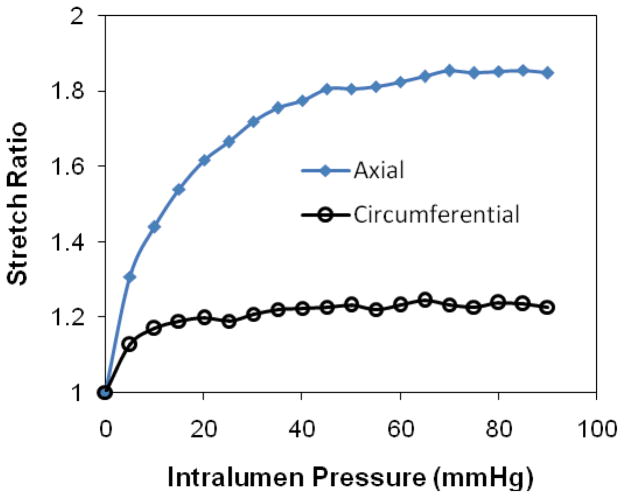
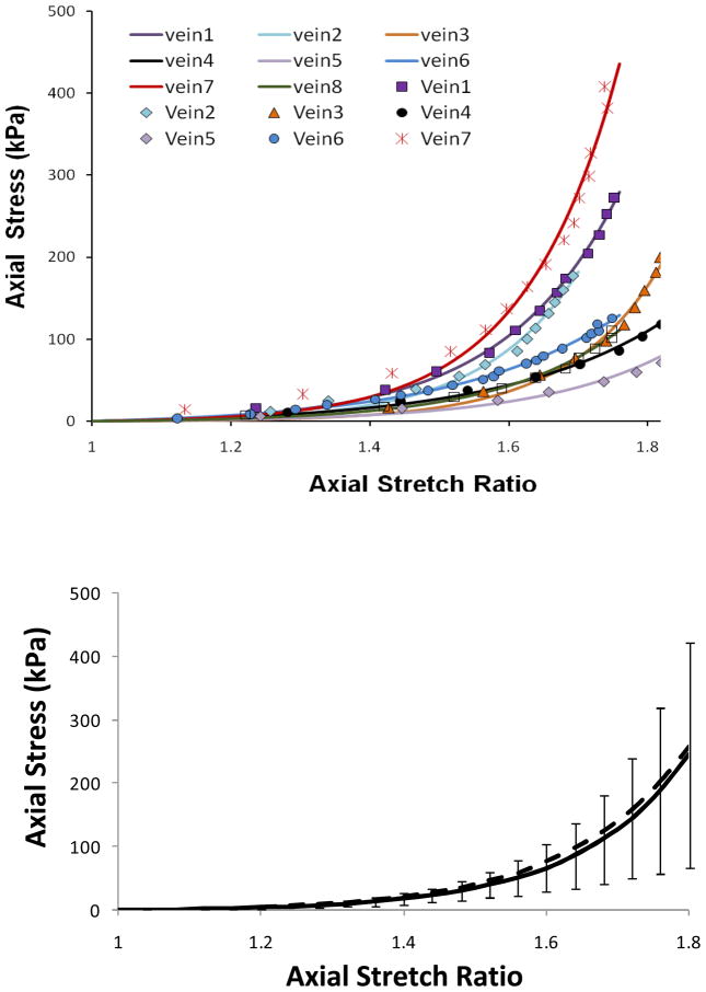
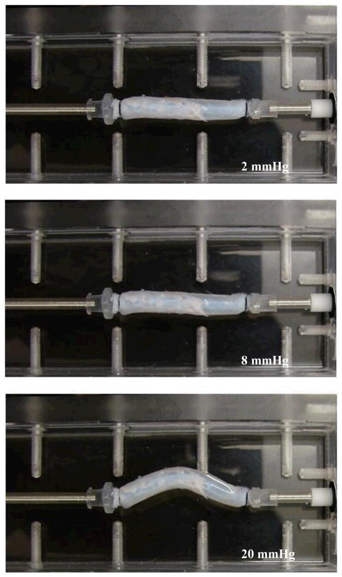
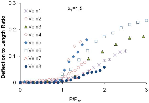
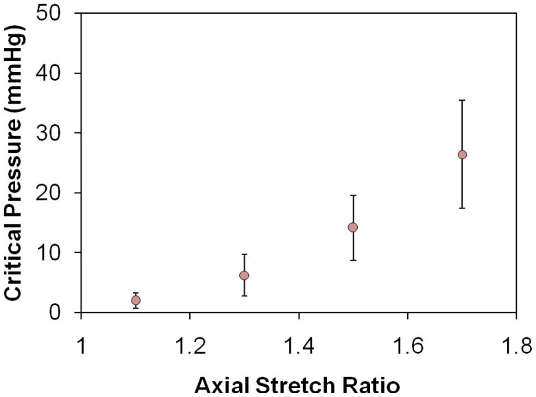
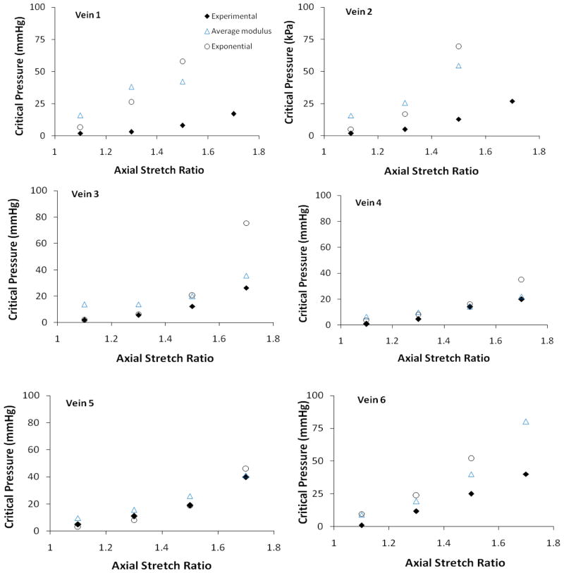
Venous tortuosity is associated with multiple disease states and is often thought to be a consequence of venous hypertension and chronic venous disease. However, the underlying mechanisms of vein tortuosity are unclear. We hypothesized that increased pressure causes vein buckling that leads to a tortuous appearance. The specific aim of this study was to determine the critical buckling pressure of veins. We determined the buckling pressure of porcine jugular veins and measured the mechanical properties of these veins. Our results showed that the veins buckle when the transmural pressure exceeds a critical pressure that is strongly related to the axial stretch ratio in the veins. The critical pressures of the eight veins tested were 14.2 +/- 5.4 and 26.4 +/- 9.0 mmHg at axial stretch ratio 1.5 and 1.7, respectively. In conclusion, veins buckle into a tortuous shape at high lumen pressures or reduced axial stretch ratios. Our results are useful in understanding the development of venous tortuosity associated with varicose veins, venous valvular insufficiency, diabetic retinopathy, and vein grafts.
Figures
Similar articles
-
Twist buckling of veins under torsional loading.J Biomech. 2017 Jun 14;58:123-130. doi: 10.1016/j.jbiomech.2017.04.018. Epub 2017 May 5. J Biomech. 2017. PMID: 28526174 Free PMC article.
-
A biomechanical model of artery buckling.J Biomech. 2007;40(16):3672-8. doi: 10.1016/j.jbiomech.2007.06.018. Epub 2007 Aug 8. J Biomech. 2007. PMID: 17689541 Free PMC article.
-
Nonlinear buckling of blood vessels: a theoretical study.J Biomech. 2008 Aug 28;41(12):2708-13. doi: 10.1016/j.jbiomech.2008.06.012. Epub 2008 Jul 23. J Biomech. 2008. PMID: 18653191
-
[Modeling of elastic deformation and vascular resistance of arterial and venous vasa vasorum].J Mal Vasc. 1998 Oct;23(4):282-8. J Mal Vasc. 1998. PMID: 9827409 Review. French.
-
Pathogenesis of varicose veins - lessons from biomechanics.Vasa. 2014 Mar;43(2):88-99. doi: 10.1024/0301-1526/a000335. Vasa. 2014. PMID: 24627315 Review.
Cited by
-
Twisted blood vessels: symptoms, etiology and biomechanical mechanisms.J Vasc Res. 2012;49(3):185-97. doi: 10.1159/000335123. Epub 2012 Mar 14. J Vasc Res. 2012. PMID: 22433458 Free PMC article. Review.
-
Effect of Axial Stretch on Lumen Collapse of Arteries.J Biomech Eng. 2016 Dec 1;138(12):1245031-6. doi: 10.1115/1.4034785. J Biomech Eng. 2016. PMID: 27657334 Free PMC article.
-
Haemodynamic Recovery Properties of the Torsioned Testicular Artery Lumen.Sci Rep. 2017 Nov 14;7(1):15570. doi: 10.1038/s41598-017-15680-3. Sci Rep. 2017. PMID: 29138449 Free PMC article.
-
Effects of elastin degradation and surrounding matrix support on artery stability.Am J Physiol Heart Circ Physiol. 2012 Feb 15;302(4):H873-84. doi: 10.1152/ajpheart.00463.2011. Epub 2011 Dec 9. Am J Physiol Heart Circ Physiol. 2012. PMID: 22159998 Free PMC article.
-
Mechanical instability of normal and aneurysmal arteries.J Biomech. 2014 Dec 18;47(16):3868-3875. doi: 10.1016/j.jbiomech.2014.10.010. Epub 2014 Oct 27. J Biomech. 2014. PMID: 25458146 Free PMC article.
References
-
- Balaz P, Rokosny S, Klein D, Adamec M. Aneurysmorrhaphy is an easy technique for arteriovenous fistula salvage. J Vasc Access. 2008;9(2):81–4. - PubMed
-
- Baumann UA, Marquis C, Stoupis C, Willenberg TA, Takala J, Jakob SM. Estimation of central venous pressure by ultrasound. Resuscitation. 2005;64(2):193–9. - PubMed
-
- Bergan JJ, Pascarella L, Schmid-Schonbein GW. Pathogenesis of primary chronic venous disease: Insights from animal models of venous hypertension. J Vasc Surg. 2008;47(1):183–92. - PubMed
-
- Beris AE, Soucacos PN, Touliatos AS. Experimental evaluation of the length of microvenous grafts under normal tension. Microsurgery. 1992;13(4):195–9. - PubMed
-
- Blankenship JC, Narke JA, Roberts JW. Transluminal extraction catheter atherectomy in long saphenous vein grafts. Cathet Cardiovasc Diagn. 1995;35(4):368–72. - PubMed
Publication types
MeSH terms
Grants and funding
LinkOut - more resources
Full Text Sources
Other Literature Sources
