. 2010 Feb 25;53(4):1799-809.
doi: 10.1021/jm901647p.

Structure-based discovery of novel chemotypes for adenosine A(2A) receptor antagonists

Affiliations

Structure-based discovery of novel chemotypes for adenosine A(2A) receptor antagonists

Vsevolod Katritch et al. J Med Chem. .

Abstract

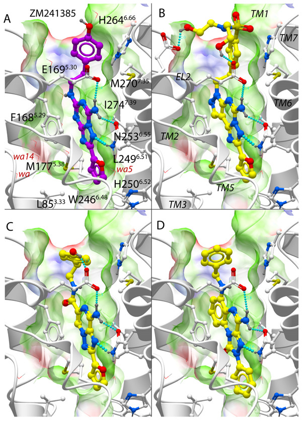
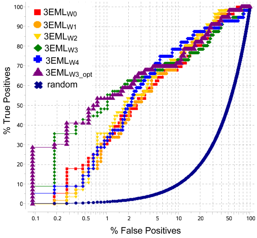
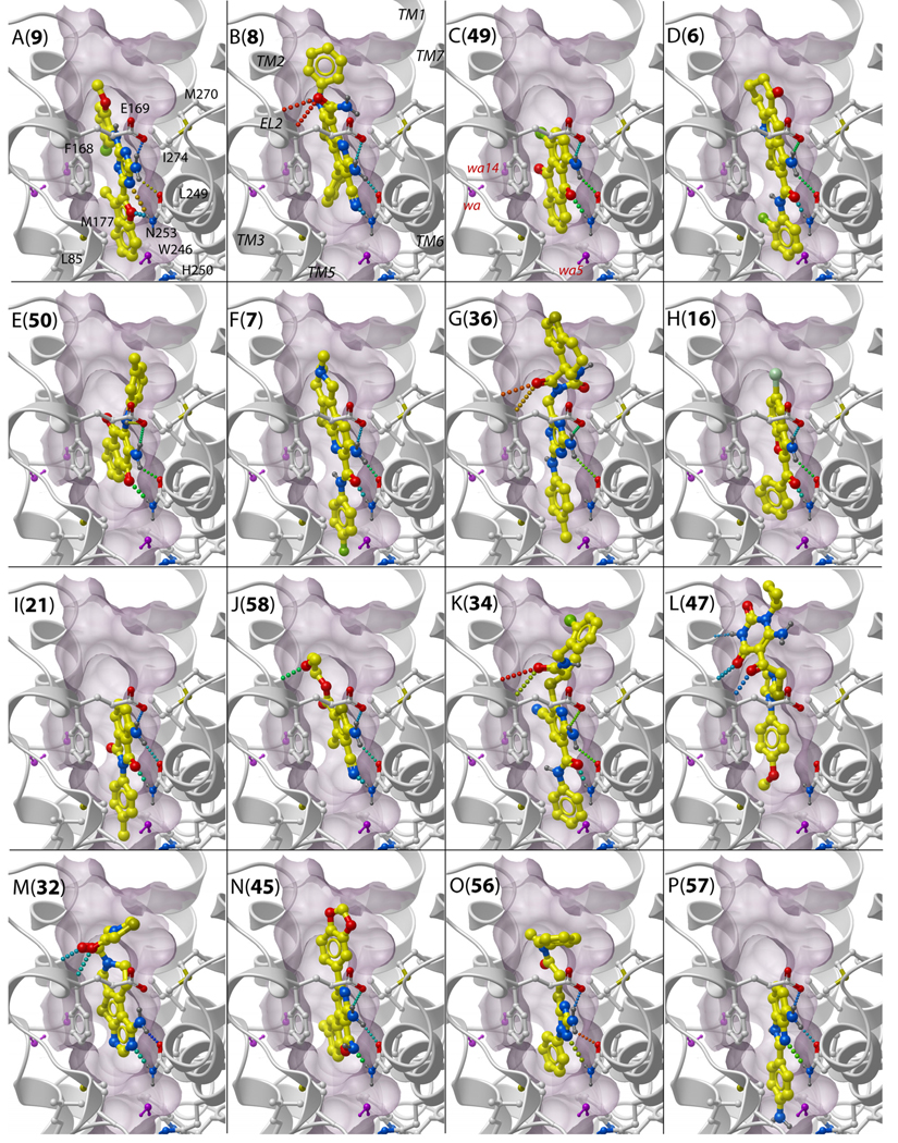
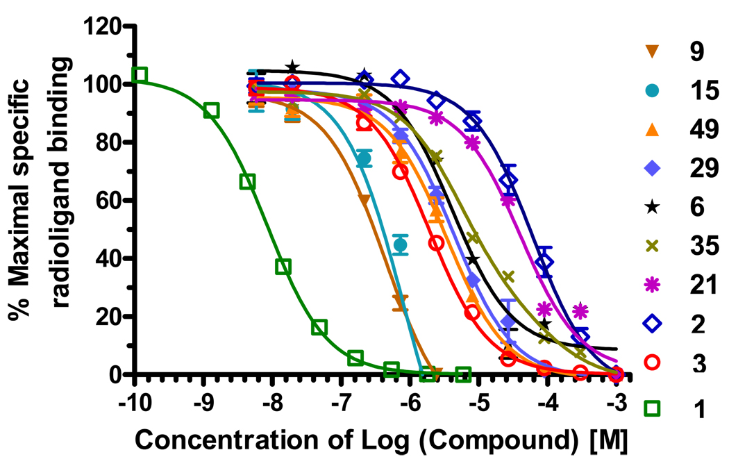
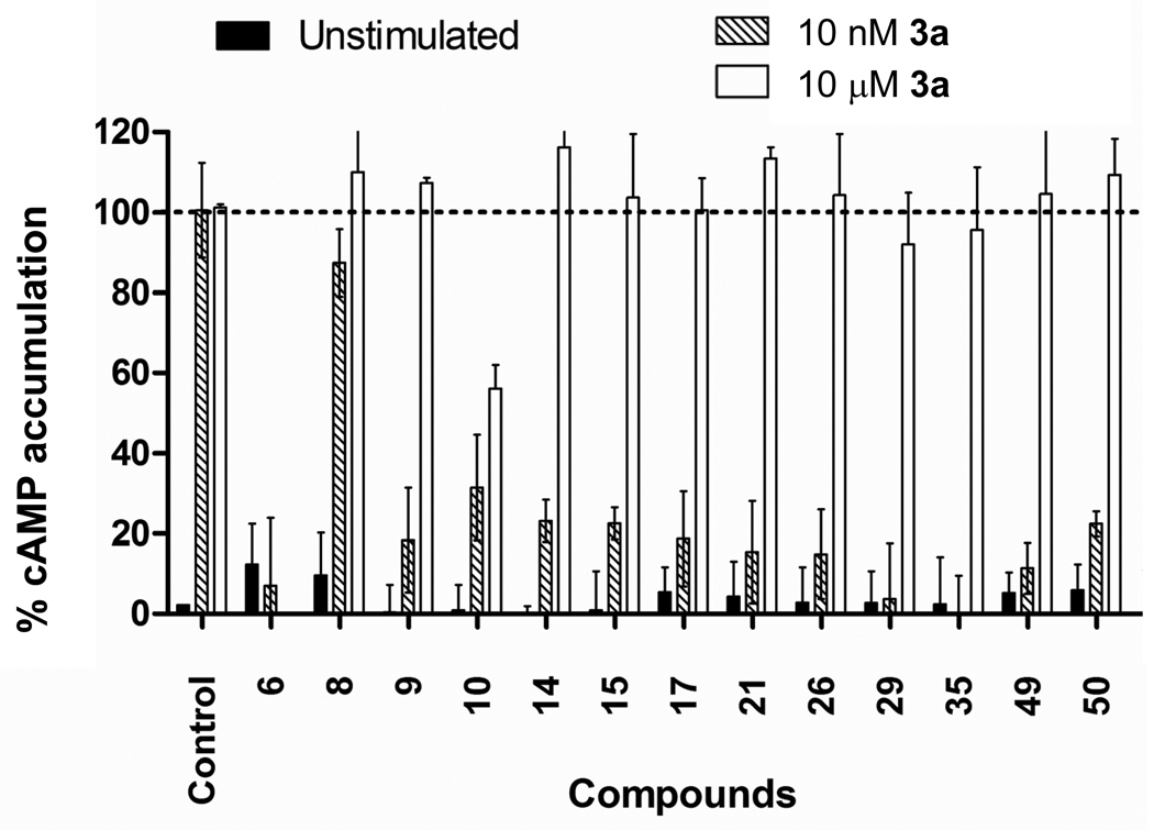
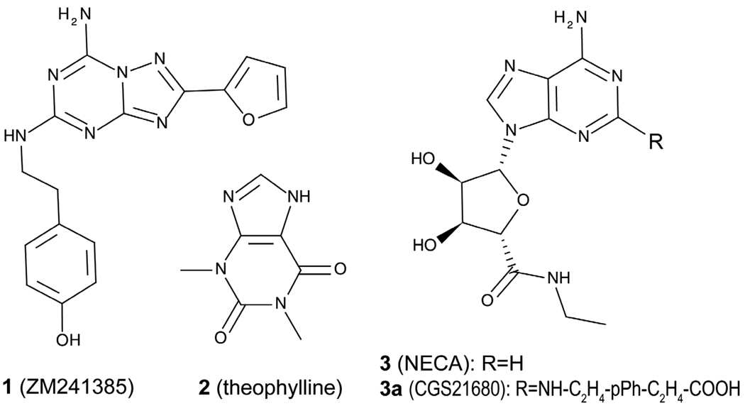
The recent progress in crystallography of G-protein coupled receptors opens an unprecedented venue for structure-based GPCR drug discovery. To test efficiency of the structure-based approach, we performed molecular docking and virtual ligand screening (VLS) of more than 4 million commercially available "drug-like" and ''lead-like'' compounds against the A(2A)AR 2.6 A resolution crystal structure. Out of 56 high ranking compounds tested in A(2A)AR binding assays, 23 showed affinities under 10 microM, 11 of those had sub-microM affinities and two compounds had affinities under 60 nM. The identified hits represent at least 9 different chemical scaffolds and are characterized by very high ligand efficiency (0.3-0.5 kcal/mol per heavy atom). Significant A(2A)AR antagonist activities were confirmed for 10 out of 13 ligands tested in functional assays. High success rate, novelty, and diversity of the chemical scaffolds and strong ligand efficiency of the A(2A)AR antagonists identified in this study suggest practical applicability of receptor-based VLS in GPCR drug discovery.


Figures

Figure 1
Figure 1
Binding of known antagonists into human adenosine A2A receptor models. (A) Co-crystal structure with compounds 1 as in PDB entry 3EML, ligand carbons are shown in magenta color. (B–D) Optimized model 3EMLW3_opt with 3 top scoring compounds from the benchmark set (B) mantri(n), (C) mantri(f) (D) mantri(k). For all ligands, the A2A binding pocket is shown as transparent skin colored by properties (green: hydrophobic, red: acceptor, blue: donor of H-bond). Water molecules are shown by thin red lines and with labels for the three structured waters with lowest B-factors. Hydrogen bonds are shown by cyan spheres.
Figure 2
Figure 2
Performance of A2AAR screening models with different number of structured water molecules (W0 to W4) and with conformational optimization (W3_opt). Receiver Operating Characteristic (ROC) curves are shown in semi-logarithmic scale.
Figure 3
Figure 3
Examples of binding poses in the 3EMLW3_opt receptor model for predicted A2AAR candidate ligands representing 16 different chemical clusters. The letters (A to P) show cluster names, the numbers in brackets indicate compound IDs. Labels for the (i) contact side chains, (ii) TM domains and (iii) structured waters of the receptor model are shown in panels A to C. Side chains of the receptor are shown by sticks with white carbon atoms, in ligands the carbon atoms are colored yellow. Structured water molecules in the model are shown by purple sticks. Hydrogen bonds are colored according to their predicted strength, from blue (strongest) to red (weakest). The binding pocket surface is shown by purple skin, which is clipped in foreground for better view.
Figure 4
Figure 4
Examples of competition binding curves for the identified A2AAR ligands, as compared for known A2AAR antagonists 1, 2, and agonist 3. Tritiated compound 1 was used as the radioligand.
Figure 5
Figure 5
Inhibition of agonist-induced cAMP production by the A2AAR hit compounds. Activity is measured at 10 µM concentration of the compounds, as compared to no compound in the control. Data is normalized where 0 % represents the unstimulated condition (black bars) and 100 % represents the accumulation of intracellular cAMP observed for stimulation with compound 3a at 10 nM or 10 µM concentrations (shaded and open bars respectively).
Chart 1
Chart 1
Chemical structures of representative antagonists (1 and 2) and agonists (3 and 3a) of A2A adenosine receptor.

References

    1. Tyndall JD, Sandilya R. GPCR agonists and antagonists in the clinic. Med Chem. 2005;1:405–421. - PubMed
    1. Lagerstrom MC, Schioth HB. Structural diversity of G protein-coupled receptors and significance for drug discovery. Nat Rev Drug Discov. 2008;7:339–357. - PubMed
    1. Jacobson KA. Introduction to adenosine receptors as therapeutic targets. Handb Exp Pharmacol. 2009:1–24. - PMC - PubMed
    1. Morelli M, Carta AR, Jenner P. Adenosine A(2A) Receptors and Parkinson's Disease. Handb Exp Pharmacol. 2009:589–615. - PubMed
    1. Sebastiao AM, Ribeiro JA. Adenosine receptors and the central nervous system. Handb Exp Pharmacol. 2009:471–534. - PubMed

Publication types

MeSH terms

LinkOut - more resources