

Improved function of diabetic wound-site macrophages and accelerated wound closure in response to oral supplementation of a fermented papaya preparation
- PMID: 20095880
- PMCID: PMC2935338
- DOI: 10.1089/ars.2009.3039
Improved function of diabetic wound-site macrophages and accelerated wound closure in response to oral supplementation of a fermented papaya preparation
Abstract
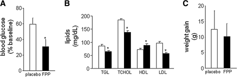
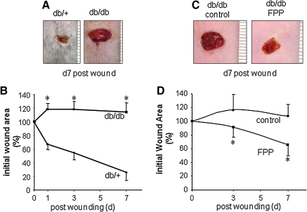
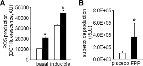
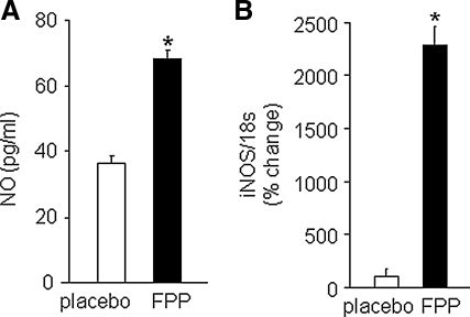
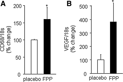
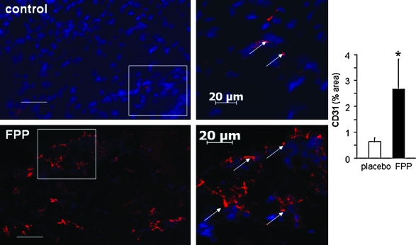
Carica papaya Linn is widely known as a medicinal fruit. We sought to study a standardized fermented papaya preparation (FPP) for its effects on wound healing in adult obese diabetic (db/db) mice. FPP blunted the gain in blood glucose and improved the lipid profile after 8 weeks of oral supplementation. However, FPP did not influence weight gain during the supplementation period. FPP (0.2 g/kg body weight) supplementation for 8 weeks before wounding was effective in correcting wound closure. Studies on viable macrophages isolated from the wound site demonstrated that FPP supplementation improved respiratory-burst function as well as inducible NO production. Reactive oxygen species support numerous aspects of wound healing; NO availability in diabetic wounds is known to be compromised. Diabetic mice supplemented with FPP showed a higher abundance of CD68 as well as CD31 at the wound site, suggesting effective recruitment of monocytes and an improved proangiogenic response. This work provides the first evidence that diabetic-wound outcomes may benefit from FPP supplementation by specifically influencing the response of wound-site macrophages and the subsequent angiogenic response. Given that FPP has a long track record of safe human consumption, testing of the beneficial effects of FPP on diabetic wound-related outcomes in a clinical setting is warranted.
Figures
Similar articles
-
Correction of aberrant NADPH oxidase activity in blood-derived mononuclear cells from type II diabetes mellitus patients by a naturally fermented papaya preparation.Antioxid Redox Signal. 2012 Aug 1;17(3):485-91. doi: 10.1089/ars.2011.4292. Epub 2012 Apr 26. Antioxid Redox Signal. 2012. PMID: 22369197 Free PMC article.
-
May Dietary Supplementation Augment Respiratory Burst in Wound-Site Inflammatory Cells?Antioxid Redox Signal. 2018 Feb 10;28(5):401-405. doi: 10.1089/ars.2017.7304. Epub 2017 Oct 16. Antioxid Redox Signal. 2018. PMID: 28810801 Free PMC article.
-
Does oral supplementation of a fermented papaya preparation correct respiratory burst function of innate immune cells in type 2 diabetes mellitus patients?Antioxid Redox Signal. 2015 Feb 1;22(4):339-45. doi: 10.1089/ars.2014.6138. Epub 2014 Nov 10. Antioxid Redox Signal. 2015. PMID: 25268638 Free PMC article. Clinical Trial.
-
Applications and bioefficacy of the functional food supplement fermented papaya preparation.Toxicology. 2010 Nov 28;278(1):6-16. doi: 10.1016/j.tox.2010.09.006. Epub 2010 Sep 24. Toxicology. 2010. PMID: 20870007 Review.
-
Diabetes as a risk factor to cancer: functional role of fermented papaya preparation as phytonutraceutical adjunct in the treatment of diabetes and cancer.Mutat Res. 2014 Oct;768:60-8. doi: 10.1016/j.mrfmmm.2014.04.007. Epub 2014 Apr 24. Mutat Res. 2014. PMID: 24769427 Review.
Cited by
-
In Vitro Effects of Some Botanicals with Anti-Inflammatory and Antitoxic Activity.J Immunol Res. 2016;2016:5457010. doi: 10.1155/2016/5457010. Epub 2016 Aug 15. J Immunol Res. 2016. PMID: 27597982 Free PMC article.
-
Fermented Carica papaya and Morinda citrifolia as Perspective Food Supplements for the Treatment of Post-COVID Symptoms: Randomized Placebo-Controlled Clinical Laboratory Study.Nutrients. 2022 May 25;14(11):2203. doi: 10.3390/nu14112203. Nutrients. 2022. PMID: 35684003 Free PMC article. Clinical Trial.
-
Oral Administration of Fermented Papaya (FPP®) Controls the Growth of a Murine Melanoma through the In Vivo Induction of a Natural Antioxidant Response.Cancers (Basel). 2019 Jan 20;11(1):118. doi: 10.3390/cancers11010118. Cancers (Basel). 2019. PMID: 30669508 Free PMC article.
-
Beneficial Effects of Fermented Papaya Preparation (FPP®) Supplementation on Redox Balance and Aging in a Mouse Model.Antioxidants (Basel). 2020 Feb 7;9(2):144. doi: 10.3390/antiox9020144. Antioxidants (Basel). 2020. PMID: 32046112 Free PMC article.
-
Myo-Inositol in Fermented Sugar Matrix Improves Human Macrophage Function.Mol Nutr Food Res. 2022 Apr;66(8):e2100852. doi: 10.1002/mnfr.202100852. Epub 2022 Feb 26. Mol Nutr Food Res. 2022. PMID: 35073444 Free PMC article.
References
-
- Albina JE. Mills CD. Barbul A. Thirkill CE. Henry WL., Jr Mastrofrancesco B. Caldwell MD. Arginine metabolism in wounds. Am J Physiol. 1988;254:E459–E467. - PubMed
-
- Amadeu TP. Costa AM. Nitric oxide synthesis inhibition alters rat cutaneous wound healing. J Cutan Pathol. 2006;33:465–473. - PubMed
-
- Amer J. Goldfarb A. Rachmilewitz EA. Fibach E. Fermented papaya preparation as redox regulator in blood cells of beta-thalassemic mice and patients. Phytother Res. 2008;22:820–828. - PubMed
-
- American Diabetes Association. ADA; 2007. Direct and indirect costs of diabetes in the United States.
-
- Anuar NS. Zahari SS. Taib IA. Rahman MT. Effect of green and ripe Carica papaya epicarp extracts on wound healing and during pregnancy. Food Chem Toxicol. 2008;46:2384–2389. - PubMed
Publication types
MeSH terms
Substances
Grants and funding
LinkOut - more resources
Full Text Sources
Other Literature Sources
Medical
Miscellaneous
