. 2010 Jan 22:5:3.
doi: 10.1186/1749-8104-5-3.

Brn3a regulates neuronal subtype specification in the trigeminal ganglion by promoting Runx expression during sensory differentiation

Affiliations

Brn3a regulates neuronal subtype specification in the trigeminal ganglion by promoting Runx expression during sensory differentiation

Iain M Dykes et al. Neural Dev. .

Abstract

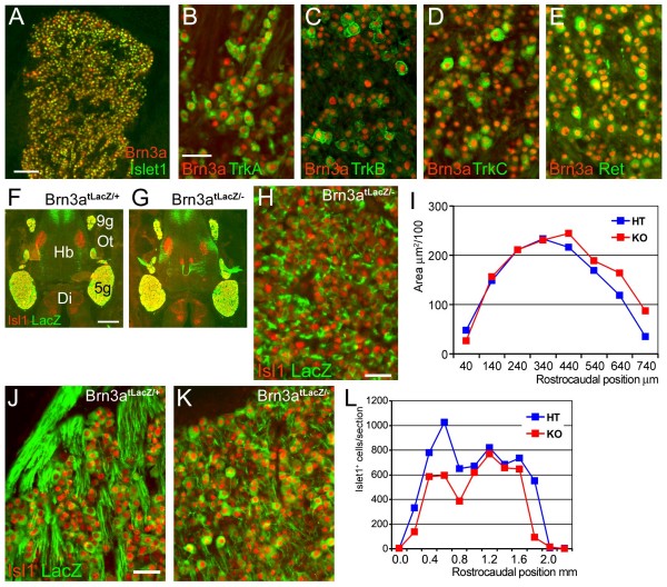
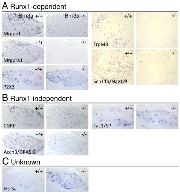
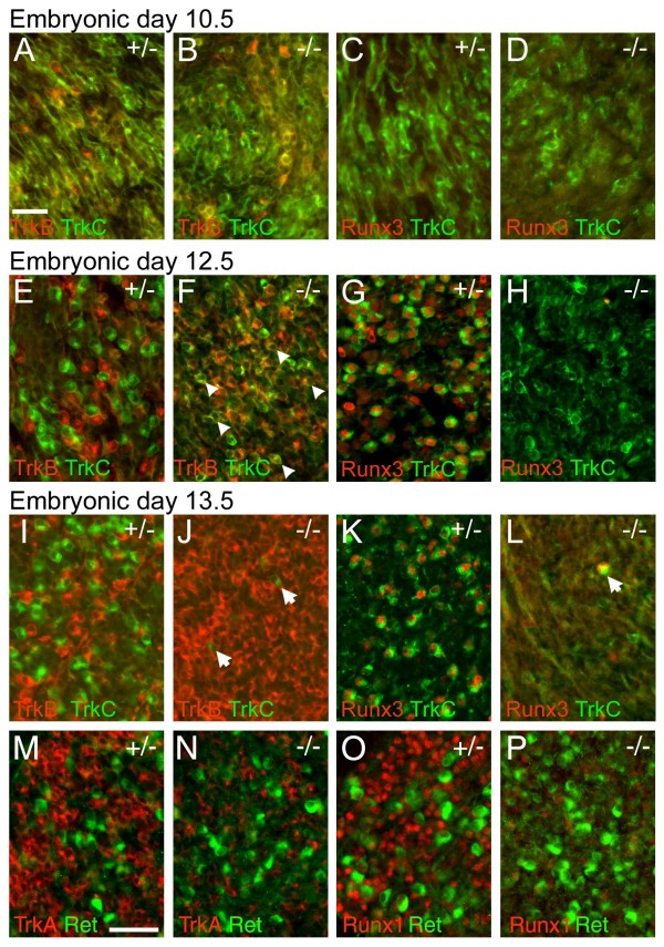
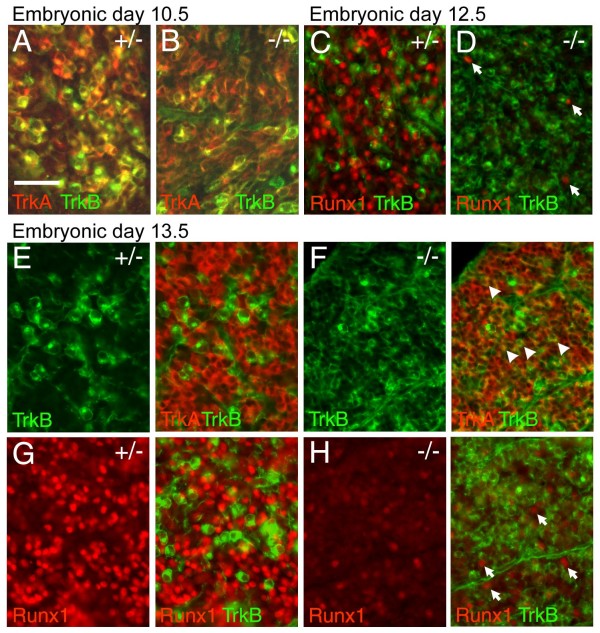
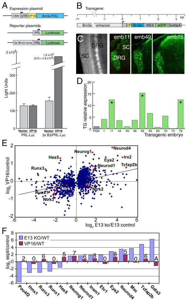
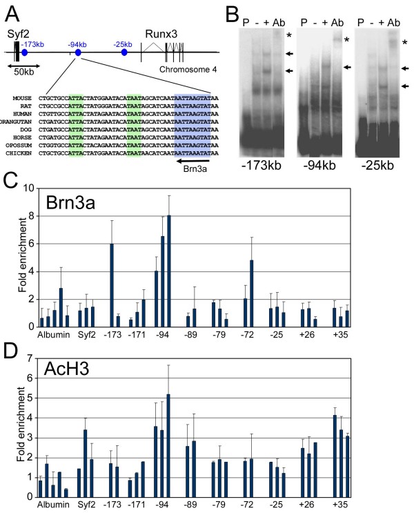
The transcription factor Brn3a, product of the pou4f1 gene, is expressed in most sensory neurons throughout embryogenesis. Prior work has demonstrated a role for Brn3a in the repression of early neurogenic genes; here we describe a second major role for Brn3a in the specification of sensory subtypes in the trigeminal ganglion (TG). Sensory neurons initially co-express multiple Trk-family neurotrophin receptors, but are later marked by the unique expression of TrkA, TrkB or TrkC. Maturation of these sensory subtypes is known to depend on the expression of Runx transcription factors. Newborn Brn3a knockout mice fail to express TrkC, which is associated in the TG with mechanoreceptors, plus a set of functional genes associated with nociceptor subtypes. In embryonic Brn3a-/- ganglia, the normal expression of Runx3 is never initiated in TrkC+ neurons, and Runx1 expression is greatly attenuated in TrkA+ nociceptors. These changes are accompanied by expanded expression of TrkB in neurons that abnormally express multiple Trks, followed by the loss of TrkC and TrkA expression. In transgenic embryos expressing a Brn3a-VP16 dominant transactivator, Runx3 mRNA expression is increased, suggesting that it is a direct regulatory target of Brn3a. Chromatin immunoprecipitation confirms that Brn3a binds in vivo to a conserved upstream enhancer element within histone H3-acetylated chromatin in the Runx3 locus. Together these data show that Brn3a acts upstream of the Runx factors, which then repress TrkB expression to allow establishment of the non-overlapping Trk receptor profiles and correct terminally differentiated phenotypes.


Figures

Figure 1
Figure 1
Pan-sensory expression of Brn3a and survival of a majority of trigeminal neurons in Brn3a knockout mice. (A-E) Co-localization of Brn3a with specific sensory markers in the TG in transverse sections of P0 wild-type ganglia. Brn3a is co-localized with the pan-sensory marker Islet1 and the subtype-specific markers TrkA, TrkB, TrkC, and Ret. (F-I) Islet1 expression and LacZ expressed from the Brn3a locus (Brn3atLacZ allele) were used to assess TG morphology and marker expression at E13.5. Ganglion morphology is grossly normal (F, G), and Islet1 continues to be expressed in LacZ+ neurons in Brn3a knockout embryos (H). No significant difference in the volumes of Brn3atLacZ/- and Brn3atLacZ/+ control TG was observed at this stage (I). The zero coordinate is the rostral pole of the ganglion. Paired T test for comparison of coronal section areas across the rostrocaudal extent of the ganglion: n = 8, P = 0.12, not significant. (J-L) At P0.5 Islet1 expression continued to identify all or nearly all LacZ+ neurons in Brn3atLacZ/- knockout and Brn3atLacZ/+ control TG (J, K, transverse section). Islet1 immunoreactive nuclei were counted in serial sections across the rostrocaudal extent of the TG (L), and mean cell number was reduced from 696 to 501 nuclei per section (72% remaining), with cell loss concentrated in the rostral pole of the ganglion. Paired T-test for comparison cell number in matched coronal sections across the entire rostrocaudal extent of the ganglion: n = 12, P = 0.01. 5 g, trigeminal ganglion; 9 g, geniculate ganglion; Di, diencephalon; Hb, hindbrain; HT, heterozygote control; KO, knockout; Ot, otic region.
Figure 2
Figure 2
Loss of Brn3a profoundly affects the major sensory subtypes at birth. Expression of markers of known sensory subtypes were examined at P0 in transverse sections of Brn3a knockout and heterozygote control TG. Heterozygote controls are phenotypically normal and have been previously shown to have minimal changes in global gene expression [26]. (A-F) Expression of TrkC, Runx3, and Etv1. TrkC and Runx3 immunoreactive neurons are reduced to less than 10% of controls. Small Etv1 immunoreactive neurons, most of which co-express Islet2, are unaffected. (G, H) Neurons expressing TrkB persist in Brn3a-/- mice at P0, but the distribution of the protein is markedly altered, with increased expression in axons and less expression in discrete cell bodies. (I-R) Expression of nociceptor markers Runx1, TrkA and Ret. The absolute number of Runx1 positive neurons is significantly decreased across the rostrocaudal extent of the ganglion (K, paired t-test, P = 0.001), and this loss is in excess of the overall decrease in cell number as assayed by Islet1 expression (L, paired t-test, P = 0.0004). TrkA expression is markedly diminished in the knockout (M, N). Small diameter neurons co-expressing Runx1 and Ret (O, arrows) are absent from the knockout, but a population of large diameter Ret+ neurons that are negative for Runx1 and TrkA, and may represent a class of mechanoreceptors, persist (O, P, arrowheads). HT, heterozygote control; KO, knockout.
Figure 3
Figure 3
Expression of Runx1-dependent and Runx1-independent transcripts for receptors, channels and neuropeptides in the Brn3a-/- TG. The TG of Brn3a-/- and control mice were examined for the expression of multiple sensory markers at P0, the last stage available due to the neonatal death of the knockout mice. Coronal sections are shown except for Trpm8 and Scn11a, which are horizontal sections. (A) Expression of transcripts showing Runx1-dependence in the DRG. (B) Expression of transcripts independent of Runx1 in the DRG. (C) Expression of transcripts for which Runx1-dependence has not been characterized.
Figure 4
Figure 4
Early defects in specification of TrkC neurons in the Brn3a-/- TG. The TG of Brn3a knockout and control embryos were examined at various stages for the expression of TrkB, TrkC and Runx3. (A-D) At E10.5 TrkB and TrkC are extensively co-expressed in both genotypes. Runx3 was not detected at this stage in either genotype. (E-H) At E12.5 TrkB and TrkC expression identify discrete subsets of neurons in control ganglia (E), but in Brn3a knockout ganglia co-expression persists (F), and examples of the numerous co-expressing neurons are indicated by arrowheads. Runx3 and TrkC are co-expressed in control ganglia (G), but Runx3 expression is not initiated in the knockout TG, and TrkC expression is diminished (H). (I-L) At E13.5, TrkB expression is markedly expanded in knockout ganglia (J), and TrkC and Runx3 expression is nearly absent (J, L). Rare remaining TrkC+/Runx3+ neurons in the knockout TG are indicated by arrows (J, L). (M-P) Early Ret+ neurons at this stage do not co-express TrkA or Runx1 in the control or knockout ganglia, and persist in Brn3a-/- ganglia. Ret+ neurons at this stage are likely to represent a subset of mechanoreceptors; Ret+ nociceptors are not detected until the perinatal period. Scale bar = 50 μM.
Figure 5
Figure 5
Defects in nociceptor specification in the Brn3a-/- TG. The TG of Brn3a knockout and control embryos were examined at various stages for the expression of TrkA, TrkB, and Runx1. (A, B) At E10.5, TrkA and TrkB are extensively co-expressed in control (A) and Brn3a knockout (B) ganglia. Runx1 is not expressed at this stage. (C, D) At E12.5, Runx1 is expressed in control ganglia (C) but is markedly diminished in the knockout (D). Arrows mark rare Runx1+ cells. (E, F) At E13.5, TrkA and TrkB expressing neurons normally segregate into discrete populations (E) but fail to do so in Brn3a knockout ganglia (F). Exposure time for TrkA in (F) is increased to reveal co-expression; see (I, J) for equal exposures. Arrowheads in (F) indicate examples of numerous abnormal TrkA+/TrkB+ cells. (G, H) At E13.5, Runx1 expression is markedly diminished in the knockout. The few Runx1+ neurons detected at this stage do not express TrkB (H, arrows). Scale bar = 50 μM.
Figure 6
Figure 6
Sensory-specific expression of a VP16-Brn3a constitutive transcriptional activator reveals possible direct regulatory targets. (A) Activation of transcription by VP16-Brn3a in transfection assays. Increased expression requires both the VP16-Brn3a protein and the presence of a consensus Brn3a binding site (3× Brn3a cons) in the reporter gene construct. (B) Structure of the VP16-Brn3a transgene. A dicistronic message encoding VP16-Brn3a and enhanced green fluorescent protein (eGFP) is driven by an 11-kb sensory enhancer from the Brn3a locus. (C) Expression of eGFP in the E13.5 dorsal root ganglia (DRG) of VP16-Brn3a transgenic founders by direct GFP fluorescence (whole mount) and immunofluorescence (sections). Appropriate axonal projections to the dorsal spinal cord (SC) are evident. The TG of these embryos were harvested for array analysis. (D) RT-PCR assays of Brn3a-VP16 expression in TG dissected from transgenic embryos, normalized to phosphoglycerate kinase (Pgk) expression. Embryos 11, 49, and 76 (asterisks) were selected for microarray analysis. (E) Microarray analysis of changes in gene expression in VP16-Brn3a transgenic embryos at E13.5. Changes in gene expression in the VP16-Brn3a transgenic TG were plotted (y-axis) against changes in the E13.5 Brn3a knockout (x axis). Only data for transcripts showing significantly increased or decreased expression in the Brn3a-/- TG relative to controls are shown, thus data points are missing from around the origin. For the entire group, the correlation was weak (slope = 0.126, R2 = 0.147), suggesting that many Brn3a targets are indirectly regulated. The microarray analyses of Brn3a-/- and control ganglia used for comparison purposes will be presented in detail elsewhere (Lanier et al., in preparation). (F) Changes in transcription factor expression in E13.5 Brn3a knockout ganglia and VP16-Brn3a ganglia, and the number of change calls (out of nine possible comparisons) for VP16-Brn3a versus control ganglia. 'A' indicates absent call. Neurod1 and Neurod4, previously shown to be directly repressed by Brn3a, were activated by VP16-Brn3a. Runx3 and Hes5 are candidates for direct positive regulation by Brn3a (expression increased by VP16-Brn3a, decreased in knockout), Neurog1, Insm1, Eya2, and Tcaf2b are further candidates for direct negative regulation (expression increased by VP16, increased in knockout).
Figure 7
Figure 7
Brn3a binds to regulatory sequences within the Runx3 locus. (A) Map of a 300-kb region of mouse chromosome 4 including the Runx3 and Syf2 loci. The location of three sites (-173 kb, -94 kb and -25 kb) showing strong in vitro Brn3a binding are indicated (blue circles). Also shown is an interspecies sequence alignment of a region within the proposed -94-kb enhancer, which contains a conserved consensus Brn3a binding site (blue box) and additional conserved TAAT motifs (green boxes) that represent additional potential homeodomain transcription factor binding sites. (B) EMSA for Brn3a binding to sites identified by sequence search; assays for the three sites exhibiting strongest binding are shown. P, probe only; -, probe incubated with protein extract from untransfected HEK cells; +, probe incubated with protein extract from HEK cells transfected with a Brn3a expression construct; Ab, Brn3a-transfected cell extract plus supershifting anti-Brn3a antibody; arrow, shifted band; asterisk, supershifted band. Multiple shifted bands may indicate cooperative binding of Brn3a to adjacent sites [56]. (C) ChIP profiling of in vivo Brn3a binding to the Runx3 locus in E13.5 TG. Two or three primer pairs were made to the regions containing each of the nine potential Brn3a binding sites identified by sequence search (Additional file 4). Quantitative PCR assays were performed on immunoprecipitated chromatin selected with an anti-Brn3a antibody from three independent samples. Fold enrichment for selected/unselected was calculated using the cycle threshold difference method (Methods). Graph shows the mean + standard error for three assays of independent samples. As expected, binding is not observed at the promoter region of the Alb (serum albumin) locus, which is not transcribed in neurons and does not contain Brn3a binding sites [14]. Unpaired t-test for selection at -94-kb site: n = 8 experimental assays, n = 13 alb locus controls, P = 7 × 10⁻⁵. (D) ChIP profiling of histone H3 acetylation at the Runx3 locus. Assays were performed as described in (C), with an antibody recognizing lysine 9/14 acetyl histone H3. Increased acetylation in the vicinity of the Brn3a binding site at -94 kb suggests an open conformation of chromatin at this location.

Similar articles

Cited by

References

    1. Hasegawa H, Wang F. Visualizing mechanosensory endings of TrkC-expressing neurons in HS3ST-2-hPLAP mice. J Comp Neurol. 2008;511:543–556. doi: 10.1002/cne.21862. - DOI - PMC - PubMed
    1. Woolf CJ, Ma Q. Nociceptors - noxious stimulus detectors. Neuron. 2007;55:353–364. doi: 10.1016/j.neuron.2007.07.016. - DOI - PubMed
    1. Marmigere F, Ernfors P. Specification and connectivity of neuronal subtypes in the sensory lineage. Nat Rev Neurosci. 2007;8:114–127. doi: 10.1038/nrn2057. - DOI - PubMed
    1. Ma Q, Chen Z, Barrantes I, de la Pompa J, Anderson D. Neurogenin1 is essential for the determination of neuronal precursors for proximal sensory ganglia. Neuron. 1998;20:469–482. doi: 10.1016/S0896-6273(00)80988-5. - DOI - PubMed
    1. Fode C, Gradwohl G, Morin X, Dierich A, LeMeur M, Goridis C, Guillemot F. The bHLH protein Neurogenin2 is a determination factor for epibranchial placode-derived sensory neurons. Neuron. 1998;20:483–494. doi: 10.1016/S0896-6273(00)80989-7. - DOI - PubMed

Publication types

MeSH terms