. 2010 May;38(2):226-36.
doi: 10.1016/j.nbd.2010.01.010. Epub 2010 Jan 21.

Dominant connexin26 mutants associated with human hearing loss have trans-dominant effects on connexin30

Affiliations

Dominant connexin26 mutants associated with human hearing loss have trans-dominant effects on connexin30

Sabrina W Yum et al. Neurobiol Dis. 2010 May.

Abstract

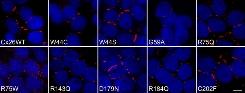
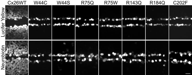
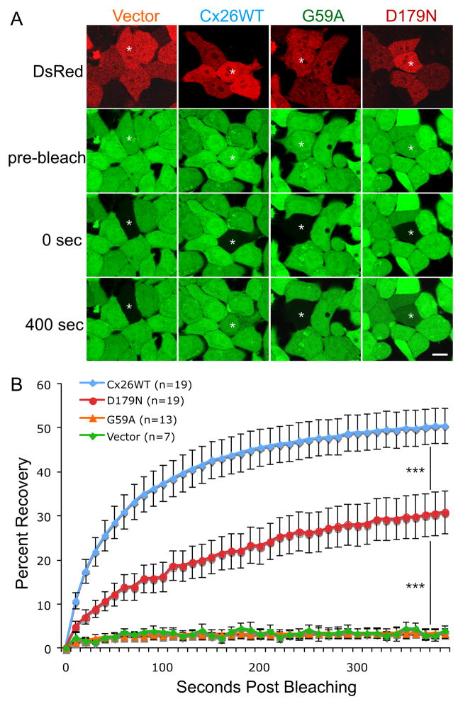
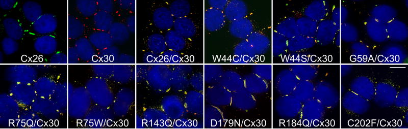
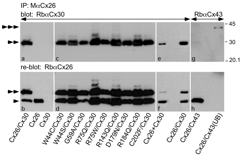
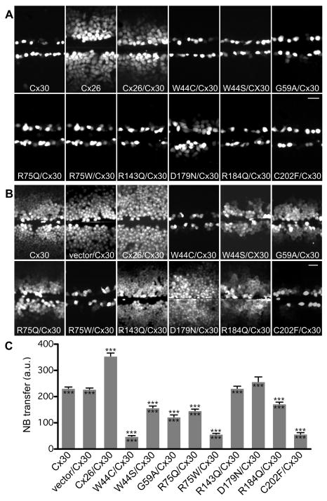
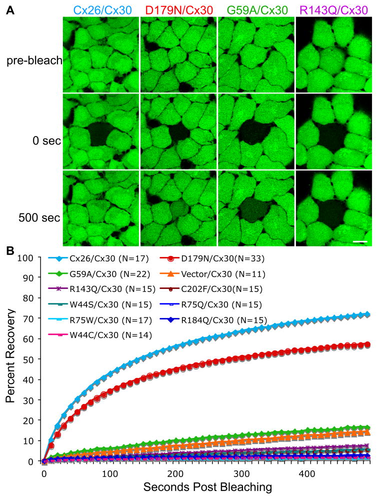
Dominant mutations in GJB2, the gene encoding the human gap junction protein connexin26 (Cx26), cause hearing loss. We investigated whether dominant Cx26 mutants interact directly with Cx30. HeLa cells stably expressing nine dominant Cx26 mutants, six associated with non-syndromic hearing loss (W44C, W44S, R143Q, D179N, R184Q and C202F) and three associated with hearing loss and palmoplantar keratoderma (G59A, R75Q and R75W), individually or together with Cx30, were analyzed by immunocytochemistry, co-immunoprecipitation, and functional assays (scrape-loading and/or fluorescence recovery after photobleaching). When expressed alone, all mutants formed gap junction plaques, but with impaired intercellular dye transfer. When expressed with Cx30, all mutants co-localized and co-immunoprecipitated with Cx30, indicating they likely co-assembled into heteromers. Furthermore, 8/9 Cx26 mutants inhibited the transfer of neurobiotin or calcein, indicating that these Cx26 mutants have trans-dominant effects on Cx30, an effect that may contribute to the pathogenesis of hearing loss.


Figures

Figure 1
Figure 1. Dominant Cx26 mutants form gap junction plaques
These are deconvolved images of bulk-selected HeLa cells that express wild type human Cx26 (Cx26WT) or the indicated Cx26 mutant. The cells were labeled with a rabbit antiserum against the C-terminus of Cx26 (red) and counterstained with DAPI (blue). All cell lines had gap junction plaques at the cell borders; the R75W mutant had smaller plaques and more intracellular staining than the other mutants. Scale bar: 10 μm.
Figure 2
Figure 2. Dominant Cx26 mutants do not form functional gap junction channels
These are digital fluorescence images of confluent bulk-selected HeLa cells that stably express wild type Cx26 (Cx26WT) or the indicated Cx26 mutants. The cells were incubated in 0.1% Lucifer Yellow (LY, upper row) or 2% neurobiotin (NB, lower row), imaged (LY) or fixed (NB, then visualized with TRITC-conjugated avidin) ~15 min after being wounded with a scalpel blade. Note that the wounded cells picked up LY or NB in all cases, but only cells expressing Cx26WT showed extensive transfer of LY or NB to neighboring cells. Scale bar: 50 μm.
Figure 3
Figure 3. The D179N mutant forms partially functional gap junction channels
HeLa cells that were transiently transfected to express wild type Cx26 (Cx26WT), G59A, D179N, or the vector alone (vector). Two days later, the cells were incubated in calcein AM to fill the cytoplasm with calcein (green), and selected cells expressing DsRed (red) that were in close contact with at least three other DsRed-positive cells were photobleached, and then the green fluorescence signal was measured every 10 sec for 400 sec. Panel A shows examples of cells immediately before (pre-bleach), immediately after (0 sec), and 400 seconds after bleaching. Single asterisk (*) indicates the cells selected for bleaching. Scale bar: 10 μm. Panel B summarizes the FRAP data for many individual cells, by normalizing the fluorescent signal present in each cell immediately prior to and immediately after photobleaching to 100% and 0%, respectively. For each cell line, the curves connect the mean percent recovery at each time point from many individual cells as indicated; the vertical bars represent the means standard error. The recovery of the calcein signal in the bleached cells expressing D179N is more than that in cells expressing vector alone (p < 0.0001), but less than that in cells expressing Cx26WT (p < 0.0001). Triple asterisks (***) denotes a significance level of p < 0.0001.
Figure 4
Figure 4. Cx26 mutants co-localize with Cx30
These are deconvolved images of bulk-selected HeLa cells that express wild type Cx26 or wild type Cx30 alone, or co-express wild type Cx30 and wild type Cx26 (Cx26/Cx30) or the indicated Cx26 mutant. The cells were double-labeled with a rabbit antiserum against the C-terminus of Cx30 (red) and a mouse monoclonal antibody against the C-terminus of Cx26 (green), and counterstained with DAPI (blue). In these merged images, Cx26 and Cx30 are largely co-localized, forming (orange to yellow) gap junction plaques at cell borders in all cases. Scale bar: 10 μm.
Figure 5
Figure 5. Cx26 mutants co-immunoprecipitate Cx30
Lysates were prepared from bulk-selected HeLa cells that stably express wild type Cx26, Cx30, or both Cx30 and Cx26 (Cx26/Cx30) or one of the Cx26 mutants as indicated (Cx26 mutation/Cx30), co-culture of stable Cx26 cells and Cx30 cells (Cx26+Cx30), as well as HeLa cells that were transiently transfected to express both Cx26 and Cx43 (Cx26/Cx43). The lysates were immunoprecipitated with a mouse monoclonal antibody against Cx26 (M Cx26), and the bound fractions or the unbound fraction were probed (upper panels) with a rabbit antiserum against Cx30 (Rb Cx30) or Cx43 (Rb Cx43), then reprobed (lower panels) with a rabbit antiserum against Cx26 (Rb Cx26). Note that M Cx26 co-immunoprecipitated Cx30 (double arrowhead), but not Cx43 (triple arrowheads), which is present in the unbound fraction (UB). Reprobing the blot reveals Cx26 in the appropriate samples (single arrowheads); the signal for Cx30 remains because the blot was not stripped before reprobing. Size markers (in kDa) are shown.
Figure 6
Figure 6. Dominant Cx26 mutants inhibit dye transfer of cells co-expressing Cx30
These are digital fluorescence images of confluent, bulk-selected HeLa cells that stably express wild type Cx30, or co-express Cx30 and an “empty vector” (vector/Cx30), as well as Cx26 (Cx30/Cx26) or the indicated Cx26 mutant. The cells were incubated in 0.1% LY (A) or 2% NB (B) for 5 min, and imaged either directly (LY) or following fixation (NB; visualized with TRITC-conjugated avidin) ~15 min after being wounded with a scalpel blade. In panel A, note that the wounded cells picked up LY in all cases, and, in contrast to cells expressing Cx30 alone, cells expressing Cx26 alone showed extensive transfer of LY to neighboring cells. None of the cells co-expressing a Cx26 mutant showed transfer of LY to neighboring cells except for cells co-expressing D179N, but this was less than in cells co-expressing wild type Cx26. Scale bar: 50 μm. In panel B, note that NB transferred extensively in cells expressing Cx30 alone, or co-expressing Cx30 and wild type Cx26, R143Q, or D179N, less extensively in cells co-expressing Cx30 and W44S, G59A, R75Q or R184Q, and not at all in cells co-expressing Cx30 and W44C, R75W, or C202F. Scale bar: 50 μm. In panel C, quantitative analysis of intercellular NB transfer after scrape-loading is shown. The columns represent the mean distance of NB transfer from the scrape line to the point where the fluorescence intensity dropped to 1.5x the background intensity. For each cell line, this was measured by acquiring at least 8 images from each of two to three different plates of cells. The error bars indicate ± s.e.m. Triple asterisks (***) above the error bars denote a significance level of p < 0.0001 when compared to vector/Cx30 expressing cells, Triple asterisks (***) below the error bars denote a significance level of p < 0.0001 when compared to Cx26/Cx30 expressing cells.
Figure 7
Figure 7. FRAP analysis of Cx26 and/or Cx30 gap junction channels
(A&B) Bulked-selected HeLa cells stably expressing wild type Cx30 or Cx26 alone, or co-expressing wild type Cx30 and Cx26 (Cx26/Cx30) or individual Cx26 mutants were incubated in calcein AM to fill the cytoplasm with calcein (green). Selected cells were photobleached, and the green fluorescence signal was measured every 10 sec for 500 sec. Panel A shows examples of cells immediately before (pre-bleach), immediately after (0 sec), and 500 seconds after bleaching. Scale bar: 10 μm. Panel B summarizes the data for many cells from individual cell lines, by normalizing the fluorescent signal present in each cell immediately prior to and immediately after photobleaching to 100% and 0%, respectively. The curves connect the mean percent recovery at each time point; standard errors are not shown. Note the robust recovery of the calcein signal in cells co-expressing Cx26/Cx30 or D179N/Cx30, but not in the bleached cells co-expressing other Cx26 mutants or the vector alone. The statistical comparisons are shown in Table S1.

References

    1. Ahmad S, et al. Connexins 26 and 30 are co-assembled to form gap junctions in the cochlea of mice. Biochem Biophys Res Commun. 2003;307:362–368. - PubMed
    1. Ahmad S, et al. Restoration of connexin26 protein level in the cochlea completely rescues hearing in a mouse model of human connexin30-linked deafness. Proc Natl Acad Sci U S A. 2007;104:1337–41. - PMC - PubMed
    1. Bakirtzis G, et al. Targeted epidermal expression of mutant Connexin 26(D66H) mimics true Vohwinkel syndrome and provides a model for the pathogenesis of dominant connexin disorders. Hum Mol Genet. 2003;12:1737–1744. - PubMed
    1. Beltramello M, et al. Permeability and gating properties of human connexins 26 and 30 expressed in HeLa cells. Biochem Biophys Res Commun. 2003;305:1024–1033. - PubMed
    1. Beltramello M, et al. Impaired permeability to Ins(1,4,5)P3 in a mutant connexin underlies recessive hereditary deafness. Nat Cell Biol. 2005;7:63–69. - PubMed

Publication types