. 2010 Feb 12;86(2):229-39.
doi: 10.1016/j.ajhg.2009.12.014. Epub 2010 Jan 21.

Association of JAG1 with bone mineral density and osteoporotic fractures: a genome-wide association study and follow-up replication studies

Affiliations

Association of JAG1 with bone mineral density and osteoporotic fractures: a genome-wide association study and follow-up replication studies

Annie W C Kung et al. Am J Hum Genet. .

Abstract

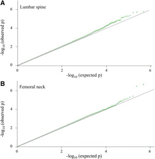
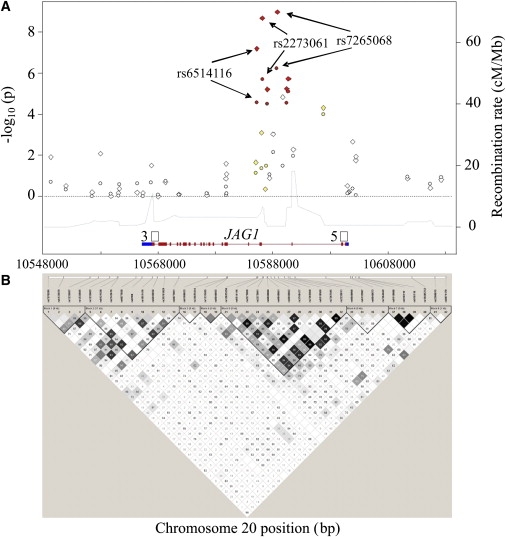
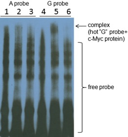
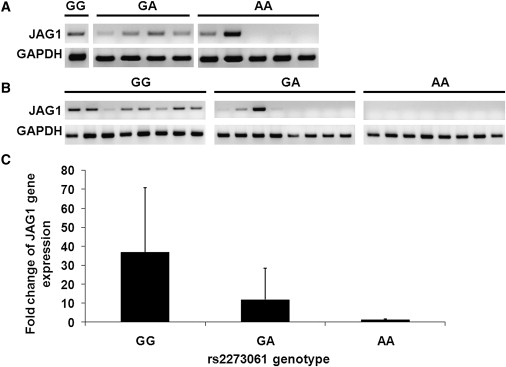
Bone mineral density (BMD), a diagnostic parameter for osteoporosis and a clinical predictor of fracture, is a polygenic trait with high heritability. To identify genetic variants that influence BMD in different ethnic groups, we performed a genome-wide association study (GWAS) on 800 unrelated Southern Chinese women with extreme BMD and carried out follow-up replication studies in six independent study populations of European descent and Asian populations including 18,098 subjects. In the meta-analysis, rs2273061 of the Jagged1 (JAG1) gene was associated with high BMD (p = 5.27 x 10(-8) for lumbar spine [LS] and p = 4.15 x 10(-5) for femoral neck [FN], n = 18,898). This SNP was further found to be associated with the low risk of osteoporotic fracture (p = 0.009, OR = 0.7, 95% CI 0.57-0.93, n = 1881). Region-wide and haplotype analysis showed that the strongest association evidence was from the linkage disequilibrium block 5, which included rs2273061 of the JAG1 gene (p = 8.52 x 10(-9) for LS and 3.47 x 10(-5) at FN). To assess the function of identified variants, an electrophoretic mobility shift assay demonstrated the binding of c-Myc to the "G" but not "A" allele of rs2273061. A mRNA expression study in both human bone-derived cells and peripheral blood mononuclear cells confirmed association of the high BMD-related allele G of rs2273061 with higher JAG1 expression. Our results identify the JAG1 gene as a candidate for BMD regulation in different ethnic groups, and it is a potential key factor for fracture pathogenesis.


Figures

Figure 1
Figure 1
Quantile-Quantile Plots of Discovery Test Statistics for the Genome-wide Associations of Single Nucleotide Polymorphisms with Bone Mineral Density y axis is the observed –log10 (p) values. x axis is the expected –log10 (p) values under the null distribution of no association for studied genome-wide SNPs, which would be expected to follow the solid black line. (A) BMD at lumbar spine (LS); (B) BMD at femoral neck (FN). The green and gray dots are p values without and with correction of the factor for chi-square statistics of the genomic control method (λ = 1.007 for LS BMD and λ = 1.003 for FN BMD), respectively.
Figure 2
Figure 2
Characteristics for the 20 kb Downstream and Upstream Regions around the Jagged 1 Gene (A) The fine scale recombination rate (right scale) across the studied region (HapMap rel22_B36); and the −log10 (p) values (left scale) plot for single marker association results of BMD at lumbar spine (LS, diamond dots) and femoral neck (FN, circle dots). The colors of dots are coded according to the degree of linkage disequilibrium (LD) with SNP rs2273061 identified originally in the genome-wide association discovery stage (r2 ≥ 0.8 red; 0.5 ≤ r2 < 0.8 orange; 0.2 ≤ r2 < 0.5 yellow; r2 < 0.2 white). The solid horizontal line at the bottom represents the JAG1 gene region (NCBI_B36) and its vertical bars represent exons. (B) LD plot for the studied region based on the r2 statistic. Blocks connecting SNP pairs are shaded based on the LD strength between SNPs by using the disequilibrium coefficient r2, which are ranged from 0 (white) to 1.0 (black).
Figure 3
Figure 3
Electrophoretic Mobility Shift and Competition Assays with c-Myc Recombinant Protein and Allelic Variants of SNP rs2273061 in JAG1 The allele A probe, corresponding to JAG1 intron 3 sequences centering rs2273061 (underlined in the following sequences) was prepared by annealing of the Biotin-labeled oligonucleotide 5′-GACAACCTGTTACCACTTATTTACCTTCTTTA-3′ with the complementary sequence 5′-TAAAGAAGGTAAATAAGTGGTAACAGGTTGTC-3′; the allele G probe, prepared by annealing the Biotin-labeled oligonucleotide 5′-GACAACCTGTTACCACTTGTTTACCTTCTTTA-3′ with the complementary sequences 5′-TAAAGAAGGTAAACAAGTGGTAACAGGTTGTC-3′. Electrophoretic mobility shift assay (EMSA) was conducted with a commercial kit (Panomics, Fremont, CA). 1 ng of Biotin-labeled probe was incubated with 0.36 μg of c-Myc recombinant protein for 30 min at 15°C in a 10 μl reaction volume containing 2 μl 5 × binding buffer and 1 μg poly d(I-C). For competition reactions, the unlabeled probe was used at 660-fold molar excess of the labeled probe. After incubation, the samples were separated by electrophoresis on a 6% nondenaturing polyacrylamide gel with 0.5 × TBE buffer. DNA-protein complexes were electroblotted to Pall Biodyne B nylon membrane (Pall Corp., Pensacola, FL) and visualized by exposure to Chemiluminescent Detection Film (Agfa, Shanghai, China). Lanes: 1, labeled A probe; 2, labeled A probe + c-Myc; 3, labeled A probe + c-Myc + unlabeled A probe; 4, labeled G probe; 5, labeled G probe + c-Myc; 6, labeled G probe + c-Myc + unlabeled G probe. Binding was not observed with oligonucleotide containing A allele (lane 2) but was present with oligonucleotide containing the G allele (lane 5). Binding to the G allele resulted in a complex that was specifically competed by unlabeled mutant probe containing the G allele (lane 6).
Figure 4
Figure 4
JAG1 Gene Expression in Subjects with Different rs2273061 Genotypes (A and B) JAG1 mRNA expression normalized to GAPDH by reverse transcription PCR (RT-PCR) in human bone-derived cells (HBDCs, A) and peripheral blood mononuclear cells (PBMCs, B) from individual subjects with GG, GA, and AA genotype of rs2273061. (C) The relative quantity of JAG1 mRNA expression normalized to ribosomal RNA by quantitative real-time PCR (q-PCR) in PBMCs from individuals with different rs2273061 genotypes (n = 20 per genotype, results are mean ± SD). HBDCs and PBMCs from healthy subjects were isolated as described previously. Total RNA was extracted from HBDCs and PBMCs with Trizol reagent according to the manufacturer's instruction (Invitrogen, CA). 0.5 μg RNA was used for cDNA synthesis by M-MLV Reverse Transcriptase according to the manufacturer's protocol (Invitrogen, CA). (A) and (B) showed JAG1 gene expression with RT-PCR with primers (5′-TCGAGTTGGAGATCCTGTCC-3′) and (5′-GGGTGTGGGATGCACTTATC-3′) spanning exon 2 to 4 with annealing temperature at 59°C. Results were normalized to GAPDH gene expression with primers (5′-GCCTCCTGCACCACCACC-3′) and (5′-CCGTTCAGCTCAGGGATGA-3′). (C) showed q-PCR results with Taqman one-step PCR master mix kit with gene-specific target primers and 3-FAM probes of JAG1 gene (ABI gene expression assay ID number Hs00164982_m1). Each sample was simultaneously quantified with eukaryotic 18S primers and target probes (ABI gene expression assay 4319413E). Amplification was performed in an ABI Prism 7000 sequence detector, with standard settings of 40 cycles with an annealing temperature of 60°C. All samples were assayed in duplicates. Results were expressed as fold-change of JAG1 gene expression of GG and GA genotypes relative to AA genotype by comparative Ct method (−2ΔΔCT). q-PCR in PBMCs revealed the GG genotype group had approximately 30-fold higher JAG1 mRNA expression levels than the AA genotype group (p = 0.037, t test).

Similar articles

Cited by

References

    1. Dequeker J., Nijs J., Verstraeten A., Geusens P., Gevers G. Genetic determinants of bone mineral content at the spine and radius: a twin study. Bone. 1987;8:207–209. - PubMed
    1. Arden N.K., Baker J., Hogg C., Baan K., Spector T.D. The heritability of bone mineral density, ultrasound of the calcaneus and hip axis length: A study of postmenopausal twins. J. Bone Miner. Res. 1996;11:530–534. - PubMed
    1. Ng M.Y., Sham P.C., Paterson A.D., Chan V., Kung A.W. Effect of environmental factors and gender on the heritability of bone mineral density and bone size. Ann. Hum. Genet. 2006;70:428–438. - PubMed
    1. Ioannidis J.P., Ng M.Y., Sham P.C., Zintzaras E., Lewis C.M., Deng H.W., Econs M.J., Karasik D., Devoto M., Kammerer C.M. Meta-analysis of genome-wide scans provides evidence for sex- and site-specific regulation of bone mass. J. Bone Miner. Res. 2007;22:173–183. - PMC - PubMed
    1. Uitterlinden A.G., Ralston S.H., Brandi M.L., Carey A.H., Grinberg D., Langdahl B.L., Lips P., Lorenc R., Obermayer-Pietsch B., Reeve J., APOSS Investigators. EPOS Investigators. EPOLOS Investigators. FAMOS Investigators. LASA Investigators. Rotterdam Study Investigators. GENOMOS Study The association between common vitamin D receptor gene variations and osteoporosis: A participant-level meta-analysis. Ann. Intern. Med. 2006;145:255–264. - PubMed

Publication types

MeSH terms