. 2010 Feb 9;20(3):253-8.
doi: 10.1016/j.cub.2009.12.021. Epub 2010 Jan 21.

Endocytosis is required for efficient apical constriction during Xenopus gastrulation

Affiliations

Endocytosis is required for efficient apical constriction during Xenopus gastrulation

Jen-Yi Lee et al. Curr Biol. .

Abstract

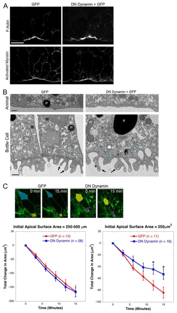
Coordinated apical constriction (AC) in epithelial sheets drives tissue invagination [1, 2] and is required for diverse morphogenetic movements such as gastrulation [3], neurulation [4, 5], and organogenesis [6]. We showed previously that actomyosin contractility drives AC in Xenopus laevis bottle cells [7]; however, it remained unclear whether it does so in concert with other processes. Here we report that endocytosis-driven membrane remodeling is required for efficient AC. We found endosomes exclusively in bottle cells in the early gastrula. Disrupting endocytosis with dominant-negative dynamin or rab5 perturbed AC, with a significant decrease in constriction rate late in the process, suggesting that endocytosis operates downstream of actomyosin contractility to remove excess membrane. Additionally, disrupting endocytosis during neurulation inhibits AC in hingepoint cells, resulting in neural tube closure defects. Thus, membrane remodeling during AC could be a general mechanism to achieve efficient invagination in embryos.


Figures

Figure 1
Figure 1
Biotinylated vesicles are present only in bottle cells in Xenopus early gastrula stage embryos. (A) Vegetal view (left, epifluorescence), midsagittal section (center, confocal), and higher magnification of midsagittal section (right) of Stage 10 embryos labeled with NHS-LC-sulfo biotin and anti-DM1α (tubulin). Arrow indicates bottle cells. Scale bar = 25μm. (B) Confocal midsagittal sections of embryos stained with anti-EEA1 and biotin. (C) Confocal midsagittal sections of embryos injected with Rab5 EGFP mRNA, then fixed and stained with anti-GFP and biotin. Arrow(s) in B and C indicate colocalization of biotin with early endosome markers. (D) Confocal midsagittal sections of embryos following pulse-chase labeling with biotin. Embryos were fixed at the indicated timepoints and stained with anti-DM1α and streptavidin. Arrows indicate endosomes that appear to be on or near the basolateral membrane. In all midsagittal sections, images are oriented vegetal to the lower left; scale bars for B,C,D = 50μm. See also Figure S1.
Figure 2
Figure 2
Dominant Negative (DN) Dynamin and DN Rab5 disrupt bottle cell formation by affecting apical constriction. (A) Epifluorescence images of stage 10.5 embryos, vegetal views. All embryos were injected with membrane-EGFP mRNA, plus DN Dynamin, WT Dynamin, or WT + DN. Arrows point to blastopore forming in the DMZ. Scale bar = 250μm. (B) Confocal midsagittal sections of embryos, all injected with membrane-EGFP, plus DN, WT, or WT + DN. Embryos were stained with anti-GFP and streptavidin. Scale bar = 50μm. (C) Quantification of blastopore depth and bottle cell morphometrics in DN Dynamin-injected embryos. *, p<0.01; **, p<0.001; ‡, p<0.05 for GFP versus rescue, p<0.001 for DN versus rescue. Each bar represents the mean of five experiments. For all graphs, error bars represent ± SEM. (D) Confocal midsagittal sections of uninjected control embryos or embryos injected with DN Rab5 mRNA and stained with anti-DM1α tubulin and streptavidin. Scale bar = 50μm. Bottom panels (D’) show higher magnification of biotin-labeled membrane. Scale bar = 10μm. (E) Quantification of blastopore depth (n=88 control embryos; 53 DN Rab5 embryos) and bottle cell morphometrics (n=187 control cells; 113 DN Rab5 cells). *, p<0.001. Each bar represents the mean of six experiments. See also Figures S2 and S3.
Figure 3
Figure 3
Apical membrane remodeling is required for efficient apical constriction downstream of actomyosin contractility. (A) Apical accumulation of F-actin and Myosin do not require endocytosis. Confocal midsagittal sections of embryos injected with membrane-EGFP alone or with membrane-EGFP plus DN Dynamin, then stained with phalloidin to visualize F-actin or anti-pMLC. Scale bar = 50μm. (B) Transmission electron micrographs of GFP and DN Dynamin-injected embryos. Animal cells do not have microvilli, while bottle cells have microvilli. Arrows, microvilli; m, mitochondria; P, pigment granule; Y, yolk platelet. Vesicles are pseudocolored in blue. Scale bars = 0.5μm. (C) Rate of constriction is the same between wild-type and DN Dynamin bottle cells until cells become very constricted. Top panels are stills from two timepoints (0 minutes, 15 minutes) of time-lapse movies (Movies 1 and 2). Larger cells (>250μm2) pseudocolored in blue and mustard; smaller cells (<250μm2) in orange and purple. Line graphs indicate the total decrease in apical surface area over 15 minutes. n, number of cells measured; *, p < 0.05. See also Table S1.
Figure 4
Figure 4
Endocytosis occurs the apically constricting dorsal lateral hingepoint cells during neurulation, and perturbing endocytosis results in apical constriction and neural tube closure defects. (A) Model of apical membrane dynamics during bottle cell apical constriction. (B) Endocytosis occurs in the neural tube. Cross-section of a stage 18 embryo, dorsal side up, stained with streptavidin. Scale bar = 50μm. (C) Injection of DN Dynamin results in a range of neural tube closure defects. Top panels show unilateral injection of membrane-EGFP in whole stage 24 embryos, dorsal views, anterior to the left. Bottom panels show higher magnification of the anterior neural tube, anterior up. Asterisks indicate injected side. (D) DN Dynamin-injected hingepoint cells appear less apically constricted than GFP-injected cells. Stage 18 embryos were stained with anti-DM1α (tubulin) and anti-GFP. Scale bar = 50μm. See also Figure S4 and Movie 3.

Similar articles

Cited by

References

    1. Lewis W. Mechanics of Invagination. Anat Rec. 1947;97:139–156. - PubMed
    1. Odell GM, Oster G, Alberch P, Burnside B. The mechanical basis of morphogenesis. I. Epithelial folding and invagination. Dev Biol. 1981;85:446–462. - PubMed
    1. Kimberly EL, Hardin J. Bottle cells are required for the initiation of primary invagination in the sea urchin embryo. Dev Biol. 1998;204:235–250. - PubMed
    1. Burnside B. Microtubules and microfilaments in newt neuralation. Dev Biol. 1971;26:416–441. - PubMed
    1. Haigo SL, Hildebrand JD, Harland RM, Wallingford JB. Shroom induces apical constriction and is required for hingepoint formation during neural tube closure. Curr Biol. 2003;13:2125–2137. - PubMed

Publication types