Review
. 2010 May 17;584(10):1948-55.
doi: 10.1016/j.febslet.2010.01.032. Epub 2010 Jan 21.

Mitochondrial ryanodine receptors and other mitochondrial Ca2+ permeable channels

Affiliations
Review

Mitochondrial ryanodine receptors and other mitochondrial Ca2+ permeable channels

Shin-Young Ryu et al. FEBS Lett. .

Abstract

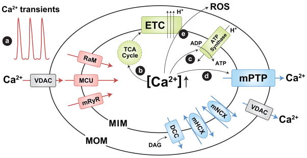
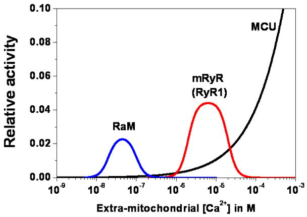
Ca(2+) channels that underlie mitochondrial Ca(2+) transport first reported decades ago have now just recently been precisely characterized electrophysiologically. Numerous data indicate that mitochondrial Ca(2+) uptake via these channels regulates multiple intracellular processes by shaping cytosolic and mitochondrial Ca(2+) transients, as well as altering the cellular metabolic and redox state. On the other hand, mitochondrial Ca(2+) overload also initiates a cascade of events that leads to cell death. Thus, characterization of mitochondrial Ca(2+) channels is central to a comprehensive understanding of cell signaling. Here, we discuss recent progresses in the biophysical and electrophysiological characterization of several distinct mitochondrial Ca(2+) channels.


Figures

Figure 1
Figure 1
Mitochondrial Ca2+ channels/transporters and role in mitochondrial function. Mitochondrial Ca2+ uptake is determined by the mitochondrial Ca2+ uniporter (MCU), rapid mode of uptake (RaM), and ryanodine receptor (mRyR, or RyR1). The mitochondrial permeability transition pore (mPTP), Na+/Ca2+ exchanger (mNCX), H+/Ca2+ exchanger (mHCX, encoded by Letm1), and DAG activated cation channels (DCC) contribute to Ca2+ efflux. Mitochondrial Ca2+ uptake contributes to (a) shaping cytosolic Ca2+ signals and triggering metabolic coupling by enhancing mitochondrial ATP synthesis: (b) stimulation of Ca2+ dependent dehydrogenases of the TCA cycle [34] to increase NADH/FADH production used to feed electrons through the electron transport chain (ETC) and (c) activation of the ATP synthase [35]. However, mitochondrial Ca2+ overload can trigger (d) mPTP activation, (e) ROS generation, and cell death. Voltage dependent anion-selective channels (VDAC) provide a pathway for Ca2+ and metabolite transport across the mitochondrial outer membrane. MIM and MOM; mitochondrial inner and outer membranes, respectively.
Figure 2
Figure 2
Ca2+ dependence of the major mitochondrial Ca2+ influx pathways. Relative activity of RaM (blue), mRyR (red), and the MCU (black) is estimated based on respective Ca2+ dependencies assuming a constant membrane potential and electrochemical gradient across the mitochondrial inner membrane. MCU is modeled according to patch clamp data of Kirichok et al. [43] and fitting with a Hill equation, 1/(1+(Km/x)n), where Km is the half-maximal concentration (19 mM) for activation, x is the extra-mitochondrial Ca2+ concentration, and n is the Hill coefficient (0.6). mRyR is modeled based on the Ca2+ dependent activation and inhibition of RyR1 channels using a modified Hill equation, c1*(1/(1+(Ka/x)n))*(1−1/(1+(Ki/x)n)), where c1 is a constant (0.0045) to enforce a 5-fold faster Ca2+ transport by mRyR compared to the MCU at 1 μM extra-mitochondrial Ca2+ according to the UV flash-induced mitochondrial Ca2+ uptake experiments of Beutner et al [10], Ka (2 μM) is the half-maximal concentration for Ca2+ dependent activation, Ki (20 μM) is the half-maximal concentration for Ca2+ dependent inhibition, x is the extra-mitochondrial Ca2+ concentration, and n is the Hill coefficient (4). RaM is modeled based on the same modified Hill’s equation, c2*(1/(1+(Ka/x)n))*(1−1/(1+(Ki/x)n)), where c2 is a constant (0.00049) to enforce a 50-fold faster Ca2+ transport of RaM compared to the MCU at 50 nM extra-mitochondrial Ca2+ according to the findings of Buntinas et al [8], Ka (20 nM) is the half-maximal concentration for Ca2+ dependent activation, Ki (100 nM) is half maximal concentration for Ca2+ dependent inhibition, x is the extra-mitochondrial Ca2+ concentration, and n is the Hill coefficient (4).
Figure 3
Figure 3
Representative single channel current traces of different mitochondrial Ca2+ channels. A. Single channel current traces of MiCa recorded from a COS-7 cell mitoplast reproduced from the original recording reported by Kirichok et al. [43] (Nature, 2004, 427:360–364, supplemental figure 2a) with permission from Nature Publishing Group. The single channel activity of MiCa shows multiple conductance states ranging from 2.6–5.2 pS at −160 mV in symmetrical 105 mM CaCl2. At positive voltages (+140 mV), the single channel activity of MiCa shows fast flickering with very small subconductance states, an indication of conduction block by Ca2+. B. Single channel current trace of a mRyR channel purified from rat heart mitochondrial inner membrane and incorporated into an artificial lipid bilayer reproduced from the original recording reported by Altschafl et al. [12] (Biochimica et Biophysica Acta - Biomembranes, 2007, 1768:1784–1795, figure 7c) with permission from Elsevier. Peak unitary mRyR conductance is between 500–800 pS using a 300/50 mM, cis/trans, Cs-methanesulfonate gradient and 50 μM cytosolic Ca2+. Impera toxin A, a RyR channel modulator, induces subconductance opening of mRyR. C. Single channel current trace of a DAG activated cation channel (DCC) recorded from a brain mitoplast reproduced from the original report of Chinopoulos et al. [63] (Journal of Bioenergetics and Biomembranes, 2007, 37(4):237–247, figure 4d) with permission from Springer. The DCC current was recorded at −50 mV in symmetrical 150 mM KCl in the presence of 10 μM cytosolic Ca2+ and 100 μM 1-oleoyl-2-acetyl-sn-glycerol (OAG), a DAG analog. D. Single channel current trace of mPTP, previously referred to as the multi-conductance channel (MCC), recorded from rat heart mitoplast reproduced from the original report of Kinnally et al. [71] (Journal of Bioenergetics and Biomembranes, 24(1):99–110, figure 3) with permission from Springer. The mPTP activity was recorded at −60 mV in symmetrical 150 mM KCl. E. Single channel current traces of VDAC reproduced from the original report of Pavlov et al. [67] (Biochimica et Biophysica Acta - Bioenergetics, 2005, 1710:96–102, figure 2b & 2c) with permission from Elsevier. (a) Typical voltage dependent channel activity of VDAC with voltage ramps between ±40 mV shows anion- or cation-selective conductance states. (b) VDAC activity was recorded at 0 mV with 150/30 mM KCl gradient. Anion- and cation- selective states are shown with arrows. Solid line indicates the 0 current level. Scale bars are presented with some modification from the original article.

Similar articles

Cited by

References

    1. Deluca HF, Engstrom GW. Calcium uptake by rat kidney mitochondria. Proc Natl Acad Sci U S A. 1961;47:1744–50. - PMC - PubMed
    1. Harris EJ. The uptake and release of calcium by heart mitochondria. Biochem J. 1977;168:447–56. - PMC - PubMed
    1. Nicholls DG. Calcium transport and porton electrochemical potential gradient in mitochondria from guinea-pig cerebral cortex and rat heart. Biochem J. 1978;170:511–22. - PMC - PubMed
    1. Gunter TE, Pfeiffer DR. Mechanisms by which mitochondria transport calcium. Am J Physiol. 1990;258:C755–86. - PubMed
    1. Gunter TE, Gunter KK, Sheu SS, Gavin CE. Mitochondrial calcium transport: physiological and pathological relevance. Am J Physiol. 1994;267:C313–39. - PubMed

Publication types

Substances