

Molecular imaging in gastrointestinal endoscopy
- PMID: 20096697
- PMCID: PMC3335170
- DOI: 10.1053/j.gastro.2010.01.009
Molecular imaging in gastrointestinal endoscopy
Abstract
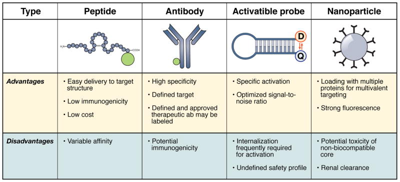
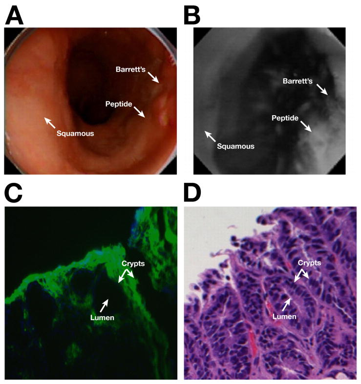
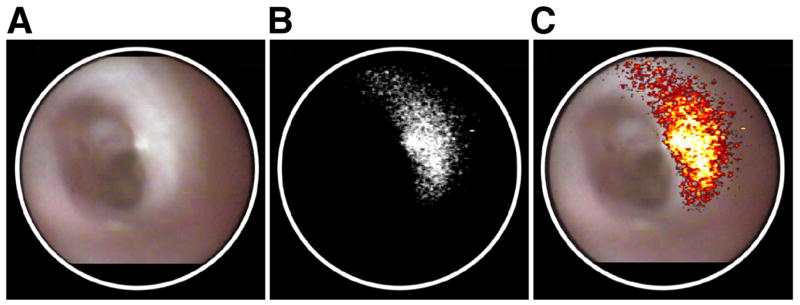
Molecular imaging is a rapidly growing new discipline in gastrointestinal endoscopy. It uses the molecular signature of cells for minimally-invasive, targeted imaging of gastrointestinal pathologies. Molecular imaging comprises wide field techniques for the detection of lesions and microscopic techniques for in vivo characterization. Exogenous fluorescent agents serve as molecular beacons and include labeled peptides and antibodies, and probes with tumor-specific activation. Most applications have aimed at improving the detection of gastrointestinal neoplasia with either prototype fluorescence endoscopy or confocal endomicroscopy, and first studies have translated encouraging results from rodent and tissue models to endoscopy in humans. Even with the limitations of the currently used approaches, molecular imaging has the potential to greatly impact on future endoscopy in gastroenterology.
Copyright 2010 AGA Institute. Published by Elsevier Inc. All rights reserved.
Figures


Similar articles
-
Molecular confocal laser endomicroscopy: a novel technique for in vivo cellular characterization of gastrointestinal lesions.World J Gastroenterol. 2014 Jun 28;20(24):7794-800. doi: 10.3748/wjg.v20.i24.7794. World J Gastroenterol. 2014. PMID: 24976717 Free PMC article.
-
Confocal laser endomicroscopy: technical advances and clinical applications.Gastroenterology. 2010 Aug;139(2):388-92, 392.e1-2. doi: 10.1053/j.gastro.2010.06.029. Epub 2010 Jun 15. Gastroenterology. 2010. PMID: 20561523 Review. No abstract available.
-
Molecular imaging: interaction between basic and clinical science.Gastroenterol Clin North Am. 2010 Dec;39(4):911-22. doi: 10.1016/j.gtc.2010.08.025. Epub 2010 Oct 8. Gastroenterol Clin North Am. 2010. PMID: 21093763 Review.
-
Confocal laser endomicroscopy in the "in vivo" histological diagnosis of the gastrointestinal tract.World J Gastroenterol. 2009 Dec 14;15(46):5770-5. doi: 10.3748/wjg.15.5770. World J Gastroenterol. 2009. PMID: 19998496 Free PMC article.
-
[Microscopy of the gastrointestinal tract: confocal endomicroscopy - clinical and scientific benefits].Zentralbl Chir. 2014 Aug;139(4):428-33. doi: 10.1055/s-0032-1327970. Epub 2013 Jul 3. Zentralbl Chir. 2014. PMID: 23824617 Review. German.
Cited by
-
Color-matched and fluorescence-labeled esophagus phantom and its applications.J Biomed Opt. 2013 Feb;18(2):26020. doi: 10.1117/1.JBO.18.2.026020. J Biomed Opt. 2013. PMID: 23403908 Free PMC article.
-
Technological advances in inflammatory bowel disease endoscopy and histology.Front Med (Lausanne). 2022 Nov 11;9:1058875. doi: 10.3389/fmed.2022.1058875. eCollection 2022. Front Med (Lausanne). 2022. PMID: 36438050 Free PMC article. Review.
-
Phase-change nanoparticles using highly volatile perfluorocarbons: toward a platform for extravascular ultrasound imaging.Theranostics. 2012;2(12):1185-98. doi: 10.7150/thno.4846. Epub 2012 Dec 23. Theranostics. 2012. PMID: 23382775 Free PMC article. Review.
-
Multi-Spectral Fluorescence Imaging of Colon Dysplasia InVivo Using a Multi-Spectral Endoscopy System.Transl Oncol. 2019 Feb;12(2):226-235. doi: 10.1016/j.tranon.2018.10.006. Epub 2018 Nov 10. Transl Oncol. 2019. PMID: 30419540 Free PMC article.
-
Endoscopic molecular imaging: status and future perspective.Clin Endosc. 2013 Nov;46(6):603-10. doi: 10.5946/ce.2013.46.6.603. Epub 2013 Nov 19. Clin Endosc. 2013. PMID: 24340252 Free PMC article. Review.
References
-
- Barrett T, Koyama Y, Hama Y, Ravizzini G, Shin IS, Jang BS, Paik CH, et al. In vivo diagnosis of epidermal growth factor receptor expression using molecular imaging with a cocktail of optically labeled monoclonal antibodies. Clin Cancer Res. 2007;13:6639–6648. - PubMed
-
- Goetz M, Ziebart A, Foersch S, Vieth M, Waldner MJ, Delaney P, Galle PR, et al. In vivo molecular imaging of colorectal cancer with confocal endomicroscopy of epidermal growth factor receptor. Gastroenterology. 2009 [Epub ahead of print] - PubMed
-
- Marten K, Bremer C, Khazaie K, Sameni M, Sloane B, Tung CH, Weissleder R. Detection of dysplastic intestinal adenomas using enzyme sensing molecular beacons in mice. Gastroenterology. 2002;122:406–414. - PubMed
Publication types
MeSH terms
Substances
Grants and funding
LinkOut - more resources
Full Text Sources
Other Literature Sources
