. 2010 May;29(4):304-16.
doi: 10.1016/j.matbio.2010.01.005. Epub 2010 Jan 22.

Reduced versican cleavage due to Adamts9 haploinsufficiency is associated with cardiac and aortic anomalies

Affiliations

Reduced versican cleavage due to Adamts9 haploinsufficiency is associated with cardiac and aortic anomalies

Christine B Kern et al. Matrix Biol. 2010 May.

Abstract

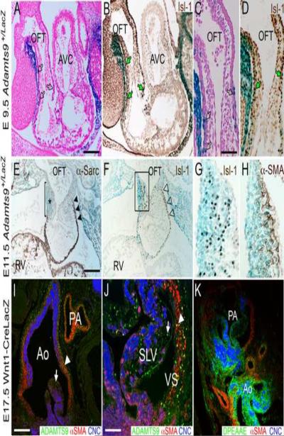
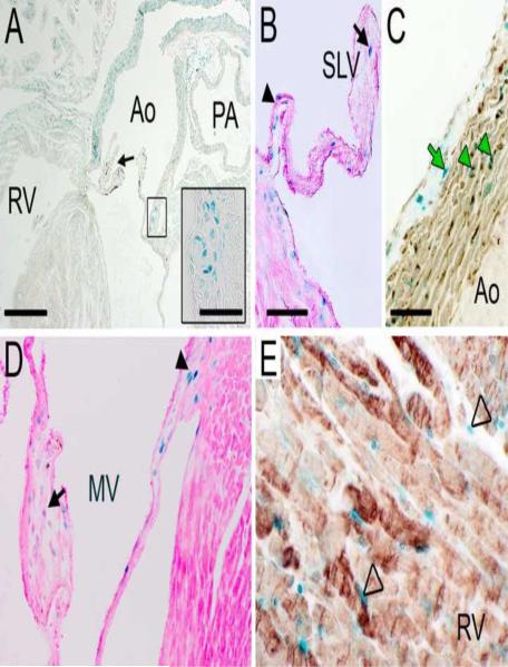
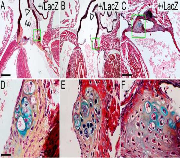
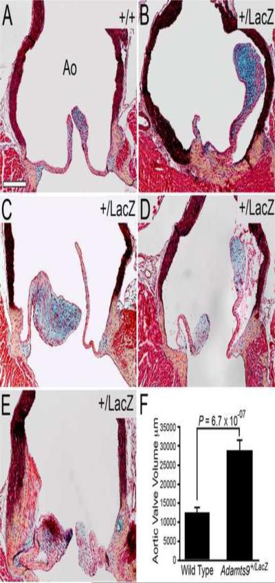
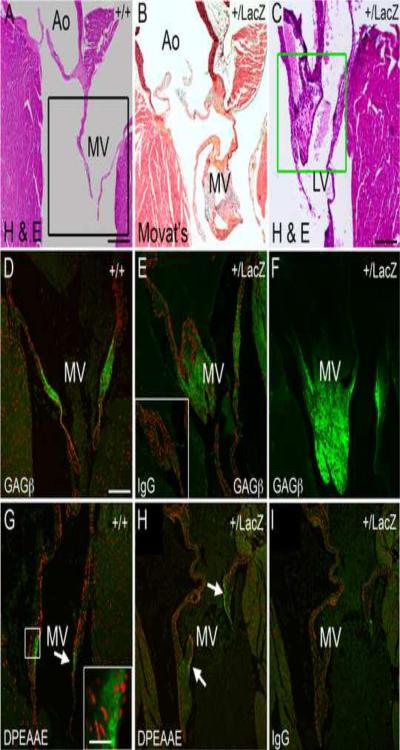
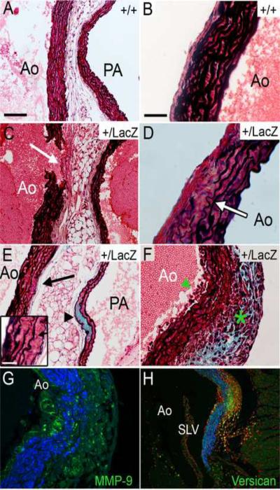
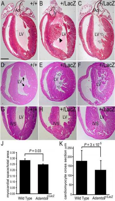
Here, we demonstrate that ADAMTS9, a highly conserved versican-degrading protease, is required for correct cardiovascular development and adult homeostasis. Analysis of Adamts9(+/LacZ) adult mice revealed anomalies in the aortic wall, valvulosinus and valve leaflets. Abnormal myocardial projections and 'spongy' myocardium consistent with non-compaction of the left ventricle were also found in Adamts9(+/LacZ) mice. During development, Adamts9 was expressed in derivatives of the Secondary Heart Field, vascular smooth muscle cells in the arterial wall, mesenchymal cells of the valves, and non-myocardial cells of the ventricles, but expression also continued in the adult heart and ascending aorta. Thus, the adult cardiovascular anomalies found in Adamts9(+/LacZ) hearts could result from subtle developmental alterations in extracellular matrix remodeling or defects in adult homeostasis. The valvular and aortic anomalies of Adamts9(+/LacZ) hearts were associated with accumulation of versican and a decrease in cleaved versican relative to WT littermates. These data suggest a potentially important role for ADAMTS9 cleavage of versican, or other, as yet undefined substrates in development and allostasis of cardiovascular extracellular matrix. In addition, these studies identify ADAMTS9 as a potential candidate gene for congenital cardiac anomalies. Mouse models of ADAMTS9 deficiency may be useful to study myxomatous valve degeneration.


Figures

Figure 1
Figure 1. Adamts9 is expressed at different stages during cardiac development in multiple cell types
Expression of Adamts9 in the developing cardiac outlet at E9.5 (A–D), E11.5 (E–H) and E17.5 (I,J). A–H show knock-in β-galactosidase (LacZ, blue) expression of Adamts9. Panels A and C show Adamts9 expression present in the transient myocardial sleeve of the OFT at E9.5 (blue, LacZ). Adamts9 is present in myocardial cells that are Islet-1 (Isl-1) positive (B,D, green arrows) suggesting that they are SHF derivatives. At E11.5 expression of Adamts9 continues predominantly in the transient myocardial sleeve (blue) (E,F) and in the right truncal cushion (* in E and box in F). α-sarcomeric actin expression is absent subjacent to the right truncal cushion revealing the loss of the transient myocardial wall (bar, E). F shows Isl-1 positive cells in the right truncal cushion, box in F magnified in panel G and shows overlapping Adamts9 expression (blue) with Isl1 (brown). Expression of α-smooth muscle actin is in the subendocardial portion of the truncal cushion and subjacent to the strongest Adamts9 expression (H). I,J show immunolocalization of ADAMTS9 (green) in the aortic and pulmonary artery walls (arrowhead), as well as in the mesenchymal cells of the semilunar valve leaflets (arrow) at E17.5. K shows a sister section with immunolocalization of the versican neo-epitope DPEAAE (green) cleavage product of versican. OFT-outflow tract; AVC-atrioventricular canal; RV-right ventricle; Ao-aorta; PA-pulmonary artery; SLV-semi-lunar valve; α-sarc- α-sarcomeric actin; α-SMA-α-smooth muscle actin; CNC- cardiac neural crest cells marked with Wnt1-Cre Rosa LacZ in panels I–K; open black arrows (A,C) Adamts9 expression in the transient myocardial sleeve of the OFT; black arrowheads- Adamts9 expression in the proximal transient myocardial sleeve; open arrowheads (F) Islet1 in the transient myocardial sleeve; Magnification bars: A= 200μm applies to B, E and F; C = 50μm and applies to D, G, H and J; I=150μm applies to K.
Figure 2
Figure 2. Adamts9 expression is maintained in connective tissues of the adult heart
Frontal section through an adult Adamts9+/LacZ heart reveals Adamts9 expressing due to β-galactosidase gene expression (LacZ, blue) in the arterial wall of the aorta (Ao). The abnormal presence of a chondrogenic-like nodule in the valvular sinus (A, black box, inset). An abnormal semi-lunar valve leaflet is also shown (A, black arrow). Adamts9 expression in the hinge region (arrowhead) of the semi-lunar valve and leaflet (arrow) (B). The right ascending aortic arterial wall immunostained with αSMA (C; αSMA, brown). αSMA- alpha smooth muscle actin; Green arrowheads-colocalization of αSMA with Adamts9; Green arrow- Adamts9 expressing cells in the adventitia. Adamts9 expression in the VIC of the mitral valve leaflet (arrow) and hinge region (arrowhead) (D). Expression of Adamts9 in noncardiomyocytes within the ventricular myocardium (E, open arrowheads); cardiomyocytes are identified with anti-alpha sarcomeric actin (E, brown). PA- pulmonary artery; MV- mitral valve. Mag bars: A = 475μm, inset = 8μm; B = 200μm applies to D; C = 30μm applies to E.
Figure 3
Figure 3. Chondrogenic-like nodules develop in the valvular sinus of Adamts9+/LacZ heterozygous hearts
Frontal sections stained with Movat pentachrome of Adamts9+/LacZ heterozygous hearts ages 9 (A,D), 15 (B,E) and 24 (C,F) weeks are shown. The areas within the green boxes in A, B, C are shown at higher magnification in D, E, and F respectively. The cells surrounded by blue matrix, indicative of sulfated proteoglycans, are in large lacunae, surrounding the lacunae is collagen (yellow). Boxes in G,H shown magnified in the respective inset. Arrowheads (A, B) denote fractured aortic arterial wall observed in all Adamts9+/LacZ heterozygous hearts examined (n=15). Ao-aorta; SLV- semi-lunar valve; At-atrium. Magnification bars: A = 475μm applies to B; C = 200μm; D = 30μm and applies to E and F.
Figure 4
Figure 4. Abnormal aortic valves are seen in Adamts9+/LacZ hearts
WT (A) and Adamts9+/LacZ heterozygous mouse heart sections (B–E) were stained with the Movat pentachrome stain. Abnormal aortic leaflets (B–E) with an increase in blue staining indicative of an increase in proteoglycans are shown (B, C and D, blue staining overlapped with versican immunoreactivity, data not shown). Quantification of the area of adult WT (n=6) and Adamts9+/LacZ heterozygous (n=8) aortic valves is shown in panel F. Area measurements were performed in frontal sections (e.g. Figure 2A and Figure 8A–C) through the widest point of the aorta spanning a minimum of 40 μm width in 9 Adamts9+/LacZ hearts and 3 WT hearts. The total area of valves in pixels was compared and the P value determined by Student t-test (2 tailed, type 2) was P = 6.7 × 10−07. The penetrance of the aortic valve phenotype was 8/15 at 53%. The Adamts9+/LacZ hearts with the aortic valve phenotype were analyzed in this analysis. The Y axis represents the total number of positive pixels of the semilunar valves measured. Magnification bar in A = 200μm applies to B–E.
Figure 5
Figure 5. Adamts9+/LacZ heterozygous mice develop myxomatous mitral valves
Frontal sections of adult WT (A–C) and Adamts9+/LacZ heterozygous hearts (B, C, E, F, H, I) containing abnormal, myxomatous mitral valves. Movat pentachrome stain shows an increase in proteoglycan staining (blue) in an example of an Adamts9+/LacZ heterozygous mouse heart (B). Additional examples of Adamts9+/LacZ heterozygous hearts with large and irregularly shaped mitral valves (C, E). Sections of WT (D) and Adamts9+/LacZ heterozygous hearts (E, F) were immunostained with the αGAGβ antibody for intact versican. IgG antibody control is presented in the inset of E. Black box in A denotes a sister section in D immunostained with αGAGβ antibody. The Green box in F is stained for αGAGβ antibody in F. The N-terminal ADAMTS versican cleavage fragment is localized (G, H) using the αDPEAAE antibody that immunolocalizes the neo-epitope of versican after ADAMTS cleavage (G box and arrow). The box in G is shown in higher magnification in the inset. Magnification bars: A = 475μm applies to B; C = 200μm; D = 150μm and applies to E–I. Inset in G = 10μm.
Figure 6
Figure 6. Adamts9+/LacZ heterozygous mice develop anomalies of the aortic wall
Movat pentachrome-stained sections of the aortic wall. Examples of WT aortic walls (A, left side; B right side). Aortic walls from Adamts9+/LacZ heterozygous hearts (C–H). Adventitia thickening (white arrow) adjacent to the aortic break* (C). Irregular and thinning elastic lamellae, containing breaks within the arterial wall from Adamts9+/LacZ hearts (D, E-inset, F). Adamts9+/LacZ heterozygous aorta shows an increase in the blue staining consistent with an increase in proteoglycans within the pulmonary artery (PA) wall (E, arrowhead). Adamts9+/LacZ aortas containing only 6 elastic lamellae (D, E inset), decreased from the normal 8–11 in the ascending aorta (A, B). An Adamts9+/LacZ heterozygous heart with an increase in cells associated with the intima (F, green arrowhead) and adventitia (F, green asterisk) with a concomitant increase in proteoglycans (blue). This tunica media shows thinning of elastic lamellae (F). A sister section of panel F was stained with versican (α-GAGβ, green) and αSMA (blue) showing the increase in versican in the region of cell infiltration (G). Sister section of G stained with propidium iodide associated to label cell nuclei immunostained with α-MMP-9 (green) and αSMA (blue) in an Adamts9+/LacZ heterozygous heart (H). Magnification bars: A = 100μm applies to C,E,G; Inset E = 8μm; B=30μm and applies to D, F, H). *The thickness of the adventitia was measured in the 5/17 Adamts9+/LacZ hearts that displayed the anomaly compared to 4 WT hearts. The average thickness of the Adamts9+/LacZ hearts was 36.5 μm (with a standard deviation of the mean +/− 7.37 μm) to 14.78 μm (+/− 5.03) WT hearts with a P value 3.0 × 10⁻¹⁰.
Figure 7
Figure 7. Immunoreactivity of the cleaved form of versican, DPEAAE, is reduced in the aortic wall of Adamts9+/LacZ heterozygous mice
WT (A,C) and Adamts9+/LacZ heterozygous (B,D) adult aortas immunostained with the DPEAAE antibody detecting the N-terminal cleaved fragment of versican (green). Quantification of expression using Amira™, normalized to area in adult WT (n=4) and Adamts9+/LacZ heterozygous (n=4) hearts showed an average of 4.6 fold higher levels of DPEAAE reactivity in WT hearts.
Figure 8
Figure 8. Ventricular myocardium of Adamts9+LacZ heterozygous mice have abnormal myocardial projections
WT (A,D,G) and Adamts9LacZ heterozygous (B,C,E,F,H,I) adult hearts containing abnormal myocardial projections in the left ventricle (open arrows, solid arrowhead). Frontal sections of adult hearts stained with Movat pentachrome (A–C). Cross sections stained with H&E (D–F). Solid arrowhead in B shows an abnormal interventricular myocardial protrusion. Solid arrows-normal papillary muscles. Adamts9LacZ heterozygous (H, I) frontal adult heart sections stained with H&E showing a wide IVS at the apex and `spongy' appearance. Morphometric analysis of the myocardial space versus the myocardial tissue from WT and Adamts9LacZ heterozygous hearts P = 0.03 (J). A total of 9 Adamts9+/LacZ hearts and 3 WT hearts were analyzed, the approximate lower half of the ventricular myocardium toward the apex as shown in G–I. The resulting P value using Student's t-test 2 tailed, type 2 was P=0.03. Error bars represent one standard deviation of the average presented in the graph (J). Quantification of the individual size of cross sections from WT and Adamts9LacZ heterozygous is also shown; P = 3 × 10⁻⁵ (K) using a minimum of 75 Adamts9LacZ and WT cardiomyocytes from 4 different animals. Ao-aorta; LV-left ventricle; IVS-interventricular septum. Magnification bars: A = 475μm applies to B–I.

Similar articles

Cited by

References

    1. Apte SS. A disintegrin-like and metalloprotease (reprolysin type) with thrombospondin type 1 motifs: the ADAMTS family. Int J Biochem Cell Biol. 2004;36:981–5. - PubMed
    1. Apte SS. A disintegrin-like and metalloprotease (reprolysin-type) with thrombospondin type 1 motif (ADAMTS) superfamily-functions and mechanisms. J Biol Chem. 2009 - PMC - PubMed
    1. Aspberg A, Adam S, Kostka G, Timpl R, Heinegard D. Fibulin-1 is a ligand for the C-type lectin domains of aggrecan and versican. Journal of Biological Chemistry. 1999;274:20444–9. - PubMed
    1. Azeloglu EU, Albro MB, Thimmappa VA, Ateshian GA, Costa KD. Heterogeneous transmural proteoglycan distribution provides a mechanism for regulating residual stresses in the aorta. Am J Physiol Heart Circ Physiol. 2008;294:H1197–205. - PubMed
    1. Brooke BS, Habashi JP, Judge DP, Patel N, Loeys B, Dietz HC., 3rd Angiotensin II blockade and aortic-root dilation in Marfan's syndrome. N Engl J Med. 2008;358:2787–95. - PMC - PubMed

Publication types

MeSH terms