

Purification and assay of mitotic motors
- PMID: 20096785
- PMCID: PMC3715878
- DOI: 10.1016/j.ymeth.2010.01.019
Purification and assay of mitotic motors
Abstract
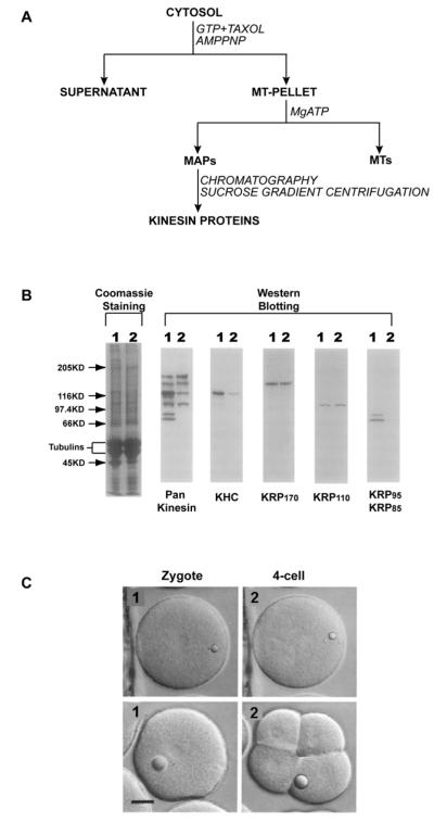
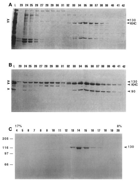
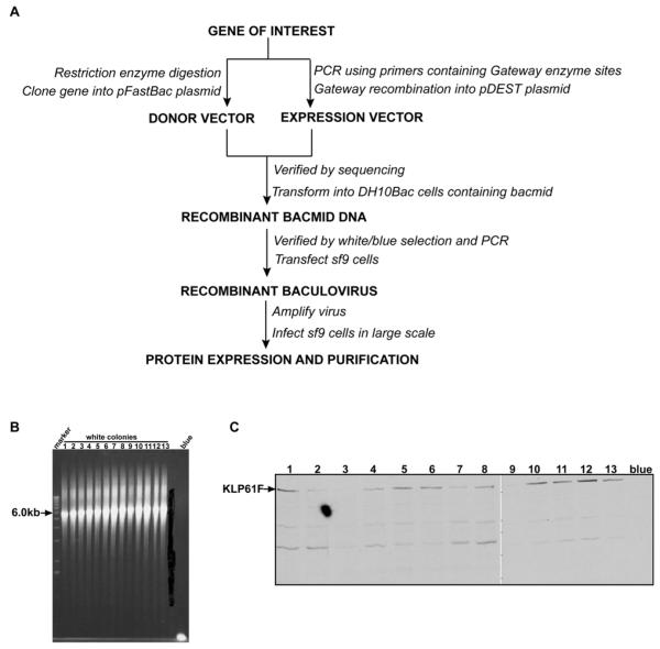
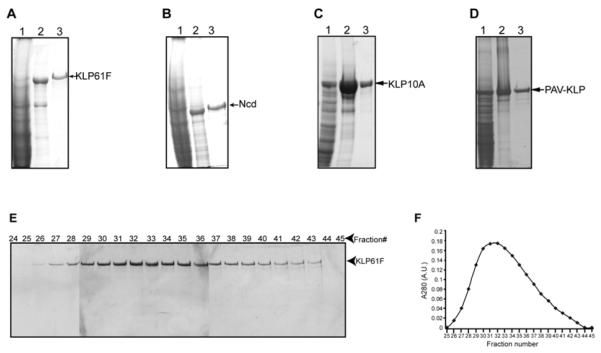
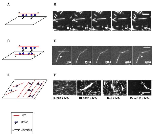
To understand how mitotic kinesins contribute to the assembly and function of the mitotic spindle, we need to purify these motors and analyze their biochemical and ultrastructural properties. Here we briefly review our use of microtubule (MT) affinity and biochemical fractionation to obtain information about the oligomeric state of native mitotic kinesin holoenzymes from eggs and early embryos. We then detail the methods we use to purify full length recombinant Drosophila embryo mitotic kinesins, using the baculovirus expression system, in sufficient yields for detailed in vitro assays. These two approaches provide complementary biochemical information on the basic properties of these key mitotic proteins, and permit assays of critical motor activities, such as MT-MT crosslinking and sliding, that are not revealed by assaying motor domain subfragments.
Figures
Similar articles
-
Purification of novel kinesins from embryonic systems.Methods Enzymol. 1998;298:133-54. doi: 10.1016/s0076-6879(98)98015-6. Methods Enzymol. 1998. PMID: 9751878
-
How kinesin motor proteins drive mitotic spindle function: Lessons from molecular assays.Semin Cell Dev Biol. 2010 May;21(3):260-8. doi: 10.1016/j.semcdb.2010.01.018. Epub 2010 Jan 28. Semin Cell Dev Biol. 2010. PMID: 20109570 Free PMC article. Review.
-
The mitotic kinesin-14 Ncd drives directional microtubule-microtubule sliding.Nat Cell Biol. 2009 Jun;11(6):717-23. doi: 10.1038/ncb1877. Epub 2009 May 10. Nat Cell Biol. 2009. PMID: 19430467
-
Molecular design principles for bipolar spindle organization by two opposing motors.Proc Natl Acad Sci U S A. 2025 Mar 25;122(12):e2422190122. doi: 10.1073/pnas.2422190122. Epub 2025 Mar 21. Proc Natl Acad Sci U S A. 2025. PMID: 40117309 Free PMC article.
-
Bidirectional motility of kinesin-5 motor proteins: structural determinants, cumulative functions and physiological roles.Cell Mol Life Sci. 2018 May;75(10):1757-1771. doi: 10.1007/s00018-018-2754-7. Epub 2018 Feb 3. Cell Mol Life Sci. 2018. PMID: 29397398 Free PMC article. Review.
Cited by
-
In vitro Microtubule Bundling Assay under Physiological Conditions.Bio Protoc. 2017 Apr 5;7(7):e2217. doi: 10.21769/BioProtoc.2217. eCollection 2017 Apr 5. Bio Protoc. 2017. PMID: 34541221 Free PMC article.
-
Centralspindlin-mediated transport of RhoGEF positions the cleavage plane for cytokinesis.Sci Signal. 2023 Jul 4;16(792):eadh0601. doi: 10.1126/scisignal.adh0601. Epub 2023 Jul 4. Sci Signal. 2023. PMID: 37402224 Free PMC article.
-
Kinesin-3 motors are fine-tuned at the molecular level to endow distinct mechanical outputs.BMC Biol. 2022 Aug 10;20(1):177. doi: 10.1186/s12915-022-01370-8. BMC Biol. 2022. PMID: 35948971 Free PMC article.
-
Gliding Assay to Analyze Microtubule-based Motor Protein Dynamics.Bio Protoc. 2017 Apr 5;7(7):e2210. doi: 10.21769/BioProtoc.2210. eCollection 2017 Apr 5. Bio Protoc. 2017. PMID: 34541218 Free PMC article.
-
Tum/RacGAP functions as a switch activating the Pav/kinesin-6 motor.Nat Commun. 2016 Apr 19;7:11182. doi: 10.1038/ncomms11182. Nat Commun. 2016. PMID: 27091402 Free PMC article.
References
-
- Brust-Mascher I, Scholey JM. Mitotic spindle dynamics in Drosophila. Int Rev Cytol. 2007;259:139–72. - PubMed
-
- Mitchison TaES. Mitosis: a history of division. Nat Cell Biol. 2001;3:E17–21. - PubMed
-
- Walczak CE, Heald R. Mechanisms of mitotic spindle assembly and function. Int Rev Cytol. 2008;265:111–58. - PubMed
-
- Mogilner A, Oster G. Polymer motors: pushing out the front and pulling up the back. Curr Biol. 2003;13:R721–33. - PubMed
-
- Sharp DJ, Rogers GC, Scholey JM. Microtubule motors in mitosis. Nature. 2000;407:41–7. - PubMed
Publication types
MeSH terms
Substances
Grants and funding
LinkOut - more resources
Full Text Sources
Molecular Biology Databases
