Review
. 2010 Feb;20(1):128-37.
doi: 10.1016/j.sbi.2009.12.015. Epub 2010 Jan 22.

Bridging the solution divide: comprehensive structural analyses of dynamic RNA, DNA, and protein assemblies by small-angle X-ray scattering

Affiliations
Review

Bridging the solution divide: comprehensive structural analyses of dynamic RNA, DNA, and protein assemblies by small-angle X-ray scattering

Robert P Rambo et al. Curr Opin Struct Biol. 2010 Feb.

Abstract

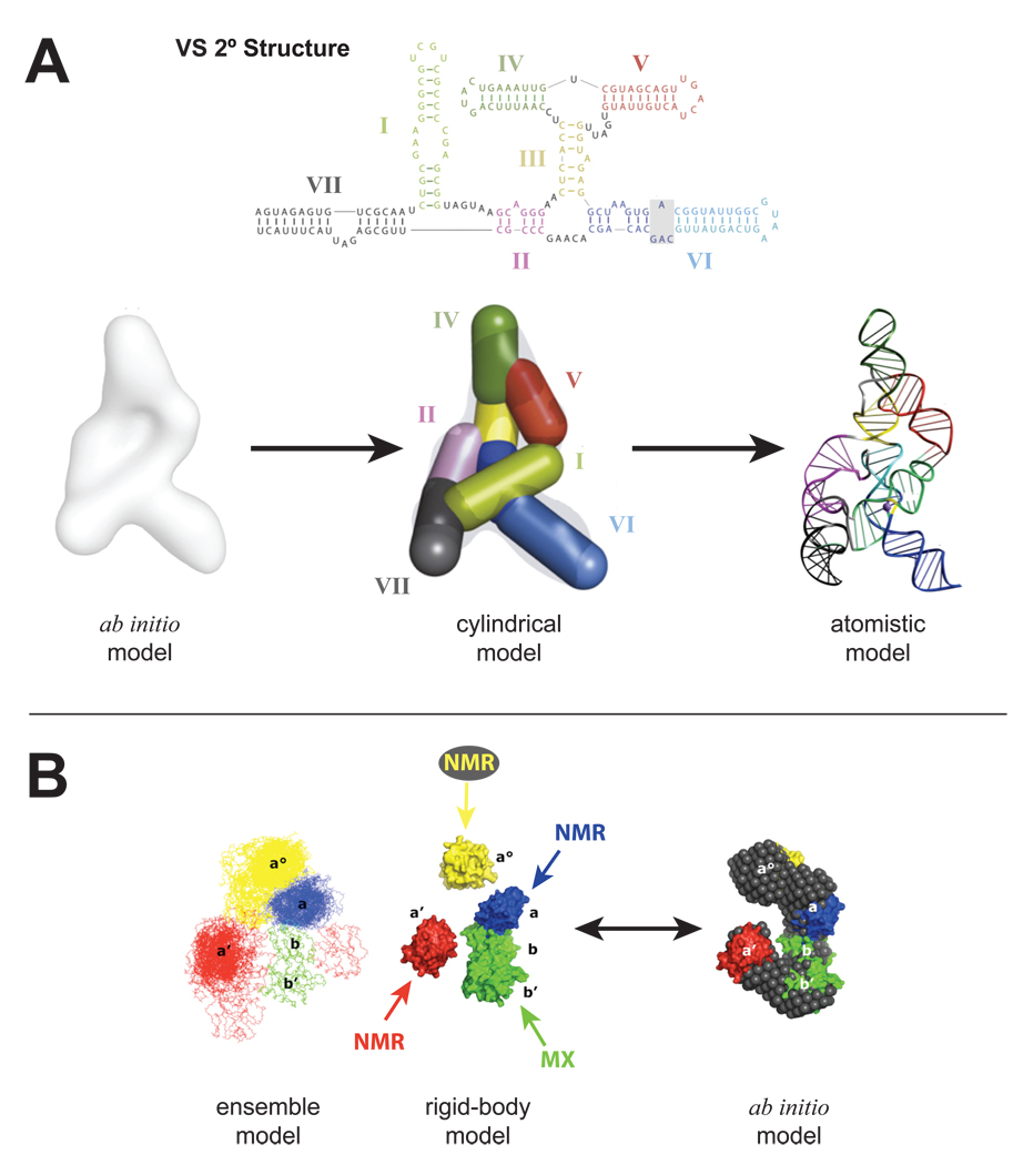
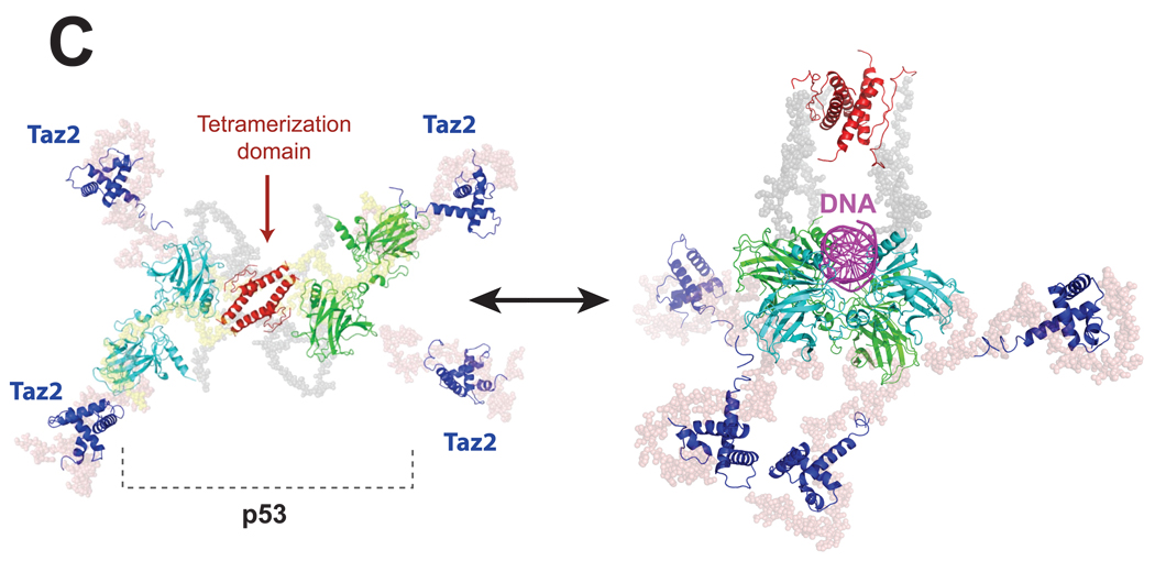
Small-angle X-ray scattering (SAXS) is changing how we perceive biological structures, because it reveals dynamic macromolecular conformations and assemblies in solution. SAXS information captures thermodynamic ensembles, enhances static structures detailed by high-resolution methods, uncovers commonalities among diverse macromolecules, and helps define biological mechanisms. SAXS-based experiments on RNA riboswitches and ribozymes and on DNA-protein complexes including DNA-PK and p53 discover flexibilities that better define structure-function relationships. Furthermore, SAXS results suggest conformational variation is a general functional feature of macromolecules. Thus, accurate structural analyses will require a comprehensive approach that assesses both flexibility, as seen by SAXS, and detail, as determined by X-ray crystallography and NMR. Here, we review recent SAXS computational tools, technologies, and applications to nucleic acids and related structures.


Figures

Figure 1
Figure 1
SAXS provides accurate information on folding and conformation in solution for both rigid and flexible macromolecules and their complexes. A. The calculation of a SAXS profile from an atomic model (CRYSOL, XPLOR-NIH, or IMP) is defined by the Debye equation and defines a relationship between the reciprocal space SAXS observations (orange) and the real space P(r)-distribution (cyan). Data in either reciprocal or real space can be used as a target for bead modeling with such programs as DAMMIN/F [46], DALAI_GA [47] or GASBOR [48]. Glucose isomerase is presented as an example above. B. Kratky plot of the lysine riboswitch in the presence and absence of Mg2+ [14]. The q2 • I(q) vs q plot demonstrates, for a folded particle, a parabolic convergence of the data at high q. Such a property fulfills the Porod invariant, Q, where the area under the curve approaches a constant value allowing for the calculation of the Porod volume. This is not true for an unfolded particle, as its scattering curve does not capture a defined area in a Kratky plot.
Figure 2
Figure 2
SAXS results suggest that flexibility in apo-receptors is reduced upon ligand binding for many RNA and protein receptors. A. Fit of the calculated (red) SAM-I riboswitch SAXS profile to the experimental (black) SAXS data, SAM-free. The crystal structure of SAM-I [49] was determined in the presence of ligand and the poor fit of the data to the model implies an alternate state of the riboswitch in solution. Further inspection of the Kratky plot demonstrates partial unfolding or increased flexibility. B. Identification of the correct dimer model using SAXS. Three possible dimers can be inferred from the crystal packing of the PYR1 protein based on surface-area contacts designated α-α (orange and blue), β-β (yellow and green), and α-β (blue and yellow) [15]. Though each subunit and dimer is identical chemically, SAXS clearly identifies the correct dimer (yellow curve) from incorrect (blue).
Figure 3
Figure 3
SAXS extends modeling of most large multi-domain macromolecular machines and suggests that flexibility and unstructured regions are generally critical for function. A. Construction of the VS ribozyme solution structure [21••] started with the ab initio model which was used to place cylindrical elements that corresponded to the helical elements described by its secondary structure. The resulting cylindrical model was then converted to a residue specific model and subject to cycles of energy minimization refinement to produce the final atomistic solution model of the entire VS ribozyme. B. The entire ERp72 solution structure [22] was achieved by refining the SAXS data of ERp72 against a starting model composed of its individual domains that were previously determined by a combination of NMR and MX. 380 separate rigid-body modeling runs converged to show a C-shape ensemble consistent with a separate ab initio reconstruction. C. The full-length p53-Taz2-DNA complex (right) [31••] was modeled from SAXS data of the all protein complex (left) [32]. The core (cyan and green) and tetramerization (red) domains were determined by MX and Taz2 was determined by NMR. The three high-resolution structures were incorporated into a rigid body analysis to produce the final cross-shaped structure showing fully extended arms held together by the tetramerization domain. Flexible regions were modeled with dummy residues (gray beads). Additional SAXS data collected in the presence of DNA (magenta) was then subject to additional rigid body modeling using an MX derived structure of the core domain bound to DNA. The binding of Taz2 to p53 and the associated p53-DNA complex facilitated visualization of the N-terminal regions of p53, an observation not seen in a previous EM study.
Figure 3
Figure 3
SAXS extends modeling of most large multi-domain macromolecular machines and suggests that flexibility and unstructured regions are generally critical for function. A. Construction of the VS ribozyme solution structure [21••] started with the ab initio model which was used to place cylindrical elements that corresponded to the helical elements described by its secondary structure. The resulting cylindrical model was then converted to a residue specific model and subject to cycles of energy minimization refinement to produce the final atomistic solution model of the entire VS ribozyme. B. The entire ERp72 solution structure [22] was achieved by refining the SAXS data of ERp72 against a starting model composed of its individual domains that were previously determined by a combination of NMR and MX. 380 separate rigid-body modeling runs converged to show a C-shape ensemble consistent with a separate ab initio reconstruction. C. The full-length p53-Taz2-DNA complex (right) [31••] was modeled from SAXS data of the all protein complex (left) [32]. The core (cyan and green) and tetramerization (red) domains were determined by MX and Taz2 was determined by NMR. The three high-resolution structures were incorporated into a rigid body analysis to produce the final cross-shaped structure showing fully extended arms held together by the tetramerization domain. Flexible regions were modeled with dummy residues (gray beads). Additional SAXS data collected in the presence of DNA (magenta) was then subject to additional rigid body modeling using an MX derived structure of the core domain bound to DNA. The binding of Taz2 to p53 and the associated p53-DNA complex facilitated visualization of the N-terminal regions of p53, an observation not seen in a previous EM study.
Figure 4
Figure 4
SAXS results suggest that conformational variation in solution is a general functional feature of macromolecular assemblies. SAXS results provide restraints for ensemble modeling that fully accounts for the scattering species in solution. The primary structure of each component is analyzed for flexible regions and regions that demarcate domain boundaries. Structures for the domains can be determined by experimental or computational (MODELER, XPLOR-NIH, SWISS-MODEL) methods and compiled into a single starting model. The initial model is used to generate the ensemble of 2,000 to 10,000 conformations. Experimental SAXS data is then used as a constraint to select the set of members from the ensemble that best describes the data (EOM or MES). Examination of the selected set should provide testable hypotheses subject to experimental methods such as limited proteolysis or chemical probing. Additional ensembles can be generated by changing the starting conditions. In the case of MES, the example shows that the minimal number of members required to explain the data is 3 and is largely described by a structure with an extended domain in multiple conformations. The graph of χ2 vs Rg shows the entire ensemble with the each χ2 goodness-of-fit calculated for each member against the experimental data.

References

    1. Fink AL. Natively unfolded proteins. Curr Opin Struct Biol. 2005;15:35–41. - PubMed
    1. Wright PE, Dyson HJ. Linking folding and binding. Curr Opin Struct Biol. 2009;19:31–38. - PMC - PubMed
    1. Putnam CD, Hammel M, Hura GL, Tainer JA. X-ray solution scattering (SAXS) combined with crystallography and computation: defining accurate macromolecular structures, conformations and assemblies in solution. Q Rev Biophys. 2007;40:191–285. - PubMed
    1. Tsuruta H, Irving TC. Experimental approaches for solution X-ray scattering and fiber diffraction. Curr Opin Struct Biol. 2008;18:601–608. - PMC - PubMed
    1. Hura GL, Menon AL, Hammel M, Rambo RP, Poole FL, 2nd, Tsutakawa SE, Jenney FE, Jr, Classen S, Frankel KA, Hopkins RC, et al. Robust, high-throughput solution structural analyses by small angle X-ray scattering (SAXS) Nat Methods. 2009;6:606–612.. This article presents an automated system and data analysis pipeline for SAXS data collection. The results on 50 samples indicate the relatively small amounts of material needed, the robust features of SAXS for efficient throughput, and the high percentage of samples for which structural data can be obtained.

Publication types