. 2010 Mar 19;28(14):2580-92.
doi: 10.1016/j.vaccine.2010.01.019. Epub 2010 Jan 22.

Genetic linkage of autologous T cell epitopes in a chimeric recombinant construct improves anti-parasite and anti-disease protective effect of a malaria vaccine candidate

Affiliations

Genetic linkage of autologous T cell epitopes in a chimeric recombinant construct improves anti-parasite and anti-disease protective effect of a malaria vaccine candidate

Balwan Singh et al. Vaccine. .

Abstract

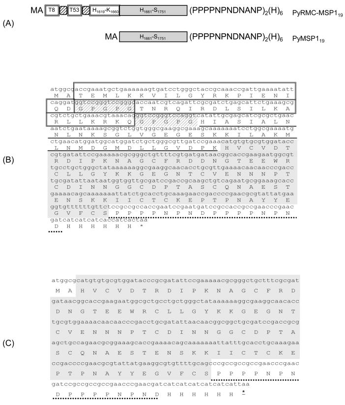
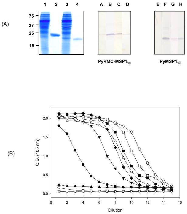
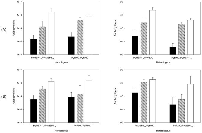
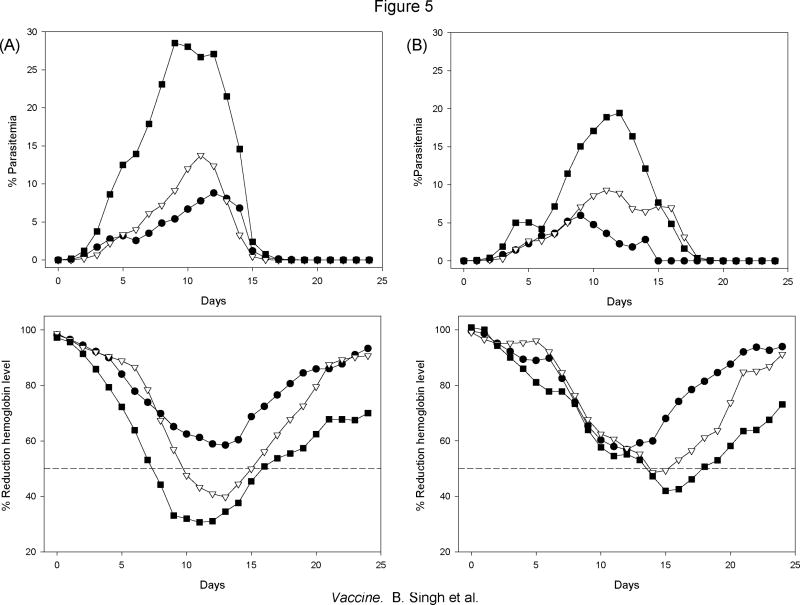
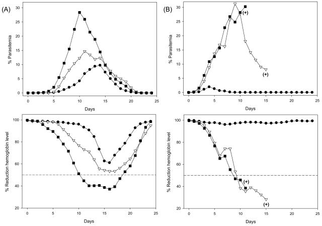
We have reported the design of polyvalent synthetic and recombinant chimeras that include promiscuous T cell epitopes as a viable delivery system for pre-erythrocytic subunit malaria vaccines. To further assess the ability of several Plasmodium T cell epitopes to enhance vaccine potency, we designed a synthetic gene encoding four Plasmodium yoelii merozoite surface protein 1 (PyMSP1) CD4(+) promiscuous T cell epitopes fused in tandem to the homologous carboxyl terminal PyMSP1(19) fragment. This Recombinant Modular Chimera (PyRMC-MSP1(19)) was tested for immunogenicity and protective efficacy in comparative experiments with a recombinant protein expressing only the PyMSP1(19) fragment. Both proteins induced comparable antibody responses. However PyRMC-MSP1(19) elicited higher anti-parasite antibody titers and more robust protection against both hyper-parasitemia and malarial anemia. Most importantly, passive transfer of anti-PyRMC-MSP1(19), but not anti-PyMSP1(19) antibodies protected against heterologous challenge. These studies show that protective efficacy can be significantly improved by inclusion of an array of autologous promiscuous T cell epitopes in vaccine constructs.


Figures

Figure 1
Figure 1
Topology of the synthetic codon optimized pyrmcmsp119 and pymsp119 genes. (A) Schematic representation of the chimeric proteins reported here. pyrmcmsp119 encoded 207 amino acids with the T cell epitopes, schematically represented by double lined boxes interspaced by five amino acid residues (striped boxes), upstream of the 19 kDa fragment of PyMSP1 fragment (GenBank accession XP_726257). PyMSP119 includes the complete amino acid sequence of the MSP119 and two carboxyl terminal tags. (B) Sequence of the PyRMC-MSP119 protein. The amino acid sequence is shown in single letter code. The sequences enclosed in boxes are the two promiscuous CD4+ T cell epitopes originally reported in the P. vivax MSP1 protein [7, 8]. The amino acid residues GPGPG were included as spacers between individual CD4+ T cell epitopes and the extended P. yoelii MSP119 protein (H1619-S1751). The underlined 42 amino acid long polypeptide, upstream from the first MSP119 EGF domain, contains two in silico predicted T cell epitopes. The carboxyl terminal P. berghei CSP repeat tag sequence PPPPNPNDPPPPNPND, was included for biochemical characterization of the antigenic integrity and to provide optional affinity purification tag is underlined with a dotted line. A carboxyl terminal (H)6 tag was included for protein purification. (C) Sequence of the MSP119 control protein includes amino acid H1661 to S1751, it also included the PbCSP repeat tag.
Figure 2
Figure 2
Biochemical and immunological characterization of the chimeric proteins described here. (A) SDS -PAGE (left panel) and western blot (center and right panels) of the purified P. yoelii PyRMC-MSP119 and PyMSP119 proteins expressed in E. coli BL21 (DE3). Left panel shows Coomassie stain after SDS-PAGE separation, total bacterial lysate and column purified proteins are shown for PyRMC-MSP119 (lanes 1 and 2) and PyMSP119 (lanes 3 and 4). Western blot analysis of the purified PyRMC-MSP119 (central panel) and PyMSP119 (right panel) incubated with sera samples from mice immunized with a synthetic peptide representing the promiscuous T cell epitope PyT53 (T1154-Q1173) (lane A and E), mice immunized with the corresponding recombinant protein (lane B and F), the monoclonal antibody 3D11 that recognize the P. berghei tag sequence [79] (lane C and G), or an anti-6X His tag monoclonal antibody (lane D and H). The molecular weight markers (BioRad) are indicated to the left. (B) Antigenicity of the P. yoelii chimeric proteins determined by ELISA, PyRMC-MSP119 closed symbols, PyMSP119, open symbols. Recombinant chimeras were tested using anti-PyT53 (●○), anti-PyRMC-MSP119 (▼;), anti- PyMSP119 (△), 3D11 (◆◇) or anti-His tag (■□). The monoclonal antibody MRA-94 that recognized the P. falciparum MSP1 was included as control of antigen specificity (▲▽). Data are presented as the geometric mean O.D. obtained at different concentrations of the corresponding monoclonal antibodies (1 μg to 0.06 ng/ml) or the reciprocal of the sera dilution obtained from mice immunized with P. yoelii PyRMC-MSP119 (dilutions 1:1000 to 1:16384000) or mice immunized with the synthetic peptide PyT53 (dilutions 1:100 to 1: 4096000). Antigen specificity was confirmed using pre-immune sera samples as a control (data not shown).
Figure 3
Figure 3
Kinetics of the anti- PyRMC-MSP119 or anti-PyMSP119 antibody responses in BALB/c and C57BL6 mice immunized with the homologous or heterologous antigen determined by ELISA. Antibody titers were measured 20 days after each immunization of BALB/c (A) or C57BL6 mice (B) with the PyRMC-MSP119 or anti-PyMSP119 and expressed as reciprocal geometric mean antibody titers ± standard deviation (n=6). Sera from mice immunized with the corresponding protein were tested with the homologous (left panel) or heterologous (right panel) antigen on day 20 (black bars), day 40 (stripped bars) and day 60 (white bars).
Figure 4
Figure 4
Reactivity of the antibodies elicited in rabbits with native P. yoelii proteins. (A) Western blot analysis of late stage parasite extracts. The samples were subjected to SDS-PAGE on a 5 to 15% polyacrylamide gradient gel, blotted onto nitrocellulose and then probed with purified IgG from rabbits immunized with PyRMC-MSP119 (lanes 1 and 2), purified IgG from rabbits immunized with PyMSP119 (lanes 3 and 4) or IgG from normal rabbits (lanes 5 and 6) at 0.6 μg/ml. The antibodies react with protein extract derived from P. yoelii 17X merozoites (lanes 1, 3 and 5) or P. yoelii nigeriensis N67 (lanes 2, 4 and 6). The mobility of molecular mass markers is indicated. (B) Recognition of soluble protein extract determined by ELISA using P. yoelii 17X, closed symbols, or P. yoelii nigeriensis N67 parasite extracts, open symbols. The protein extract were tested using purified IgG from rabbits immunized with PyRMC-MSP119 (●○), purified IgG from rabbits immunized with PyMSP119 (▼;△) or purified IgG from normal rabbits (■□). Data are presented as O.D. values at 405 nm using different concentrations of purified IgG preparations. (C) Characteristic immunofluorescence pattern of purified IgG from rabbits immunized with PyRMC-MSP119 on erythrocytes infected with P. yoelii 17X (upper panel) or P. yoelii nigeriensis N67 (lower panel). Left panels show parasite nuclei stained with DAPI, central panels show the anti-PyRMC-MSP119 antibody reactivity using AlexaFluor 488 goat anti-rabbit IgG-specific. The right panels are a merge of the blue and green fluorescence channels. A weak specific fluorescence was detected on erythrocytes infected with P. yoelii 17X using PyMSP119 (data not shown). No specific fluorescence was detected on erythrocytes infected with P. yoelii nigeriensis N67 with purified IgG rabbits immunized with PyMSP119 (data not shown). No specific fluorescence was found with purified IgG derived from normal rabbits using either homologous or heterologous strains.
Figure 5
Figure 5
Effects of immunization with either PyRMC-MSP119 or PyMSP119 on the time course curves after experimental challenge with P. yoelii 17X. BALB/c (A) or C57BL6 (B) mice were immunized three times subcutaneously using Montanide ISA 51 as adjuvant. Placebo control mice received adjuvant alone. The top panels summarize the area under the parasitemia time curves and the bottom panels the area under the hemoglobin time curves, obtained using the trapezoidal method [48, 49], in mice immunized with PyRMC-MSP119 (●), PyMSP119 (▽) or placebo (■). For parasitemia values, the total areas under the parasitemia change versus time curves were calculated and the mean change values expressed as percentage of parasitemia (n=6). For hemoglobin values, the total areas under the hemoglobin change versus time curves were calculated. Subsequently the mean change values in hemoglobin concentration per day were determined by comparison with baseline concentrations and expressed as percentage of reduction in hemoglobin levels. Data are representative of two independent experiments. 50% reduction in hemoglobin concentration relative to baseline is indicated with broken lines.
Figure 6
Figure 6
Effects of passive immunization on the time course curves after experimental challenge with P. yoelii 17X (A) or P. yoelii nigeriensis N67 (B). The top panels summarize the area under the parasitemia time curves and the bottom panels the area under the hemoglobin time curves, obtained using the trapezoidal method [48, 49], in mice immunized with PyRMC-MSP119 (●), PyMSP119 (▽) or placebo (■). For parasitemia values, the total areas under the parasitemia change versus time curves were calculated and the mean change values expressed as percentage of parasitemia (n=10). For hemoglobin values, the total areas under the hemoglobin change versus time curves were calculated. Subsequently the mean change values in hemoglobin concentration per day were determined by comparison with baseline concentrations and expressed as percentage of reduction in hemoglobin levels. Data are representative of two independent experiments. 50% reduction in hemoglobin concentration relative to baseline is indicated with broken lines. (+) Indicate animals that were euthanized due to severe anemia.

Similar articles

Cited by

References

    1. Organization WH, editor. WHO. World malaria report 2008. Geneva: WHO Press; 2008.
    1. Langhorne J, Ndungu FM, Sponaas AM, Marsh K. Immunity to malaria: more questions than answers. Nat Immunol. 2008;9(7):725–32. - PubMed
    1. Conway DJ, Cavanagh DR, Tanabe K, Roper C, Mikes ZS, Sakihama N, et al. A principal target of human immunity to malaria identified by molecular population genetic and immunological analyses. Nat Med. 2000;6(6):689–92. - PubMed
    1. Bejon P, Lusingu J, Olotu A, Leach A, Lievens M, Vekemans J, et al. Efficacy of RTS,S/AS01E vaccine against malaria in children 5 to 17 months of age. N Engl J Med. 2008;359(24):2521–32. - PMC - PubMed
    1. Kester KE, Cummings JF, Ockenhouse CF, Nielsen R, Hall BT, Gordon DM, et al. Phase 2a trial of 0, 1, and 3 month and 0, 7, and 28 day immunization schedules of malaria vaccine RTS,S/AS02 in malaria-naive adults at the Walter Reed Army Institute of Research. Vaccine. 2008;26(18):2191–202. - PubMed

Publication types

MeSH terms