

Differential expression of hepatic fibrosis mediators in sick and spontaneously recovered mice with experimental biliary atresia
- PMID: 20097372
- PMCID: PMC2844460
- DOI: 10.1016/j.jss.2009.10.038
Differential expression of hepatic fibrosis mediators in sick and spontaneously recovered mice with experimental biliary atresia
Abstract
Background: Hepatic fibrosis leading to cirrhosis is the major morbidity in patients with biliary atresia (BA). This fibrosis is due to an imbalance in extracellular matrix (ECM) breakdown and deposition. We have previously demonstrated increased mRNA expression for inhibitors of ECM breakdown without increased expression for mediators of ECM deposition in our animal model of BA by d 14. However, only a mild degree of hepatic fibrosis was seen at this time. We hypothesized that expression patterns for these proteins may change once more significant fibrosis had been established, and added resuscitation to the model to improve survival. Interestingly, we found that some mice spontaneously recovered at later time points with resuscitation, and thus compared expression for inhibitors of ECM breakdown and deposition in sick and recovered mice to determine the differences.
Methods: Newborn Balb/c mice received an intraperitoneal injection 1.0 x 10(6) fluorescence forming units of rhesus rotavirus 24h after birth. Mice were monitored daily for weight gain, development of jaundice, acholic stools, and bilirubinuria. Fifty muL/g of 5% dextrose in normal saline were subcutaneously injected daily to each mouse starting on d 7 until sacrifice. Mice that survived past d 14 were sacrificed at d 21 after saline or RRV infection. Livers were then harvested post-injection d 21 for histologic and immunohistochemical analysis. RNA expression of known mediators of fibrosis was evaluated using quantitative real-time PCR. Protein expression was assessed using ELISA. Weights and normally distributed data were compared using Student's t test. Histologic findings were compared using Fisher's exact test. Comparisons of gene expression and skewed data were performed by the Mann-Whitney U test. Statistical significance was assigned to any P value less than 0.05.
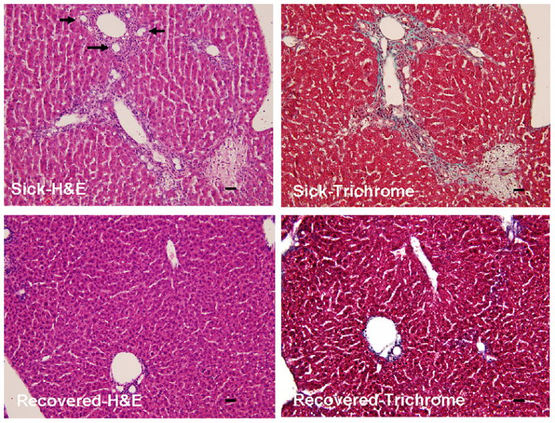
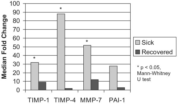
Results: Daily resuscitation resulted in a 35% (24/68) survival rate to d 21 in our model. Mice that recovered were significantly heavier than those that remained ill on d 14 (6.15 +/- 1.16 versus 4.94 +/- 0.82, P = 0.02) and 21 (7.31 +/- 1.41 versus 4.14 +/- 0.53, P < 0.001) despite the fact that there was no difference between the groups with respect to weight on d 7 (4.29 +/- 0.90 versus 3.89 +/- 0.81, P = 0.32). We found that all (10/10) animals that displayed clinical signs of biliary atresia on d 21 had moderate or severe histologic findings, while only one (1/9) of the recovered animals had liver abnormalities at sacrifice (P < 0.001 versus sick group). We also found that the sick mice had statistically significant median fold-increases of mRNA expression for TIMP-1 (31.9 versus 9.1, P = 0.041), TIMP-4 (88.1 versus 1.8, P = 0.022), and MMP-7 (51.8 versus 11.9, P = 0.006) compared with those that recovered. There was a trend toward decreased mRNA expression for PAI-1, which did not reach statistical significance (median 27.7 versus 2.19, P = 0.066). Increased protein expression for TIMP-1 and PAI-1 were also found in the sick group. The mRNA expression for the fibrillar collagens, fibronectin-1, connective tissue growth factor, snail-1, TIMP-2 and -3, and MMP-2 and MMP-9 was not different in the sick and recovered groups 21 d after RRV infection, and was not elevated from baseline gene expression.
Conclusions: With resuscitation added to the animal model of BA, some mice spontaneously recover while others progress to more significant hepatic fibrosis. Mice with hepatic fibrosis have a continued increase in mRNA expression of TIMP-1, TIMP-4, and MMP-7, with a trend toward increased mRNA expression of PAI-1 on d 21. Protein levels for TIMP-1 and PAI-1 were also increased in the sick mice. Recovered mice display mild to no hepatic parenchymal disease and a normal pattern of mRNA expression for the mediators of fibrosis tested. No increase in mRNA expression for the mediators of ECM deposition was found in either group. These data further support the notion that inhibition of ECM breakdown alone is sufficient to induce hepatic fibrosis. Modulation of this process may be a putative target for preventing liver injury in patients with BA.
Figures

Similar articles
-
Dysregulation of upstream and downstream transforming growth factor-β transcripts in livers of children with biliary atresia and fibrogenic gene signatures.J Pediatr Surg. 2013 Oct;48(10):2047-53. doi: 10.1016/j.jpedsurg.2013.03.047. J Pediatr Surg. 2013. PMID: 24094956 Free PMC article.
-
Integrin alphavbeta6 and mediators of extracellular matrix deposition are up-regulated in experimental biliary atresia.J Surg Res. 2009 Jun 1;154(1):21-9. doi: 10.1016/j.jss.2008.05.023. Epub 2008 Jun 20. J Surg Res. 2009. PMID: 19084240
-
Transcriptional basis for hepatic fibrosis in cystic fibrosis-associated liver disease.J Pediatr Gastroenterol Nutr. 2012 Mar;54(3):328-35. doi: 10.1097/MPG.0b013e3182432034. J Pediatr Gastroenterol Nutr. 2012. PMID: 22157922
-
microRNA-222 modulates liver fibrosis in a murine model of biliary atresia.Biochem Biophys Res Commun. 2014 Mar 28;446(1):155-9. doi: 10.1016/j.bbrc.2014.02.065. Epub 2014 Feb 22. Biochem Biophys Res Commun. 2014. PMID: 24569080
-
Role of myeloid differentiation factor 88 in Rhesus rotavirus-induced biliary atresia.J Surg Res. 2013 Sep;184(1):322-9. doi: 10.1016/j.jss.2013.05.032. Epub 2013 Jun 1. J Surg Res. 2013. PMID: 23768919 Free PMC article.
Cited by
-
Serum proteome profiling identifies novel and powerful markers of cystic fibrosis liver disease.PLoS One. 2013;8(3):e58955. doi: 10.1371/journal.pone.0058955. Epub 2013 Mar 14. PLoS One. 2013. PMID: 23516586 Free PMC article.
-
Role of viruses in biliary atresia: news from mice and men.Innov Surg Sci. 2018 Apr 4;3(2):101-106. doi: 10.1515/iss-2018-0009. eCollection 2018 Jun. Innov Surg Sci. 2018. PMID: 31579773 Free PMC article. Review.
-
Aetiology of biliary atresia: what is actually known?Orphanet J Rare Dis. 2013 Aug 29;8:128. doi: 10.1186/1750-1172-8-128. Orphanet J Rare Dis. 2013. PMID: 23987231 Free PMC article. Review.
-
Dysregulation of upstream and downstream transforming growth factor-β transcripts in livers of children with biliary atresia and fibrogenic gene signatures.J Pediatr Surg. 2013 Oct;48(10):2047-53. doi: 10.1016/j.jpedsurg.2013.03.047. J Pediatr Surg. 2013. PMID: 24094956 Free PMC article.
-
Increased MMP-7 expression in biliary epithelium and serum underpins native liver fibrosis after successful portoenterostomy in biliary atresia.J Pathol Clin Res. 2016 May 12;2(3):187-98. doi: 10.1002/cjp2.50. eCollection 2016 Jul. J Pathol Clin Res. 2016. PMID: 27499927 Free PMC article.
References
-
- Hsieh CS, Chuang JH, Huang CC, et al. Evaluation of matrix metalloproteinases and their endogenous tissue inhibitors in biliary atresia-associated liver fibrosis. J Pediatr Surg. 2005;40:1568. - PubMed
-
- Nadler EP, Patterson D, Violette S, et al. Integrin alphavbeta6 and mediators of extracellular matrix deposition are upregulated in experimental biliary atresia. J Surg Res. 2008 Jun 20; Epub ahead of print. - PubMed
-
- Petersen C, Biermanns D, Kuske M, et al. New aspects in a murine model for extrahepatic biliary atresia. J Pediatr Surg. 1997;32:1190. - PubMed
-
- Georges PC, Hui JJ, Gombos Z, et al. Increased stiffness of the rat liver precedes matrix deposition: implications for fibrosis. Am J Physiol Gastrointest Liver Physiol. 2007;293:G1147. - PubMed
Publication types
MeSH terms
Substances
Grants and funding
LinkOut - more resources
Full Text Sources
Medical
Research Materials
Miscellaneous
