. 2010 Mar 30;399(1):1-10.
doi: 10.1016/j.virol.2009.12.031. Epub 2010 Jan 25.

Modulation of type I IFN induction by a virulence determinant within the alphavirus nsP1 protein

Affiliations

Modulation of type I IFN induction by a virulence determinant within the alphavirus nsP1 protein

Catherine C Cruz et al. Virology. .

Abstract

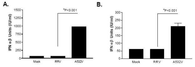
Alphaviruses are mosquito-borne viruses that cause serious human and animal diseases. Previous studies demonstrated that a determinant within the nsP1/nsP2 cleavage domain of the virulent Sindbis AR86 virus played a key role in regulating adult mouse virulence without adversely affecting viral replication. Additional characterization of this determinant demonstrated that a virus with the attenuating mutation induced more type I IFN production both in vivo and in vitro. Interestingly, this phenotype was not specific to the Sindbis AR86 virus, as a similar mutation in a distantly related alphavirus, Ross River Virus (RRV), also led to enhanced IFN induction. This effect was independent of virus-induced host shutoff, since IRF-3 phosphorylation, which occurs independently of de novo host transcription/translation, was induced more robustly in cells infected with the mutant viruses. Altogether, these results demonstrate that critical determinants within the nsP1/nsP2 cleavage domain play an important role in regulating alphavirus-induced IFN responses.


Figures

Figure 1
Figure 1. The mutant SIN T538I virus induces more type I interferon
A) Groups of six-week old (n=3) CD-1 mice were infected with diluent alone, wild type Sindbis AR86, or Sindbis T538I mutant at 1×10³ pfu via the intracranial (i.c.) route. Serum was harvested at the indicated time points (hours) and diluted (1:10) into media. B) L929 cells were either mock infected or infected with wild type AR86 or T538I mutant at an MOI of 5.0 and supernatants were harvested at 18 hours post-infection. Serum and supernatants were subjected to an interferon bioassay on L929 cells. Each bar represents the average of triplicate samples and the p values were determined by ANOVA statistical analysis. Error bars represent the standard error of the mean. The limit of detection in each bioassay was 31IU/ml.
Figure 2
Figure 2. The Sindbis T538I mutant virus shuts off host RNA transcription and protein translation with similar kinetics to the wild type AR86
L929 cells were mock infected (M) or infected with wild type Sindbis AR86 (T) or mutant T538I (I) at an MOI of 50. A) To analyze host RNA synthesis, at various times post-infection (2, 5, 8, and 16 hours), the media was replaced with media containing 20μCi/ml of 3H-Uridine and cells were labeled for a total of 3 hours. Total RNA was harvested and analyzed by agarose gel electrophoresis as previously described. B) To analyze host protein synthesis, L929 cells were labeled with 35S Met/Cys for 1 hour at the hours indicated and cell lysates (in duplicates) were analyzed by SDS-PAGE. C) Residual host cell protein synthesis in figure 2B was evaluated by measuring the amount of radioactivity detected in the protein band corresponding to actin (as marked by the arrow) and normalized to the amount of radioactivity detected in the same protein band in mock infected cells (AR86-□, T538I-▲). The data shown are representative of two independent experiments.
Figure 3
Figure 3. Characterizations of the RRV A532V mutant virus
A) Schematic diagram of the single amino acid substitutions in the Sindbis AR86 (T538I) and Ross River Virus (A532I and A532V) mutants. B) Single step growth curve. BHK cells were infected at an MOI of 5 with either RRV (■) or A532V (△) viruses. Supernatants at 1, 4, 7, 10, 13, and 25hpi were analyzed by plaque assay. C) Multi-step growth curve. BHK-21 cells were infected at an MOI of 0.01 with either RRV (■) or A532V (△) viruses. Supernatants at 4, 7, 10, 13, 16, and 25hpi were analyzed by plaque assays. Each data point represent the average of triplicate samples and significance was determined by a 2-factor ANOVA statistical analysis (*p<0.05, **p<0.01). Error bars represent the standard error of the mean.
Figure 4
Figure 4. The RRV A532V mutant virus induces more type I IFN than the wild type
L929 cells were infected with RRV and A532V viruses at an MOI of 5. A) Type I IFN bioassays were performed on the supernatants harvested at 24 hpi. The limit of detection for the bioassay is 7 IU/ml. B) and C) Total RNA was extracted at 6 hpi (B) or 12 hpi (C) and analyzed by quantitative real time PCR (Applied Biosystems) for IFN-beta message transcripts. The data are represented as the fold induction over Mock infected cells and have been normalized to 18S rRNA. Each bar above represents the average of triplicate samples and the p values were determined by ANOVA statistical analysis. Error bars represent the standard error of the mean.
Figure 5
Figure 5. The RRV mutant virus induces more IFN than the wildtype RRV in the absence of the Type I IFN αβ Receptor
A) Sv/129 MEFS and B) Sv/129 IFNR−/− MEFS were infected with RRV and A532V viruses at an MOI of 5. Type I IFN bioassays were performed on the supernatants harvested at 24 hpi. Each bar represents the average of triplicate samples and the p values were determined by ANOVA statistical analysis. Error bars represent the standard error of the mean and the limit of detection for each bioassay is 61 IU/ml).
Figure 6
Figure 6. Characterization of virus-induced shutoff by the RRV A532V mutant
L929 cells were either mock infected (M) or infected at an MOI of 50 with RRV (R) or A532V (V). A) At various times post-infection (2, 5, 8, and 16 hrs), cells were labeled with 20μCi/ml of 3H-Uridine (as previously described) for analysis of host RNA synthesis. B) L929 cells were labeled with 35S Met/Cys at the indicated times post-infection for 1 hour to monitor host protein synthesis. Total cell lysates (duplicates) were analyzed by SDS-PAGE as described in the materials and methods. C) The kinetics of cellular β-actin protein synthesis was evaluated by measuring the amount of radioactivity detected by densitometry in the β-actin protein band in the infected cells and normalized to the mock infected cells at 24hpi. D) L929 cells were either mock infected or infected with RRV or A532V mutant viruses at an MOI (50). Supernatants at 24hpi were analyzed for IFN by bioassay as previously described. The limit of detection in this assay is 2 IU/ml and each bar represents the average of triplicate samples. The p-values were determined by ANOVA statistical analysis and error bars represent the standard error of the mean
Figure 7
Figure 7. The RRV A532V mutant robustly induces IRF-3 phosphorylation compared to wild type virus
A) L929 cells were either mock infected, infected with RRV and A532V viruses at an MOI of 5, or transfected with 1ug of poly I:C with Lipofectamine 2000 (Invitrogen). Cells were lysed at the indicated hours in NP40 lysis buffers containing protease and phosphatase inhibitors. 20ug of total protein was analyzed by SDS-PAGE and probed with an anti-IRF-3 antibody (Santa Cruz, C-20) for phosphorylated murine IRF-3 (p-IRF-3). The membranes were then re-probed with an anti-β-actin antibody (Sigma). B) The phosphorylated IRF-3 bands in (A) were quantified using ImageQuant 5.0 and represented as fold over mock. The data shown are representative of three independent experiments.

Similar articles

Cited by

References

    1. Ahmed M, McKenzie MO, Puckett S, Hojnacki M, Poliquin L, Lyles DS. Ability of the matrix protein of vesicular stomatitis virus to suppress beta interferon gene expression is genetically correlated with the inhibition of host RNA and protein synthesis. J Virol. 2003;77(8):4646–57. - PMC - PubMed
    1. Ahola T, Kaariainen L. Reaction in alphavirus mRNA capping: formation of a covalent complex of nonstructural protein nsP1 with 7-methyl-GMP. Proc Natl Acad Sci U S A. 1995;92(2):507–11. - PMC - PubMed
    1. Ahola T, Laakkonen P, Vihinen H, Kaariainen L. Critical residues of Semliki Forest virus RNA capping enzyme involved in methyltransferase and guanylyltransferase-like activities. J Virol. 1997;71(1):392–7. - PMC - PubMed
    1. Andrejeva J, Childs KS, Young DF, Carlos TS, Stock N, Goodbourn S, Randall RE. The V proteins of paramyxoviruses bind the IFN-inducible RNA helicase, mda-5, and inhibit its activation of the IFN-beta promoter. Proc Natl Acad Sci U S A. 2004;101(49):17264–9. - PMC - PubMed
    1. Basler CF, Mikulasova A, Martinez-Sobrido L, Paragas J, Muhlberger E, Bray M, Klenk HD, Palese P, Garcia-Sastre A. The Ebola virus VP35 protein inhibits activation of interferon regulatory factor 3. J Virol. 2003;77(14):7945–56. - PMC - PubMed

Publication types

MeSH terms