

Review
doi: 10.1016/j.tips.2009.12.007.
Epub 2010 Jan 25.
Targeting voltage sensors in sodium channels with spider toxins
Affiliations
- PMID: 20097434
- PMCID: PMC2847040
- DOI: 10.1016/j.tips.2009.12.007
Item in Clipboard
Review
Targeting voltage sensors in sodium channels with spider toxins
Trends Pharmacol Sci.
2010 Apr.
Abstract
Voltage-activated sodium (Nav) channels are essential in generating and propagating nerve impulses, placing them amongst the most widely targeted ion channels by toxins from venomous organisms. An increasing number of spider toxins have been shown to interfere with the voltage-driven activation process of mammalian Nav channels, possibly by interacting with one or more of their voltage sensors. This review focuses on our existing knowledge of the mechanism by which spider toxins affect Nav channel gating and the possible applications of these toxins in the drug discovery process.
Figures
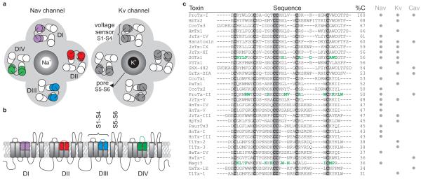
a. Cartoon representing a top view of a Nav channel (left) and a Kv channel (right). The central Na+- or K+-selective pore is surrounded by the four voltage sensors of the four domains (DI-DIV). In the Nav channel, the paddles are not identical and are therefore colored differently. In the Kv channel, the paddles are identical and therefore have the same color. b. Cartoon of a side view of a Nav channel imbedded in a lipid membrane. Each domain (DI-DIV) consists of six transmembrane segments (S1-S6) of which S1-S4 form the voltage sensor and the S5-S6 segments of each domain come together to form the Na+-selective pore of the channel. c. Sequence alignment of spider toxins with an ICK motif and three disulfide bridges that target Nav, Kv, and/or Cav channels (indicated by grey circles). Residues that have been shown to be a part of the functionally important surfaces of SGTx1, ProTx-II, and Magi5 are indicated in green. %C = % conserved residues.
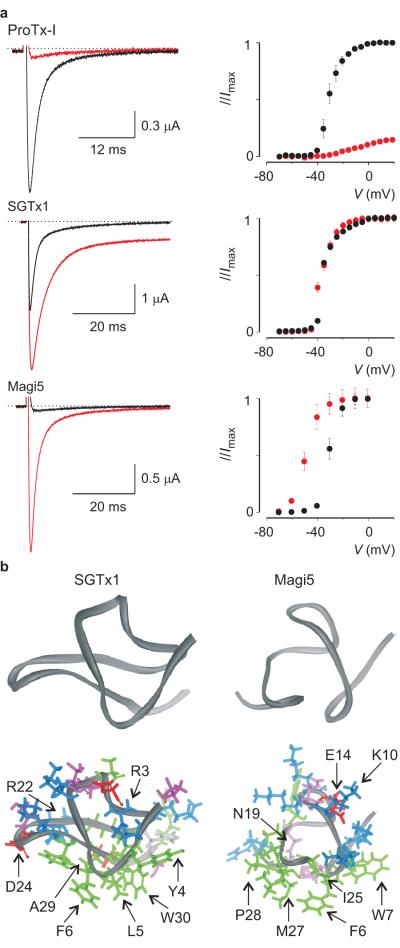
a. Effects of 100nM ProTx-I, 100nM SGTx1, and 1μM Magi5 on rNav1.2a channels expressed in Xenopus laevis oocytes and recorded with the two-electrode voltage-clamp technique. Left, sodium currents elicited by a depolarization to a suitable membrane voltage before (black) and after toxin addition (red), are shown. Right, corresponding conductance-voltage relationships are shown (n=3; error bars are s.e.m.). b. NMR solution structures of SGTx1 and Magi5. Residue coloring is as follows: blue, basic; red, acidic; green, hydrophobic; white, histidine; pink, serine/threonine/asparagine. Backbone fold is shown on top in dark grey. Images were created using DSViewer Pro and Protein Data Bank accession IDs 1LA4 for SGTx1 and 2GX1 for Magi5.
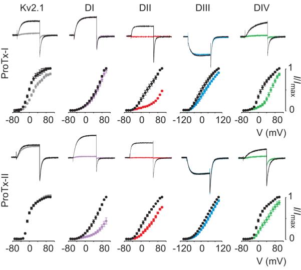
Effects of 100nM ProTx-I and ProTx-II on Kv2.1 and rNav1.2 paddle chimeras are shown where paddle motifs were transferred from each voltage-sensing domain from rNav1.2a into Kv2.1. For each toxin and construct, potassium currents were elicited by depolarizations near the foot of the voltage-activation curve (top). Currents are shown before (black) and in the presence of toxin (colored). Normalized tail current voltage-activation relationships are also shown (bottom), where tail current amplitude (I/Imax) is plotted against test voltage before (black) and in the presence of toxins (colored). n=3-5; error bars are s.e.m.

Ribbon representation of the X-ray structure of a paddle chimera between the Kv2.1 and Kv1.2 channel viewed from the external side of the membrane (top view) and from within the membrane (side view). The S3b-S4 paddle motif is colored blue, the pore domain (S5-S6) is colored yellow and possible lipid molecules are colored grey. Basic residues in S4 are shown as stick representations (Protein Data Bank accession ID is 2R9R). The side view of the chimeric channel shows the S1–S4 voltage-sensing domain and its interface with the pore domain together with the possible location of lipid molecules.
Similar articles
-
Spider-venom peptides that target voltage-gated sodium channels: pharmacological tools and potential therapeutic leads.Toxicon. 2012 Sep 15;60(4):478-91. doi: 10.1016/j.toxicon.2012.04.337. Epub 2012 Apr 20. Toxicon. 2012. PMID: 22543187 Review.
-
Insect-selective spider toxins targeting voltage-gated sodium channels.Toxicon. 2007 Mar 15;49(4):490-512. doi: 10.1016/j.toxicon.2006.11.027. Epub 2006 Dec 5. Toxicon. 2007. PMID: 17223149 Review.
-
Deconstructing voltage sensor function and pharmacology in sodium channels.Nature. 2008 Nov 13;456(7219):202-8. doi: 10.1038/nature07473. Nature. 2008. PMID: 19005548 Free PMC article.
-
Two tarantula peptides inhibit activation of multiple sodium channels.Biochemistry. 2002 Dec 17;41(50):14734-47. doi: 10.1021/bi026546a. Biochemistry. 2002. PMID: 12475222
-
Four novel tarantula toxins as selective modulators of voltage-gated sodium channel subtypes.Mol Pharmacol. 2006 Feb;69(2):419-29. doi: 10.1124/mol.105.015941. Epub 2005 Nov 2. Mol Pharmacol. 2006. PMID: 16267209
Cited by
-
Animal toxins influence voltage-gated sodium channel function.Handb Exp Pharmacol. 2014;221:203-29. doi: 10.1007/978-3-642-41588-3_10. Handb Exp Pharmacol. 2014. PMID: 24737238 Free PMC article. Review.
-
The tarantula toxin β/δ-TRTX-Pre1a highlights the importance of the S1-S2 voltage-sensor region for sodium channel subtype selectivity.Sci Rep. 2017 Apr 20;7(1):974. doi: 10.1038/s41598-017-01129-0. Sci Rep. 2017. PMID: 28428547 Free PMC article.
-
Optimizing Nav1.7-Targeted Analgesics: Revealing Off-Target Effects of Spider Venom-Derived Peptide Toxins and Engineering Strategies for Improvement.Adv Sci (Weinh). 2024 Nov;11(42):e2406656. doi: 10.1002/advs.202406656. Epub 2024 Sep 9. Adv Sci (Weinh). 2024. PMID: 39248322 Free PMC article.
-
Engineering Highly Potent and Selective Microproteins against Nav1.7 Sodium Channel for Treatment of Pain.J Biol Chem. 2016 Jul 1;291(27):13974-13986. doi: 10.1074/jbc.M116.725978. Epub 2016 Apr 22. J Biol Chem. 2016. PMID: 27129258 Free PMC article.
-
µ-Theraphotoxin Pn3a inhibition of CaV3.3 channels reveals a novel isoform-selective drug binding site.Elife. 2022 Jul 20;11:e74040. doi: 10.7554/eLife.74040. Elife. 2022. PMID: 35858123 Free PMC article.
References
-
- Catterall WA. From ionic currents to molecular mechanisms: the structure and function of voltage-gated sodium channels. Neuron. 2000;26:13–25. - PubMed
-
- Hille B. Ion channels of excitable membranes. Sinauer Associates, Inc.; 2001.
-
- Cannon SC. Pathomechanisms in Channelopathies of Skeletal Muscle and Brain. Annu Rev Neurosci. 2006;29:387–415. - PubMed
-
- Goldin AL. Resurgence of sodium channel research. Annu Rev Physiol. 2001;63:871–894. - PubMed
Publication types
MeSH terms
Substances
Grants and funding
LinkOut - more resources
Full Text Sources
Other Literature Sources
