Review
. 2010 Mar 2;9(3):237-49.
doi: 10.1016/j.dnarep.2010.01.008. Epub 2010 Jan 25.

Unwinding the functions of the Pif1 family helicases

Affiliations
Review

Unwinding the functions of the Pif1 family helicases

Matthew L Bochman et al. DNA Repair (Amst). .

Abstract

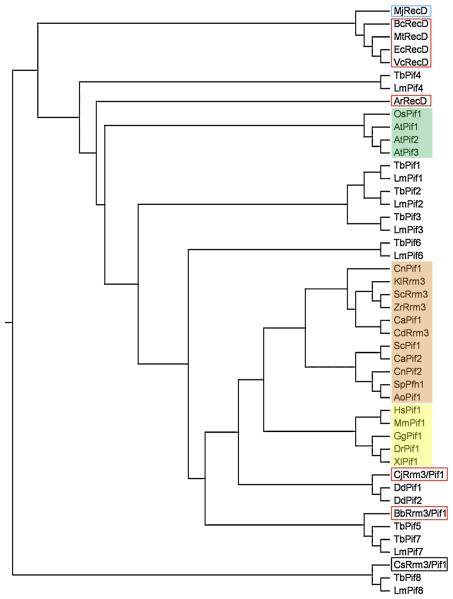
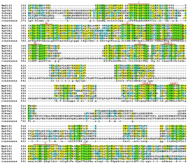
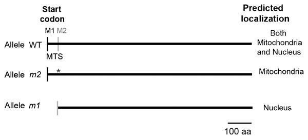
Helicases are ubiquitous enzymes found in all organisms that are necessary for all (or virtually all) aspects of nucleic acid metabolism. The Pif1 helicase family is a group of 5'-->3' directed, ATP-dependent, super family IB helicases found in nearly all eukaryotes. Here, we review the discovery, evolution, and what is currently known about these enzymes in Saccharomyces cerevisiae (ScPif1 and ScRrm3), Schizosaccharomyces pombe (SpPfh1), Trypanosoma brucei (TbPIF1, 2, 5, and 8), mice (mPif1), and humans (hPif1). Pif1 helicases variously affect telomeric, ribosomal, and mitochondrial DNA replication, as well as Okazaki fragment maturation, and in at least some cases affect these processes by using their helicase activity to disrupt stable nucleoprotein complexes. While the functions of these enzymes vary within and between organisms, it is evident that Pif1 family helicases are crucial for both nuclear and mitochondrial genome maintenance.


Figures

Figure 1
Figure 1
Evolutionary relationship among Pif1 family and RecD helicases. The indicated sequences were aligned using ClustalX [88], and the phylogenetic relationship among them was drawn as a rooted tree (using the unrelated human beta actin protein (NP_001092) as an outgroup (not shown) with TreeView v. 1.6.6. software (http://taxonomy.zoology.gla.ac.uk/rod/rod.html). Prokaryotic proteins are outlined in red, and the archaeal protein is outlined in blue. Fungal proteins are shaded pink, plant proteins are shaded green, and metazoan proteins are shaded yellow. Proteins from the following organisms were aligned: Agrobacterium radiobacter (Ar), Arabidopsis thaliana (At), Aspergillus oryzae (Ao), Bacillus cereus (Bc), Bdellovibrio bacteriovorus (Bb), Campylobacter jejuni (Cj), Candida albicans (Ca), Candida dubliensis (Cd), Clostridium sporogenes (Cs), Cryptococcus neoformans (Cn), Danio rerio (Dr), Dictyostelium discoidium (Dd), Escherichia coli (Ec), Gallus gallus (Gg), Homo sapiens (Hs), Kluveromyces lactis (Kl), Leishmania major (Lm), Methanocaldococcus jannaschii (Mj), Mus musculus (Mm), Mycobacterium tuberculosis (Mt), Oryza sativa (Os), Saccharomyces cerevisiae (Sc), Schizosaccharomyces pombe (Sp), Trypanosoma brucei (Tb), Xenopus laevis (Xl), Vibrio cholerae (Vc), and Zygosaccharomyces rouxii (Zr). The GenBank accession numbers are as follows: AoPif1, XP_001824182; ArRecD, YP_002544895; AtPif1, CAB91581; AtPif2, NP_190738; AtPif3, CAB63155; BbRrm3/Pif1, BcRecD, YP_085716; CaPif1, XP_718694; CaPif2, XP_712340; CdRrm3, XP_002421612; CjRrm3/Pif1, YP_002344343.1; CnPif1, XP_572423; CnPif2, XP_569577; CsRrm3/Pif1, ZP_02995968.1; DdPif1, XP_642006; DdPif2, XP_647539; DrPif1, NP_942102; EcRecD, AAB40466.1; GgPif1, XP_426648; HsPif1, NP_079325; KlRrm3, XP_453658; LmPif1, XP_001681501; LmPif2, XP_001681500; LmPif3, XP_001684538; LmPif4, XP_001685476; LmPif6, XP_001683071; LmPif7, XP_001681013; LmPif8, XP_001684097; MjRecD, NP_248527; MmPif1, EDL26099; MtRecD, NP_215143; OsPif1, ABB47755; ScPif1, NP_013650; ScRrm3, NP_011896; SpPfh1, NP_596488; TbPif1, XP_828762; TbPif2, XP_828763; TbPif3, XP_829242; TbPif4, XP_829537; TbPif5, XP_847187; TbPif6, XP_822349; TbPif7, XP_846907; TbPif8, XP_845724; XlPif1, Q0R4F1; VcRecD, NP_231950; and ZrRrm3, XP_002498680.
Figure 2
Figure 2
Conserved motifs in the Pif1 family helicases. The sequences of the Hs-, Mm-, and ScPif1, ScRrm3, SpPfh1, and TbPif5 helicases used to generate Figure 1 were aligned using ClustalW [88], and the BOXSHADE program in the Biology WorkBench suite (http://workbench.sdsc.edu) was used to color-code conserved residues. The completely conserved residues are in green, conserved similarities are in cyan, and identical residues are yellow; the amino acid similarity groups were defined as FYW, IVLM, RK, DE, GA, TS, and NQ. Due to spatial constraints, the divergent N- and C-termini are not shown, leaving only the highly conserved core ATPase/helicase domain. The seven conserved SFI helicase motifs (red Roman numerals; [89]) and three additional motifs with high homology to E. coli RecD (red A, B, and C; [90]) are shown. A putative Pif1 family signature motif is indicated with the dashed red box (see Section 2. for details).
Figure 3
Figure 3
Mitochondrial and nuclear isoforms of ScPif1. Schematic of the wild type, mitochondrial (m2), and nuclear (m1) PIF1 alleles with the predicted localization in the cell. M1 denotes the position of the first AUG site, and M2 marks the position of the second AUG site. Mutated M2 to alanine is represented with an asterisk. The picture is drawn to scale. MTS, mitochondrial target signal: aa, amino acids.
Figure 4
Figure 4
Replication fork movement through the tRNAY gene. Top) Cartoon of the 2D gel technique: 1N, non-replicating fragment; 2N, the nearly fully replicated fragment before sister chromatids separate; P, replication pause; BU, bubble-shaped replication intermediates. Bottom) Southern blots were probed to detect the tRNAY gene (tY[GUA]F1; EcoRV fragment, YFR012W). These images are reproduced from [58] and are reprinted following the guidelines of Cell Press's Authors' Rights statement.
Figure 5
Figure 5
SpPfh1 is detected in both the nucleus and mitochondria. Wild type cells (untagged Pfh1) and cells expressing Pfh1 fused with GFP at the C-terminus (Pfh1-GFP) are viewed by phase contrast and fluorescence microscopy. Pfh1 is visualized by GFP (green), DNA by Hoechst (blue), and mitochondria by mitotracker (red). The white arrow points out concentrated Pfh1-GFP in the nucleolus. The scale bar indicates 10 μm. This figure is adapted from the journal of Molecular and Cellular Biology, copyright © American Society for Microbiology [Molecular and Cellular Biology , Vol 28, 2008, p.6598, doi:10.1128/MCB.00191-08] [72].
Figure 6
Figure 6
The kinetoplast and kDNA. A) In the kinetoplast, the network of maxicircles and minicircles is located near the flagellar basal body. While little is known about maxicircle replication [76], research shows that minicircles (black ovals) detach from the kDNA disk and migrate to the KFZ where they replicate unidirectionally via θ-type replication. Daughter minicircles then move to the antipodal sites where replication continues (including Okazaki fragment maturation) [76]. TbPIF1 (B. Liu and P. Englund, unpublished data) and 5 [79] are located in the antipodal sites, and TbPIF8 localizes to the kDNA disk (B. Liu and P. Englund, unpublished data). Highly expressed GFP-tagged TbPIF2 (not shown) was found throughout the kinetoplast [79]. This figure is based on Figure 3 from [76]. B) Electron micrograph of part of a kDNA network from the kinetoplastid Crithidia fasciculata. C) Topoisomerase II decatenation yields 2.5 kb minicircles and 38 kb maxicircles (left, middle). Micrographs are at approximately the same magnification. The images in B) and C) and legends are adapted from [91] with kind permission of Springer Science+Business Media.

Similar articles

Cited by

References

    1. Foury F, Kolodynski J. pif mutation blocks recombination between mitochondrial rho+ and rho− genomes having tandemly arrayed repeat units in Saccharomyces cerevisiae. Proc Natl Acad Sci. 1983;80:5345–5349. - PMC - PubMed
    1. Lahaye A, Stahl H, Thines-Sempoux D, Foury F. PIF1: a DNA helicase in yeast mitochondria. EMBO J. 1991;10:997–1007. - PMC - PubMed
    1. Schulz VP, Zakian VA. The Saccharomyces PIF1 DNA helicase inhibits telomere elongation and de novo telomere formation. Cell. 1994;76:145–155. - PubMed
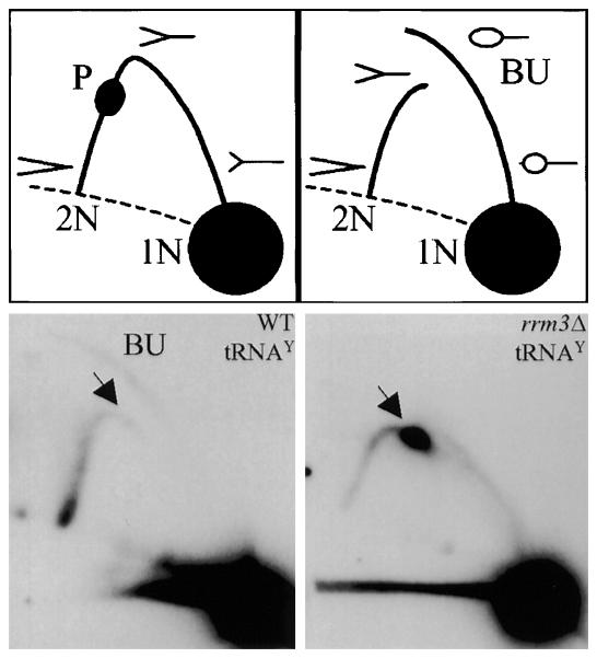
    1. Ivessa AS, Zhou J-Q, Zakian VA. The Saccharomyces Pif1p DNA helicase and the highly related Rrm3p have opposite effects on replication fork progression in ribosomal DNA. Cell. 2000;100:479–489. - PubMed
    1. Keil RL, McWilliams AD. A gene with specific and global effects on recombination of sequences from tandemly repeated genes in Saccharomyces cerevisiae. Genetics. 1993;135:711–718. - PMC - PubMed

Publication types

LinkOut - more resources