. 2010 Mar 26;285(13):9346-9356.
doi: 10.1074/jbc.M109.095505. Epub 2010 Jan 22.

Molecular identification of carnosine synthase as ATP-grasp domain-containing protein 1 (ATPGD1)

Affiliations

Molecular identification of carnosine synthase as ATP-grasp domain-containing protein 1 (ATPGD1)

Jakub Drozak et al. J Biol Chem. .

Abstract

Carnosine (beta-alanyl-L-histidine) and homocarnosine (gamma-aminobutyryl-L-histidine) are abundant dipeptides in skeletal muscle and brain of most vertebrates and some invertebrates. The formation of both compounds is catalyzed by carnosine synthase, which is thought to convert ATP to AMP and inorganic pyrophosphate, and whose molecular identity is unknown. In the present work, we have purified carnosine synthase from chicken pectoral muscle about 1500-fold until only two major polypeptides of 100 and 90 kDa were present in the preparation. Mass spectrometry analysis of these polypeptides did not yield any meaningful candidate. Carnosine formation catalyzed by the purified enzyme was accompanied by a stoichiometric formation, not of AMP, but of ADP, suggesting that carnosine synthase belongs to the "ATP-grasp family" of ligases. A data base mining approach identified ATPGD1 as a likely candidate. As this protein was absent from chicken protein data bases, we reconstituted its sequence from a PCR-amplified cDNA and found it to fit with the 100-kDa polypeptide of the chicken carnosine synthase preparation. Mouse and human ATPGD1 were expressed in HEK293T cells, purified to homogeneity, and shown to catalyze the formation of carnosine, as confirmed by mass spectrometry, and of homocarnosine. Specificity studies carried out on all three enzymes were in agreement with published data. In particular, they acted with 15-25-fold higher catalytic efficiencies on beta-alanine than on gamma-aminobutyrate. The identification of the gene encoding carnosine synthase will help for a better understanding of the biological functions of carnosine and related dipeptides, which still remain largely unknown.


Figures
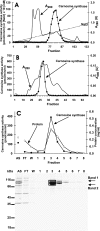
FIGURE 1.
FIGURE 1.
Purification of chicken carnosine synthase to near homogeneity. Chicken carnosine synthase was purified by chromatography on DEAE-Sepharose (A), Q-Sepharose (not shown), Superdex 200 (B), and ATP-Sepharose (C) as described under “Experimental Procedures.” Fractions were tested for carnosine synthase activity and protein concentration was determined with the Bradford assay. The indicated fractions of the ATP-Sepharose column were analyzed by SDS-PAGE and the gel was stained with Coomassie Blue. AS, applied sample; FT, flow through; W, wash. Fractions 1 and 2 were eluted with 100 mm NaCl, whereas the others were eluted with 5 mm MgATP. The indicated bands were cut out of the gel, submitted to trypsin digestion, and analyzed by mass spectrometry.
FIGURE 2.
FIGURE 2.
Time course of the changes in carnosine, ADP, and AMP concentration during carnosine synthesis. A chicken enzyme preparation (11 μg of protein) purified by chromatography on DEAE-Sepharose, Q-Sepharose, and Superdex 200 was incubated for 0, 60, and 120 min with 1 mm MgATP in the absence (empty symbols) or presence (filled symbols) of 3 mm l-histidine, 1 mm β-alanine, as well as 750 × 103 cpm of [3H]β-alanine. ΔADP values were calculated by subtracting ADP concentration values in the absence of β-alanine and l-histidine from the corresponding values in the presence of these substrates. The formation of radiolabeled carnosine was determined after chromatographic separation from β-alanine. ADP and AMP were determined by HPLC. The figure shows the mean of two experiments performed under similar conditions and yielding similar results (less than 5% variation).
FIGURE 3.
FIGURE 3.
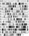
Amino acid sequence alignment of human ATPGD1 with its oyster, mouse, and chicken orthologues. Fully conserved residues are highlighted with a black background. The human (GenBank accession number NP_001159694) and mouse sequences (GenBank accession number NP_598909) have been confirmed by PCR amplification of the cDNA and sequencing. The chicken sequence has been determined in the present work (GenBank accession number GU453679). The oyster (C. virginica) sequence was reconstituted from ESTs (GenBank accession number BK007044). The peptides identified by mass spectrometry in the protein purified from chicken pectoral muscle are underlined in the chicken sequence.
FIGURE 4.
FIGURE 4.
Mass spectra of a dipeptide produced by mouse ATPGD1. Homogenous recombinant mouse ATPGD1 was incubated for 18 h with 3 mm β-alanine and 3 mm l-histidine in the absence or presence of 3 mm MgATP. The produced dipeptide was purified and submitted to mass spectrometry either as such or after acetylation with acetic anhydride. Tandem mass spectra of the putative carnosine (A) and N-acetylated carnosine (B) were acquired. The identity of the carnosine fragment ion at m/z 180 was [M + H − H2O − HCO]+.

Similar articles

Cited by

References

    1. Gulewitch W., Admiradzibi S. (1900) Ber. Dtsch. Chem. Ges. 33, 1902–1903
    1. Crush K. G. (1970) Comp. Biochem. Physiol. 34, 3–30 - PubMed
    1. Cameron J. N. (1989) J. Exp. Biol. 143, 543–548
    1. Boldyrev A. A. (2007) Carnosine and Oxidative Stress in Cells and Tissues, Nova Scientific Publisher, New York
    1. Kish S. J., Perry T. L., Hansen S. (1979) J. Neurochem. 32, 1629–1636 - PubMed

Publication types

Associated data