. 2010 Mar 19;285(12):8515-26.
doi: 10.1074/jbc.M109.079079. Epub 2010 Jan 22.

Loss of function of ATXN1 increases amyloid beta-protein levels by potentiating beta-secretase processing of beta-amyloid precursor protein

Affiliations

Loss of function of ATXN1 increases amyloid beta-protein levels by potentiating beta-secretase processing of beta-amyloid precursor protein

Can Zhang et al. J Biol Chem. .

Abstract

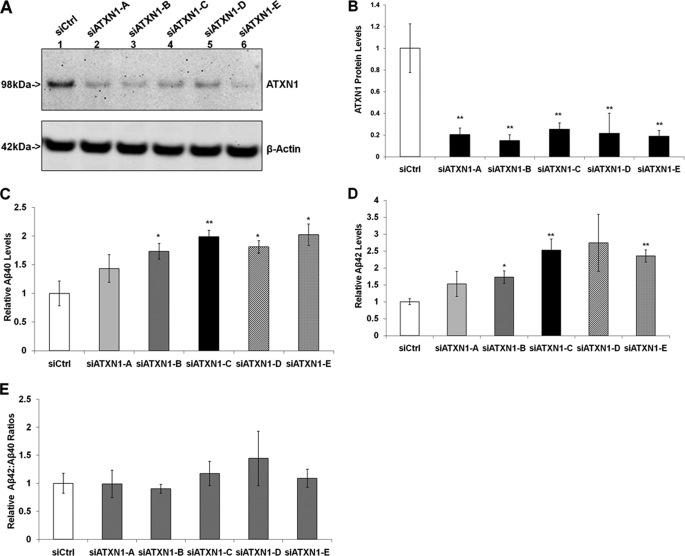
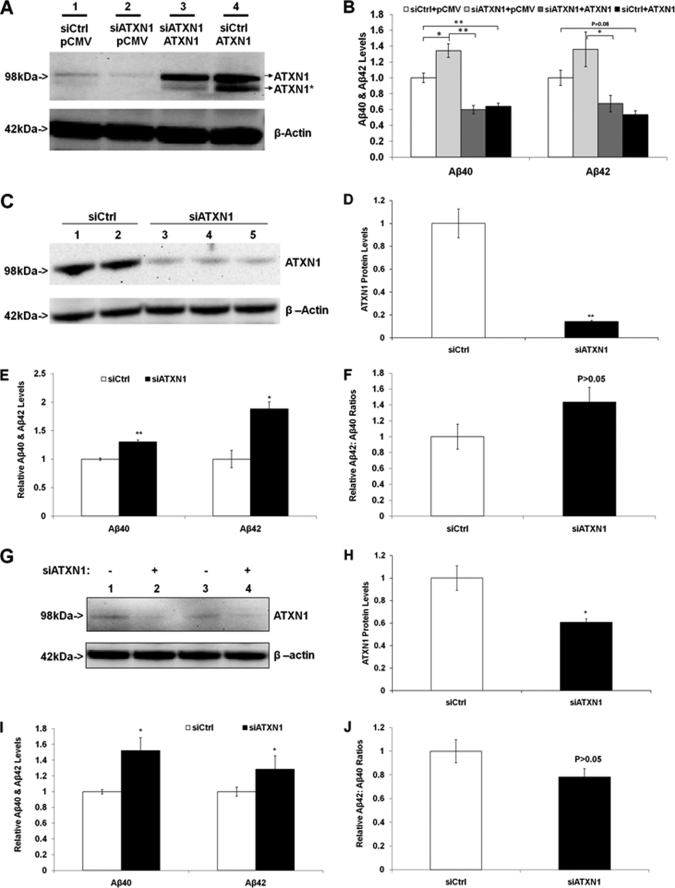
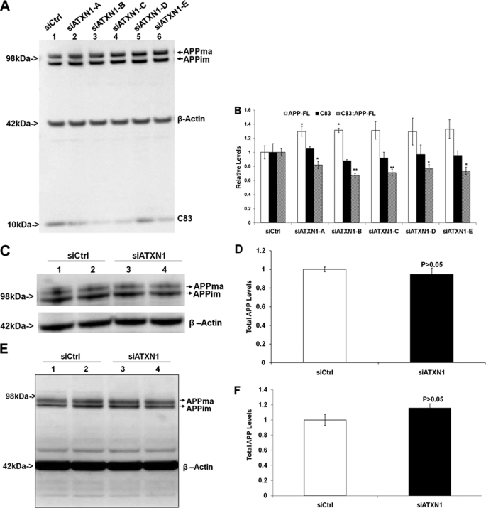
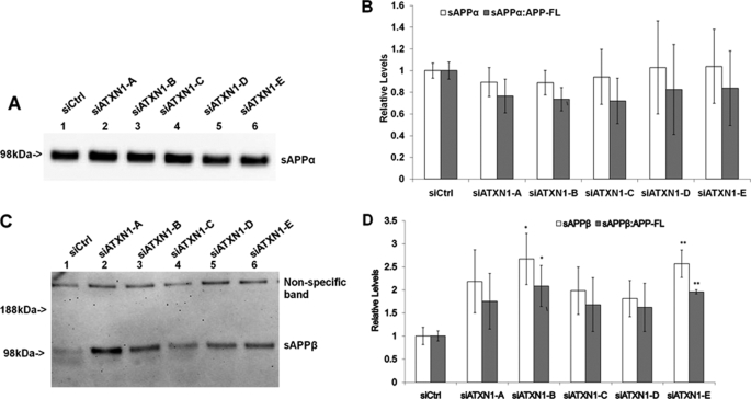
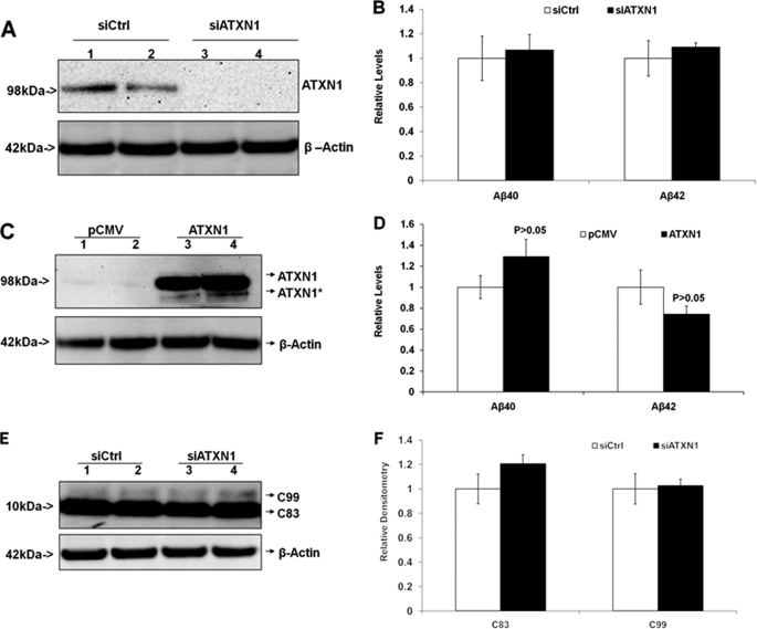
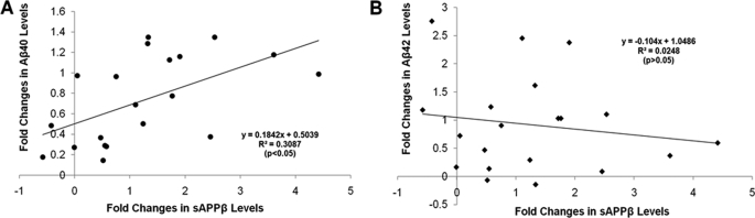
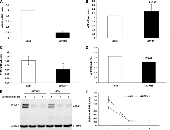
Alzheimer disease (AD) is a devastating neurodegenerative disease with complex and strong genetic inheritance. Four genes have been established to either cause familial early onset AD (APP, PSEN1, and PSEN2) or to increase susceptibility for late onset AD (APOE). To date approximately 80% of the late onset AD genetic variance remains elusive. Recently our genome-wide association screen identified four novel late onset AD candidate genes. Ataxin 1 (ATXN1) is one of these four AD candidate genes and has been indicated to be the disease gene for spinocerebellar ataxia type 1, which is also a neurodegenerative disease. Mounting evidence suggests that the excessive accumulation of Abeta, the proteolytic product of beta-amyloid precursor protein (APP), is the primary AD pathological event. In this study, we ask whether ATXN1 may lead to AD pathogenesis by affecting Abeta and APP processing utilizing RNA interference in a human neuronal cell model and mouse primary cortical neurons. We show that knock-down of ATXN1 significantly increases the levels of both Abeta40 and Abeta42. This effect could be rescued with concurrent overexpression of ATXN1. Moreover, overexpression of ATXN1 decreased Abeta levels. Regarding the underlying molecular mechanism, we show that the effect of ATXN1 expression on Abeta levels is modulated via beta-secretase cleavage of APP. Taken together, ATXN1 functions as a genetic risk modifier that contributes to AD pathogenesis through a loss-of-function mechanism by regulating beta-secretase cleavage of APP and Abeta levels.


Figures

FIGURE 1.
FIGURE 1.
Down-regulation of ATXN1 significantly increases Aβ40 and Aβ42 levels in H4-APP751 cells. Stable H4-APP751 cells were transiently transfected with control siRNA (siCtrl) or different ATXN1 siRNA constructs (siATXN1) and harvested 48 h post-transfection. Cell lysates were subjected to Western blotting analysis to assess ATXN1 protein, and conditioned medium was applied to ELISA analysis to measure Aβ40 and Aβ42 levels as described under “Experimental Procedures.” A and B, quantitative Western blotting analysis showed that all ATXN1 siRNA treatments significantly decreased ATXN1 protein levels compared with control siRNA treatment. C, ATXN1 siRNA treatment increased Aβ40 levels compared with control siRNA treatment. D, ATXN1 siRNA treatment increased the Aβ42 levels compared with control siRNA treatment. E, ATXN1 siRNA treatment did not alter the ratios of Aβ42:Aβ40 compared with control siRNA treatment (p > 0.05). n = 4 for each experiment group; mean ± S.E.; *, p < 0.05; **, p < 0.01 versus siCtrl.
FIGURE 2.
FIGURE 2.
Validation of ATXN1 loss-of-function effect on Aβ levels: it can be rescued by ATXN1 overexpression in H4-APP751 cells; and additionally ATXN1 siRNA elevated Aβ levels in naive H4 cells and mouse primary cortical neurons. A, H4-APP751 cells were transfected with control siRNA (siCtrl) and/or ATXN1 siRNA (siATXN1), as well as the empty vector (pCMV) and/or ATXN1-cDNA and applied to Western blotting analysis as described under “Experimental Procedures.” B, samples in A were applied to ELISA as described under “Experimental Procedures.” ATXN1 cDNA not only decreased Aβ40 and Aβ42 levels (comparing siCtrl/pCMV and siCtrl/ATXN1), but also rescued the ATXN1 siRNA effect on Aβ40 and Aβ42 levels (comparing siATXN1/pCMV and siATXN1/ATXN1). C–F, naive H4 cells were transfected with control siRNA (siCtrl) or ATXN1 siRNA (siATXN1) and applied to Western blotting analysis or ELISA as described under “Experimental Procedures.” ATXN1 siRNA treatment significantly decreased ATXN1 protein levels (C and D), as well as increased both Aβ40 and Aβ42 levels (E), but did not significantly change the ratios of Aβ42:Aβ40 (F). G–J, mouse primary cortical neurons were transfected with control siRNA or ATXN1 siRNA and applied to Western blotting analysis or ELISA as described under “Experimental Procedures.” ATXN1 siRNA treatment significantly decreased ATXN1 protein levels (G and H), and increased both Aβ40 and Aβ42 levels (I), but did not significantly change the ratios of Aβ42:Aβ40 (J) compared with control siRNA treatment. n = 3 for each experimental group; mean ± S.E.; *, p < 0.05; **, p < 0.01 versus control.
FIGURE 3.
FIGURE 3.
Down-regulation of ATXN1 alters APP processing activity. A, H4-APP751 cells were transfected with ATXN1 siRNA or control siRNA (siCtrl) and harvested 48 h post-transfection. Cell lysates were applied to Western blotting analysis as described under “Experimental Procedures.” B, graphic representation of data from A as described under “Experimental Procedures.” The ATXN1 siRNAs (siATXN1-C, -D, and -E) did not change full-length APP levels (p > 0.05), whereas the ATXN1 siRNA (siATXN1-A and -B) modestly increased full-length APP levels (p < 0.05). The ATXN1 siRNA treatment did not alter C83 levels alone, but decreased the ratio of C83:full-length APP compared with control siRNA treatment (n = 4). C, naive H4 cells treated with ATXN1 siRNA or control siRNA were applied to Western blotting analysis as described under “Experimental Procedures.” D, graphic representation of data from C as described under “Experimental Procedures.” ATXN1 siRNA treatment did not significantly change full-length APP levels (n = 3). E, mouse primary cortical neurons were treated with ATXN1 siRNA or control siRNA and applied to Western blotting analysis as described under “Experimental Procedures.” F, graphic representation of data from E. ATXN1 siRNA treatment did not significantly change full-length APP levels. n = 3 for each experimental group; mean ± S.E.; *, p < 0.05; **, p < 0.01 versus control.
FIGURE 4.
FIGURE 4.
Knock-down of ATXN1 potentiates β-secretase processing of APP. A, H4-APP751 cells were transfected with different ATXN1 siRNAs and control siRNA (siCtrl) and harvested 48 h post-transfection. Conditioned medium was applied to Western blotting analysis as described under “Experimental Procedures.” B, graphic representation of data from A. The sAPPα levels were divided by full-length APP levels from the same samples to represent the ratio of sAPPα:APP-FL. ATXN1 siRNA treatment did not change the sAPPα levels (p > 0.05 versus control). ATXN1 siRNA treatment did not significantly alter the ratio of sAPPα to full-length APP (p > 0.05 versus control). C, H4-APP751 cells were transfected with different ATXN1 siRNAs and control siRNA and harvested 48 h post-transfection. The sAPPβ-specific antibody was used to detect sAPPβ in the conditioned medium. D, graphic representation of data from C. The sAPPβ levels were compared with full-length APP levels from the same samples. ATXN1 siRNA treatment elevated both the sAPPβ levels and the ratio of sAPPβ to full-length APP. n = 4 for each experimental group; mean ± S.E.; *, p < 0.05; **, p < 0.01 versus corresponding controls.
FIGURE 5.
FIGURE 5.
Modulation of ATXN1 levels does not alter APP processing or Aβ levels in H4-APP-C99 cells. A and B, H4-APP-C99 cells were transfected with ATXN1 siRNA or control siRNA (siCrtl) and harvested 48 h post-transfection. Cell lysates and conditioned medium were applied to Western blotting analysis and ELISA, respectively, as described under “Experimental Procedures.” ATXN1 siRNA treatment markedly decreased the ATXN1 protein level. B, there were no differences in Aβ40 or Aβ42 levels between the cells treated with ATXN1 siRNA and those treated with control siRNA. C and D, H4-APP-C99 cells were transfected with ATXN1-cDNA or pCMV empty vector and harvested 48 h post-transfection. Cell lysates and conditioned medium were applied to Western blotting analysis and ELISA. C, ATXN1-cDNA treatment markedly increased ATXN1 protein levels. D, ATXN1-cDNA did not change either Aβ40 or Aβ42 levels (p > 0.05; versus control). E and F, cell lysates from A (H4-APP-C99 cells treated with ATXN1 or control siRNAs) were applied to Western blotting analysis, and probed with APP8717 and β-actin antibodies. F, graphic representation of data from E. There existed no significant differences in the levels of APP-C83 or APP-C99. n = 3 for each experimental groups; mean ± S.E.; *, p < 0.05; **, p < 0.01 versus corresponding controls.
FIGURE 6.
FIGURE 6.
Alteration in sAPPβ levels significantly correlates with changes in Aβ40 levels, but not Aβ42 levels. Levels of sAPPβ were plotted together with levels of Aβ40 and Aβ42 from the same samples in previous experiments. The data were represented as a percentage change by comparing the siATXN1-treated samples to control. The x axis was represented by the percentage change in sAPPβ levels, and the y axis represented by the percentage change in Aβ40 or Aβ42 levels. The line represents the linear regression for this data. There was a significant correlation between the changes in sAPPβ levels and the changes in Aβ40 levels (p < 0.05), but not Aβ42 levels (p > 0.05).
FIGURE 7.
FIGURE 7.
Knock-down of ATXN1 does not alter APP mRNA levels or protein turnover rate. A, naive H4 cells were transfected with ATXN1 siRNA or control siRNA. Cells were harvested 48 h post-transfection and applied to quantitative PCR analysis. ATXN1 siRNA treatment significantly decreased ATXN1 mRNA levels. B, APP mRNA levels in the same samples from A. ATXN1 siRNA treatment did not significantly alter APP mRNA levels. C, mouse primary cortical neurons were transfected with ATXN1 siRNA or control siRNA as described under “Experimental Procedures.” Cells were harvested 72 h post-transfection and applied to quantitative PCR analysis. ATXN1 siRNA treatment significantly decreased ATXN1 mRNA levels. D, APP mRNA levels in the same samples from C. ATXN1 siRNA treatment did not significantly alter APP mRNA levels (p > 0.05). E, H4-APP751 cells were transfected with ATXN1 siRNA or control siRNA for 42 h, and then treated with 40 μg/ml of cycloheximide for a different time period (0, 3, or 6 h). Cells were harvested and cell lysates were subjected to Western blotting analysis as described under “Experimental Procedures.” F, graphic representation of data from E. There existed no significant differences in the protein levels of full-length APP at 3 and 6 h of cycloheximide treatment. n ≥ 3 for each experimental group; mean ± S.E.; *, p < 0.05; **, p < 0.01 versus corresponding controls.

Similar articles

Cited by

References

    1. Cummings J. L. (2004) N. Engl. J. Med. 351, 56–67 - PubMed
    1. Hardy J., Selkoe D. J. (2002) Science 297, 353–356 - PubMed
    1. Bertram L., Tanzi R. E. (2008) Nat. Rev. Neurosci. 9, 768–778 - PubMed
    1. Gandy S. (2005) J. Clin. Invest. 115, 1121–1129 - PMC - PubMed
    1. Tanzi R. E., Bertram L. (2005) Cell 120, 545–555 - PubMed

Publication types

MeSH terms