. 2010 Apr;77(4):621-32.
doi: 10.1124/mol.109.062174. Epub 2010 Jan 22.

Environmental neurotoxic pesticide increases histone acetylation to promote apoptosis in dopaminergic neuronal cells: relevance to epigenetic mechanisms of neurodegeneration

Affiliations

Environmental neurotoxic pesticide increases histone acetylation to promote apoptosis in dopaminergic neuronal cells: relevance to epigenetic mechanisms of neurodegeneration

C Song et al. Mol Pharmacol. 2010 Apr.

Abstract

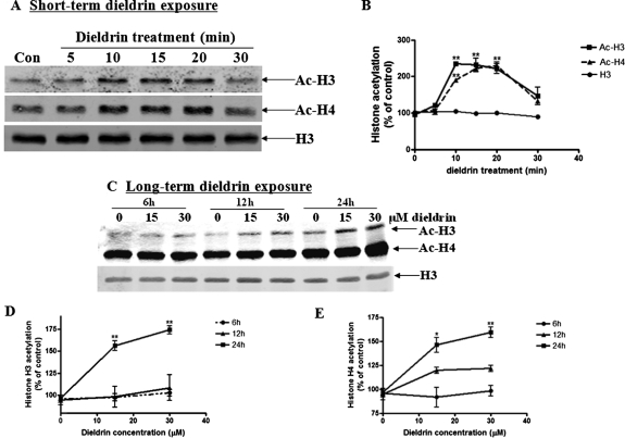
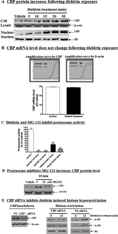
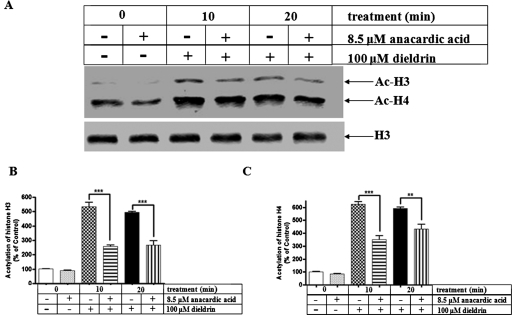
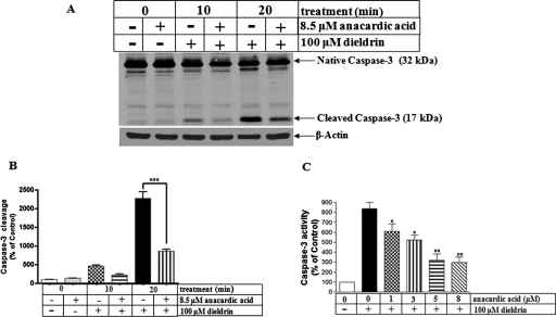
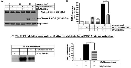
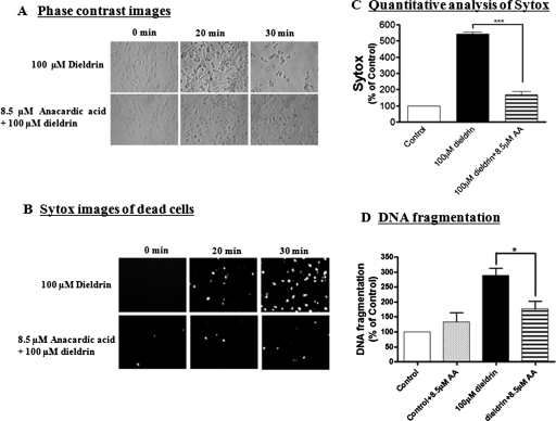
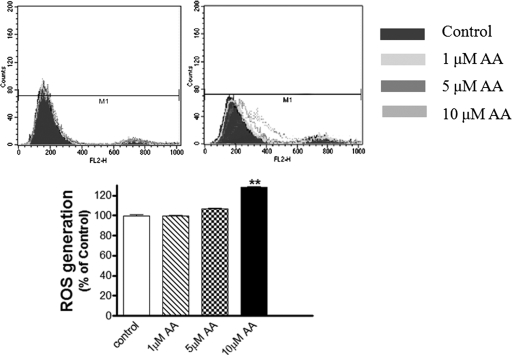
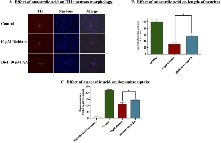
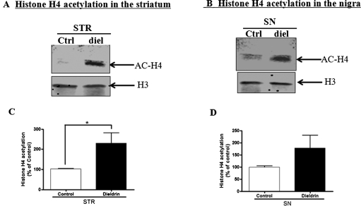
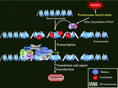
Pesticide exposure has been implicated in the etiopathogenesis of Parkinson's disease (PD); in particular, the organochlorine insecticide dieldrin is believed to be associated with PD. Emerging evidence indicates that histone modifications play a critical role in cell death. In this study, we examined the effects of dieldrin treatment on histone acetylation and its role in dieldrin-induced apoptotic cell death in dopaminergic neuronal cells. In mesencephalic dopaminergic neuronal cells, dieldrin induced a time-dependent increase in the acetylation of core histones H3 and H4. Histone acetylation occurred within 10 min of dieldrin exposure indicating that acetylation is an early event in dieldrin neurotoxicity. The hyperacetylation was attributed to dieldrin-induced proteasomal dysfunction, resulting in accumulation of a key histone acetyltransferase (HAT), cAMP response element-binding protein. The novel HAT inhibitor anacardic acid significantly attenuated dieldrin-induced histone acetylation, Protein kinase C delta proteolytic activation and DNA fragmentation in dopaminergic cells protected against dopaminergic neuronal degeneration in primary mesencephalic neuronal cultures. Furthermore, 30-day exposure of dieldrin in mouse models induced histone hyperacetylation in the striatum and substantia nigra. For the first time, our results collectively demonstrate that exposure to the neurotoxic pesticide dieldrin induces acetylation of core histones because of proteasomal dysfunction and that hyperacetylation plays a key role in dopaminergic neuronal degeneration after exposure of dieldrin.


Figures

Fig. 1.
Fig. 1.
Dieldrin induces acetylation of core histones H3 and H4 in a time-dependent manner in dopaminergic neuronal cells. A, N27 dopaminergic neuronal cells were exposed to 100 μM dieldrin, and then acetylation of histones H3 and H4 was monitored at various time points. Native H3 was used as an internal control. B, densitometric quantification of acetylated H3 band and H4 band in A. Statistical significance between the control group and each treatment group was determined by ANOVA, p < 0.01. C, N27 dopaminergic neuronal cells were exposed to dieldrin at the physical concentration (30 μM), and then acetylation of histones H3 and H4 was also monitored at various time points. Native H3 was used as an internal control. Densitometric quantification of acetylated H3 band (D) and acetylated H4 band (E) of C. Statistical significance between the control group and each treatment group was determined by ANOVA, *, p < 0.05; **, p < 0.01.
Fig. 2.
Fig. 2.
Dieldrin increases HAT CBP levels in dopaminergic cells via inhibition of ubiquitin proteasome function. CBP protein level increases during dieldrin treatment. A, N27 cells were treated with 100 μM dieldrin for 5, 10, 15, 20, and 30 min and then cell lysates and nuclear fractions were prepared as described under Materials and Methods. The CBP levels in lysates and nuclear fractions were measured by Western blot using anti-CBP antibody. Native H3 and β-actin were used as the loading control. B, CBP mRNA level does not change after dieldrin exposure. N27 cells were treated with 100 μM dieldrin for 30 min, and then mRNA was extracted. CBP mRNA level was performed by relative quantitative real-time-polymerase chain reaction with CBP specific primers. C, Dieldrin inhibits proteasome activity in dopaminergic N27 cells. Cells were treated with 100 μM dieldrin and then chymotrypsin-like proteasome activity was measured at 10 and 20 min after exposure using fluorogenic substrate. The proteasome inhibitor MG-132 was used as a positive control. All the data represent the mean ± S.E.M. for three samples in each group. Asterisks (***, p < 0.001, Student's t test) indicate statistically significant differences compared with vehicle-treated N27 cells. D, proteasome inhibitor MG-132 increases CBP protein level. N27 cells were treated with 5 or 10 μM MG-132 for 20 min, and the level of CBP was examined by Western blot using anti-CBP antibody. The membrane was reprobed with anti-β-actin antibody to show equal loading. E, CBP-specific siRNA inhibits dieldrin-induced histone hyperacetylation. N27 cells were transfected with CBP-specific siRNA or nonspecific (NS) siRNA for 24 h. After transfection, cells were treated with 100 μM dieldrin for 10 and 20 min. Histones were then extracted and acetylation level was examined by Western blot with anti-acetyl-lysine antibody.
Fig. 3.
Fig. 3.
HAT inhibitor anacardic acid attenuates dieldrin-induced H3 and H4 acetylation. A, N27 dopaminergic cells were pretreated with 8.5 μM anacardic acid for 1 h and then exposed to 100 μM dieldrin. H3 and H4 acetylation was measured from the nuclear histone extract by Western blot. Native H3 was used as an internal control. Densitometric quantification of acetylated H3 band (B) and acetylated H4 band (C). Statistical significance between the dieldrin exposed groups, with or without anacardic acid pretreatment, was determined by ANOVA, **, p < 0.01; ***, p < 0.001.
Fig. 4.
Fig. 4.
Anacardic acid attenuates dieldrin-induced caspase-3 proteolytic activation. A, N27 cells were pretreated with 8.5 μM anacardic acid for 1 h and then exposed to 100 μM dieldrin. Caspase-3 activation was measured by cleaved caspase-3 Western blot and densitometric quantification of cleaved caspase-3 band intensity (B). Statistical significance between the dieldrin exposure groups with or without anacardic acid pretreatment was determined by ANOVA, p < 0.001. C, measurement of caspase-3 enzyme activity by fluorogenic caspase-3 substrate Ac-DEVD-AFC. Asterisks (**, P < 0.01) indicate significant differences between anacardic acid pretreated and dieldrin-alone treated cells.
Fig. 5.
Fig. 5.
The HAT inhibitor anacardic acid attenuates dieldrin-induced PKCδ proteolytic cleavage and kinase activation. A, N27 cells were pretreated with 8.5 μM anacardic acid for 1 h and then exposed to 100 μM dieldrin. PKCδ cleavage was measured in the cell lysate by immunoblotting. Equal loading of protein was demonstrated by using β-actin. B, densitometric quantification of cleaved PKCδ band statistical significance between the dieldrin-exposed groups, with or without anacardic acid pretreatment, was determined by ANOVA, p < 0.01. C, after treatment, cell lysates were collected and subjected to immunoprecipitation kinase assays as described under Materials and Methods. Phosphorylated histone bands were quantified by filmless autoradiographic analysis after scanning the dried gel, and the data are expressed as percentage of control. The values represent mean ± S.E. from two separate experiments performed in triplicate. Asterisks (**, P < 0.01) indicate significant difference compared with dieldrin-alone treated cells and anacardic acid together with dieldrin-treated N27 cells.
Fig. 6.
Fig. 6.
Anacardic acid protects against dieldrin-induced cytotoxicity and DNA fragmentation. N27 cells were pretreated with 8.5 μM anacardic acid for 1 h and then exposed to 100 μM dieldrin. Phase contrast images of N27 cells treated with dieldrin in the presence and absence of anacardic acid, B, Sytox fluorescence staining in N27 cells treated with dieldrin in the presence and absence of anacardic acid. C, Sytox green fluorescence in cells treated with dieldrin was also quantified using a microplate reader. The data represent n = 6. Asterisks (***, p < 0.001) represent significant differences between the dieldrin-treated group and the cells treated with dieldrin plus anacardic acid. D, apoptotic cell death was determined by measuring DNA fragmentation in an ELISA sandwich assay as described under Materials and Methods. Asterisks (*p < 0.05; *** p < 0.001) indicate significant difference compared with dieldrin-treated cells and dieldrin plus anacardic acid-treated cells.
Fig. 7.
Fig. 7.
Effect of anacardic acid on ROS generation. N27 dopaminergic cells were pretreated with 1 to 10 μM anacardic acid for 60 min, and then ROS was measured using hydroethidine in a flow cytometer. Top, representative flow cytometric histogram. Bottom, quantitative data. **, p < 0.01; ***, p < 0.001 compared with the control group (n = 3). **, p < 0.01; ***, p < 0.001 compared with the control group (n = 3).
Fig. 8.
Fig. 8.
Neuroprotective role of anacardic acid against dieldrin-induced dopaminergic neuronal degeneration in primary mesencephalic neuronal cultures. Mesencephalic cultures were cotreated with 10 μM dieldrin and 10 μM anacardic acid for 36 h. Immunocytochemical staining by anti-TH antibody reveals the morphology of the dopaminergic neurons, B, measurement of neurites length of TH+ neuron by Metamorph software (Molecular Devices). The data represent the mean ± S.E.M. for three samples in each group. Asterisks (*, p < 0.05, Student's t test) indicate statistically significant differences compared between dieldrin treated group and the treatment group exposed by dieldrin together with anacardic acid. C, assessment of viability of dopaminergic neurons using [3H]DA uptake assay. (*, p < 0.05).
Fig. 9.
Fig. 9.
Hyperacetylation of histones in the substantia nigra and striatum in animal model of dieldrin neurotoxicity. C57 black mice were treated with 5 mg/kg dieldrin via oral gavage every other day for 30 days. B, acetylation of histones H3 and H4 was examined in the striatum and substantia nigra by Western blot with anti-acetyl-Lys antibody. Native H3 was used as an internal control. Densitometric quantification of acetylated H4 band in the striatum (C) and substantia nigra (D) are presented. Asterisks (*, p < 0.05, Student's t test) indicate statistically significant differences between dieldrin-treated group and the vehicle control group.
Fig. 10.
Fig. 10.
Schematic representation of mechanisms underlying dieldrin-induced hyperacetylation. Exposure to neurotoxic insult dieldrin inhibits proteasome dysfunction, resulting in accumulation of a major HAT CBP. Increased CBP results in greater acetylation of nuclear histones in the chromatin, which ultimately results in alterations of gene expression associated with the neurodegenerative process, including oxidative damage and apoptosis in dopaminergic neurons. The symbols used in the scheme were taken from the SABiosciences Corporation web site.

Similar articles

Cited by

References

    1. Afeseh Ngwa H, Kanthasamy A, Anantharam V, Song C, Witte T, Houk R, Kanthasamy AG. (2009) Vanadium induces dopaminergic neurotoxicity via protein kinase Cdelta dependent oxidative signaling mechanisms: relevance to etiopathogenesis of Parkinson's disease. Toxicol Appl Pharmacol 240:273–285 - PMC - PubMed
    1. Aron JL, Parthun MR, Marcucci G, Kitada S, Mone AP, Davis ME, Shen T, Murphy T, Wickham J, Kanakry C, et al. (2003) Depsipeptide (FR901228) induces histone acetylation and inhibition of histone deacetylase in chronic lymphocytic leukemia cells concurrent with activation of caspase 8-mediated apoptosis and down-regulation of c-FLIP protein. Blood 102:652–658 - PubMed
    1. Bader N, Jung T, Grune T. (2007) The proteasome and its role in nuclear protein maintenance. Exp Gerontol 42:864–870 - PubMed
    1. Balasubramanyam K, Swaminathan V, Ranganathan A, Kundu TK. (2003) Small molecule modulators of histone acetyltransferase p300. J Biol Chem 278:19134–40 - PubMed
    1. Boutillier AL, Trinh E, Loeffler JP. (2003) Selective E2F-dependent gene transcription is controlled by histone deacetylase activity during neuronal apoptosis. J Neurochem 84:814–828 - PubMed

Publication types

MeSH terms