. 2010 Mar;76(6):1926-34.
doi: 10.1128/AEM.02295-09. Epub 2010 Jan 22.

Significance of wall structure, macromolecular composition, and surface polymers to the survival and transport of Cryptosporidium parvum oocysts

Affiliations

Significance of wall structure, macromolecular composition, and surface polymers to the survival and transport of Cryptosporidium parvum oocysts

Michael B Jenkins et al. Appl Environ Microbiol. 2010 Mar.

Erratum in

  • Appl Environ Microbiol. 2010 Aug;76(15):5334

Abstract

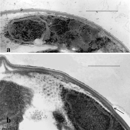
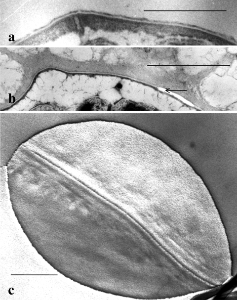
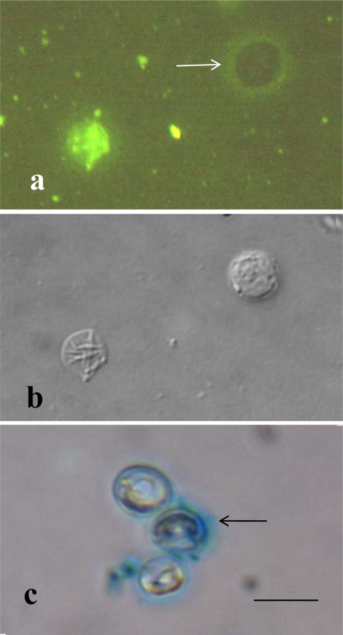
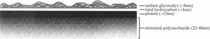
The structure and composition of the oocyst wall are primary factors determining the survival and hydrologic transport of Cryptosporidium parvum oocysts outside the host. Microscopic and biochemical analyses of whole oocysts and purified oocyst walls were undertaken to better understand the inactivation kinetics and hydrologic transport of oocysts in terrestrial and aquatic environments. Results of microscopy showed an outer electron-dense layer, a translucent middle layer, two inner electron-dense layers, and a suture structure embedded in the inner electron-dense layers. Freeze-substitution showed an expanded glycocalyx layer external to the outer bilayer, and Alcian Blue staining confirmed its presence on some but not all oocysts. Biochemical analyses of purified oocyst walls revealed carbohydrate components, medium- and long-chain fatty acids, and aliphatic hydrocarbons. Purified walls contained 7.5% total protein (by the Lowry assay), with five major bands in SDS-PAGE gels. Staining of purified oocyst walls with magnesium anilinonaphthalene-8-sulfonic acid indicated the presence of hydrophobic proteins. These structural and biochemical analyses support a model of the oocyst wall that is variably impermeable and resistant to many environmental pressures. The strength and flexibility of oocyst walls appear to depend on an inner layer of glycoprotein. The temperature-dependent permeability of oocyst walls may be associated with waxy hydrocarbons in the electron-translucent layer. The complex chemistry of these layers may explain the known acid-fast staining properties of oocysts, as well as some of the survival characteristics of oocysts in terrestrial and aquatic environments. The outer glycocalyx surface layer provides immunogenicity and attachment possibilities, and its ephemeral nature may explain the variable surface properties noted in oocyst hydrologic transport studies.


Figures

FIG. 1.
FIG. 1.
Transmission electron micrographs of thin sections of peripheral portions of glutaraldehyde-formalin-fixed C. parvum whole oocysts showing a peripheral sporozoite with a nucleus (a) and perpendicular cross sections through the oocyst wall (a and b). Note the four-layer structure of the wall and, in the outer portion of the wall, a membrane-like electron-translucent layer, which appears to be split along the axis of the translucent zone (as indicated by the arrow in panel b). Bars, 0.5 μm.
FIG. 2.
FIG. 2.
(a and b) Transmission electron micrographs of thin sections of peripheral portions of a glutaraldehyde-formalin-fixed C. parvum whole oocyst showing the wall layers with a suture complex embedded in the inner layers beneath the middle translucent layer and outer dense layer (a) and a freeze-substituted whole oocyst showing a split in the weak middle translucent layer of the wall (compare with Fig. 1b) and an extensive polymer matrix (glycocalyx) extending outward from the translucent layer of the wall (b). (c) Freeze fracture replica of a whole oocyst showing a smooth convex fracture face containing the zipper-like suture complex seen in cross section in panel a. Bars, 0.5 μm.
FIG. 3.
FIG. 3.
(a and b) Paired epifluorescence (a) and DIC (b) images of the same microscopic field treated with FITC-labeled concanavalin A. The flattened empty oocyst sac in panel b is brightly stained in panel a, indicating that concanavalin A was bound to polysaccharide material in the wall. The intact oocyst in panel b is not stained, showing that the concanavalin A did not react strongly with the outside of the oocyst. Instead, a halo of lightly stained glycocalyx material (arrow) surrounds the intact oocyst in panel a. (c) Bright-field image confirming that the oocysts stained with ethanolic Alcian Blue, which revealed a matrix of acidic polymers (arrow) surrounding some but not all oocysts. Bar, 0.5 μm.
FIG. 4.
FIG. 4.
(a) Thin-section electron micrograph of purified oocyst walls showing two parallel wall sections joined by their outer layers. Note the similarity of the purified wall profiles to those of whole oocysts in Fig. 1 and 2a. Bar, 100 nm. (b and c) Paired epifluorescence (b) and phase-contrast (c) light micrographs showing uniform staining of purified oocyst walls with Mg ANS (b) and the absence of residual bodies in the purified walls (c). Bar, 0.5 μm.
FIG. 5.
FIG. 5.
Proposed model for the C. parvum oocyst wall based on data presented in this paper.

References

    1. Abrahamsen, M. S., T. J. Templeton, S. Enomoto, J. E. Abrahante, G. Zhu. C. A. Lancto, M. Deng, C. Liu, G. Widmer, S. Tzipori, G. A. Buck, P. Xu, A. T. Bankier, P. H. Dear, B. A. Konfortov, H. F. Spriggs, L. Iyer, V. Anantharaman, L. Aravind, and V. Kapur. 2004. Complete genome sequence of the apicomplexan, Cryptosporidium parvum. Science 304:441-445. - PubMed
    1. Alugupalli, S., F. Portaels, and L. Larsson. 1994. Systematic study of the 3-hydroxy fatty acid composition of mycobacteria. J. Bacteriol. 176:2962-2969. - PMC - PubMed
    1. Anguish, L. J., and W. C. Ghiorse. 1997. Computer-assisted laser scanning and video microscopy for analysis of Cryptosporidium parvum oocysts in soil, sediment, and feces. Appl. Environ. Microbiol. 63:724-733. - PMC - PubMed
    1. Bligh, E. G., and W. J. Dyer. 1959. A rapid method of total lipid extraction and purification. Can. J. Biochem. Physiol. 37:911-917. - PubMed
    1. Brush, C. F., M. F. Walter, L. J. Anguish, and W. C. Ghiorse. 1998. Influence of pretreatment and experimental conditions on electrophoretic mobility and hydrophobicity of Cryptosporidium parvum oocysts. Appl. Environ. Microbiol. 64:4439-4445. - PMC - PubMed

Publication types

LinkOut - more resources