. 2010 Jan;22(1):234-48.
doi: 10.1105/tpc.109.066266. Epub 2010 Jan 22.

MRL1, a conserved Pentatricopeptide repeat protein, is required for stabilization of rbcL mRNA in Chlamydomonas and Arabidopsis

Affiliations

MRL1, a conserved Pentatricopeptide repeat protein, is required for stabilization of rbcL mRNA in Chlamydomonas and Arabidopsis

Xenie Johnson et al. Plant Cell. 2010 Jan.

Abstract

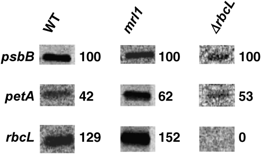
We identify and functionally characterize MRL1, a conserved nuclear-encoded regulator of the large subunit of ribulose-1,5-bisphosphate carboxylase/oxygenase. The nonphotosynthetic mrl1 mutant of Chlamydomonas reinhardtii lacks ribulose-1,5-bisphosphate carboxylase/oxygenase, and the resulting block in electron transfer is partially compensated by redirecting electrons toward molecular oxygen via the Mehler reaction. This allows continued electron flow and constitutive nonphotochemical quenching, enhancing cell survival during illumination in spite of photosystem II and photosystem I photoinhibition. The mrl1 mutant transcribes rbcL normally, but the mRNA is unstable. The molecular target of MRL1 is the 5 ' untranslated region of rbcL. MRL1 is located in the chloroplast stroma, in a high molecular mass complex. Treatment with RNase or deletion of the rbcL gene induces a shift of the complex toward lower molecular mass fractions. MRL1 is well conserved throughout the green lineage, much more so than the 10 other pentatricopeptide repeat proteins found in Chlamydomonas. Depending upon the organism, MRL1 contains 11 to 14 pentatricopeptide repeats followed by a novel MRL1-C domain. In Arabidopsis thaliana, MRL1 also acts on rbcL and is necessary for the production/stabilization of the processed transcript, presumably because it acts as a barrier to 5 ' >3 ' degradation. The Arabidopsis mrl1 mutant retains normal levels of the primary transcript and full photosynthetic capacity.


Figures

Figure 1.
Figure 1.
Phenotype of the mrl1 Mutant. (A) Fluorescence transients of Chlamydomonas wild-type and mrl1 strains in the presence or absence of DCMU (20 μ M). Inset: same traces on an expanded time scale (B) Wild-type and mrl1 cells were resuspended in water at a concentration of 104 cells mL−1 and spotted onto Petri dishes of TAP or MIN media and grown for 10 d. In the last two lines, cells were mixed with DCMU (10 μ M final concentration) before spotting onto TAP. (C) Immunoblot of total cell extracts of wild-type, mrl1, and mrl1.C (complemented mrl1 strain) strains reacted with an antibody to RbcL and RbcS (Rubisco large and small subunits) and an antibody to cytochrome f (cytf) as a control. RbcS appears as a double band (arrows) in this gel system. [See online article for color version of this figure.]
Figure 2.
Figure 2.
Total Cell RNA Hybridized with rbcL and psbA as a Control Probe. The mrl1 strain accumulates no rbcL mRNA compared with the wild type or mrl1.C. Low light is 10 μ E m−2 s−1, and high light is 200 μ E m−2 s−1.
Figure 3.
Figure 3.
The MRL1 Gene. (A) Top: Map of the MRL1 gene. Location of the aphVIII gene is indicated, together with the NheI (N) and XmaI (X) sites used in DNA gel blotting (see Supplemental Figure 1 online). Exons are shown as boxes and introns as arrowed lines. The gray bar at the 5 ′ end denotes the genomic fragment that was cloned into the EcoRI (E) site of the cDNA to generate the promoter-cDNA construct. Bottom: Map of the MRL1 protein, showing its transit peptide (TP) and four domains (with numbering of their first amino acid) and the positions of introns (ticks on lower line) and of restriction enzyme sites used in transformation experiments (M, MluI; S, SgrAI; B, BstEII). The three insertions found in the C-domain of Chlamydomonas compared with other sequences are indicated by gray shading. (B) Cladogram of MRL1 proteins. The tree was obtained with the program PhyML, based on the alignment of Supplemental Figure 2 online after truncation of the first 589 ill-aligned positions. Streptophytes appear at the bottom (Sellaginella, Physcomitrella, rice, maize, Arabidopsis, grapevine, and Populus), Prasinophytes at top left (Ostreococcus and Micromonas), and other chlorophytes at top right (Chlorella, Volvox, and Chlamydomonas). Each branch is labeled with its aLRT value.
Figure 4.
Figure 4.
Run-On Transcription Experiment. In vivo–labeled RNA from wild-type, mrl1, and ΔrbcL strains was hybridized to gene fragments separated by electrophoresis and blotted onto nitrocellulose. Numbers indicate labeling intensity, normalized to the psbB control. Specificity of the rbcL signal is indicated by its absence in the ΔrbcL strain.
Figure 5.
Figure 5.
A High Molecular Mass Ribonucleoprotein Complex Containing MRL1 Is Located in the Chloroplast Stroma in Chlamydomonas. (A) Detection of MRL1 in stromal proteins from cw15 and mrl1 by immunoblotting using the MRL1 antibody. A cross-reacting protein at 55 kD is marked by an asterisk. The psbD-specific translational activator RBP40 was used as a loading control. (B) Stromal proteins were separated by size exclusion chromatography, and fractions 1 to 15 were subjected to protein gel blot analysis using the MRL1 antibody. Samples are from cw15 (treated or not with 250 units RNase I) and from the cw15 Δ rbcL strain. Molecular masses were calculated by parallel analysis of high molecular mass calibration markers. (C) Quantitation of signal intensity in (B), with error bars calculated from three (the wild type and mrl1) or two ( ΔrbcL) independent experiments.
Figure 6.
Figure 6.
MRL1 Targets the 5 ′ Region of rbcL. (A) The wild type, mrl1, and the corresponding pRF-transformed strains containing the 5 ′ rbcL-petA chimera replacing the native petA gene were analyzed by fluorescence induction to measure photosynthetic activity. Actinic light is turned on at t = 0 and a pulse of saturating light superimposed at t = 2 s to reach Fm, after which the light is turned off. Curves are normalized to Fm. (B) RNA gel blot hybridization analysis of the strains in (A), showing accumulation of either endogenous or chimeric petA transcript and rbcL mRNAs; psbB was used as a loading control.
Figure 7.
Figure 7.
Analysis of At-mrl1 Mutants. (A) Sites of T-DNA insertions in At-MRL1. The arrowheads indicate primers used for the RT-PCR shown in (B). Gray boxes, exons; black lines, introns. (B) RT-PCR was performed using 33 cycles for MRL1 and 27 cycles for UBQ. (C) Plants of the indicated genotypes were grown on soil for 3 weeks under a 16-h-light/8-h-dark photoperiod. The morphological differences can be ascribed to the fact that mrl1-2 is in the Wassilewskija ecotype and mrl1-1 is in Col-0. (MRL1-2 is a wild-type progeny from the heterozygous mrl1-2 seed stock.) Bar = 1 cm. [See online article for color version of this figure.]
Figure 8.
Figure 8.
Analysis of At-mrl1 Mutants. (A) PSII-to-PSI ratio, measured from their respective contribution to the light-induced electrochromic shift (ECS) signal. Bars represent the se of three measurements (B) Light-driven electron transport activity, as derived from the fluorescence parameter Φ PSII (see Methods). Bars represent the se of three measurements (C) RNA gel blot analysis of rbcL and psbA mRNA. The bottom panel is the ethidium bromide–stained gel. The closed arrowhead indicates the primary transcript and the open arrowhead the processed species.
Figure 9.
Figure 9.
Analysis of At-mrl1 Mutants. (A) Primer extension analysis of the rbcL transcript, showing absence of the processed transcript (open arrowhead) in the mutants but retention of the primary transcript (closed arrowhead). (B) and (C) RLM-RACE experiments: result of the PCR step and alignment on the rbcL upstream sequence of the 5 ′ ends identified by cloning the bands marked by arrowheads. After ligation of an RNA oligonucleotide at its 5 ′ end, the mRNA was reverse transcribed and amplified. In the mutant, a single transcript was amplified (closed arrowhead), and its enhancement by pyrophosphatase (PPase) treatment indicates that it corresponds to a primary transcript. In the wild type, for reasons unclear, only the shorter form was amplified (open arrowhead). Its lack of enhancement by PPase treatment indicates a 5 ′ -monophosphate end typical of a processed RNA. In the mutant, only a very faint band (gray arrowhead) was found at this position, but its sequencing variable 5 ′ ends, all different from that of the processed transcript in the wild type.
Figure 10.
Figure 10.
Light Dependence of Φ PSII and O2 Evolution in Chlamydomonas. Light dependence of Φ PSII (A) and O2 evolution (B). Chlorophyll concentration was ∼ 150 μ g mL−1. O2 evolution was followed with a Clark electrode by increasing light intensity every 2 min and expressed as net photosynthesis (i.e., the maximum rate of photosynthesis after correction for respiration) at any given light intensity. Bars represent the se of three independent experiments.
Figure 11.
Figure 11.
Φ PSII and Respiration as a Function of Oxygen Concentration in Chlamydomonas mrl1 versus Wild-Type Cells. Oxygen concentration was varied by letting the cells respire in the dark. Cells were illuminated for 30 s before measuring the Φ PSII at 130 μ E m−2 s−1. Bars represent the se of three independent measurements.

Similar articles

Cited by

References

    1. Alonso J.M., Stepanova A.N. (2003). T-DNA mutagenesis in Arabidopsis. Methods Mol. Biol. 236: 177–188 - PubMed
    1. Anisimova M., Gascuel O. (2006). Approximate likelihood-ratio test for branches: A fast, accurate, and powerful alternative. Syst. Biol. 55: 539–552 - PubMed
    1. Anthonisen I.I., Salvador M.L., Klein U. (2001). Specific sequence elements in the 5 ′ untranslated regions of rbcL and atpB gene mRNAs stabilize transcripts in the chloroplast of Chlamydomonas reinhardtii. RNA 7: 1024–1033 - PMC - PubMed
    1. Barkan A., Goldschmidt-Clermont M. (2000). Participation of nuclear genes in chloroplast gene expression. Biochimie 82: 559–572 - PubMed
    1. Biegert A., Mayer C., Remmert M., Soding J., Lupas A.N. (2006). The MPI Bioinformatics Toolkit for protein sequence analysis. Nucleic Acids Res. 34: W335–339 - PMC - PubMed

Publication types

MeSH terms